Author
Gonobobleva Elena Anatolyevna
Leading doctor
Dentist
until February 28
Free* consultation with dentists (generalist, surgeon, orthodontist) More details All promotions
Dental computed tomography
(CT) is a modern method of radiation diagnostics that allows you to obtain a three-dimensional image (3D) of the entire maxillofacial apparatus. Dental computed tomography is currently the most informative study of the condition of the oral cavity and teeth. It helps to identify diseases in the initial stages that are difficult to detect on conventional (2D) radiographs. It is especially important to do a dental CT in preparation for implantation and prosthetics. The resulting 3D image greatly simplifies the production of a three-dimensional model of the jaw.
3D CT – what is this procedure and what is it for?
3D imaging of the jaw is one of the main dental procedures used in diagnosis. The data obtained allows us to assess the condition of a specific maxillofacial area and plan further treatment.
This diagnostic tool is universal; it is used by both therapists and orthopedists, periodontists and implantologists. Using a 3D image, the therapist assesses the condition of the roots and canals of the tooth, and clarifies the localization of the inflammatory process.
An orthopedist can examine the anatomical structure of the temporomandibular joints, an implantologist can examine the volume and density of bone tissue in the area of the upcoming operation, as well as the parameters of the maxillary sinuses (maxillary sinuses).
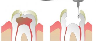
Sometimes a large part of the tooth remains, as they say, “behind the scenes”, and the dentist simply cannot see it. Then computed tomography comes to the rescue, which allows you to examine the hidden area and prevent unpleasant consequences for the patient.
Until recently, the “gold standard” for instrumental diagnostics was a panoramic image. Today there is a more advanced method in which the maxillofacial area is scanned using computer technology.
Unique visualization method
A computed tomograph produces a high-quality three-dimensional image of an individual tooth, maxillary sinuses, one or both jaws. Unlike standard panoramic images, a 3D tomogram allows the doctor to see the desired anatomical structures in a virtual section, from any angle.
During the procedure, the doctor can enlarge, rotate and study the maxillofacial area of interest at the required angle, which is unrealistic with conventional radiography.
Computed tomography is an integral and primary stage of examination before implantation. It allows you not only to assess the condition of bone tissue, but also to measure its height, width and density. Moreover, three-dimensional beams help to choose the optimal method for installing implants through preliminary virtual surgery.
3D CT is a multi-purpose and indispensable diagnostic tool that makes it possible to avoid many medical errors and complications. Thanks to this examination, the quality of treatment increases significantly and eliminates unnecessary traumatic operations.
Indications
Computed tomography is prescribed to identify:
- hidden carious lesions;
- defects in the structure of the jaw and dentition;
- fully or partially unerupted teeth;
- dystopic dental units with incorrect location or direction of growth;
- supernumerary teeth;
- damage to the dentition due to jaw fractures and other injuries;
- pathologies of the temporomandibular joint TMJ;
- tumors, cysts and other neoplasms in the jaws;
- condition of periodontal and periodontal tissues in case of gum disease, inflammation in the root area;
- number of roots, canals of teeth;
- cracks in the roots of teeth;
- features of the structure of bone tissue before jaw surgery (installation of implants, bone augmentation).
A 3D photo must be taken before dental implantation. The fact is that the jaw bone is clearly visible on a regular x-ray, but it does not allow assessing the soft tissues. On a three-dimensional tomogram, you can see in detail not only the bone, but also the nerve of the lower jaw, as well as blood vessels.
A 3D tomogram is much more informative than a panoramic image or targeted photographs of all teeth.
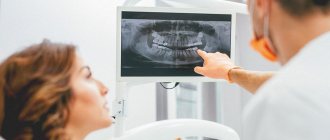
CT scan of the jaw: what it shows, how it is done, radiation doses
Dental computed tomography is an important part of diagnostic procedures in the dental field. This is a very informative method that allows you to see the structural features of the jaw, dentition, and also identify pathological processes. Previously, X-rays were used for this, but CT is a more advanced diagnostic method that allows you to get the most accurate picture.
3D dental diagnostics are carried out using a tomograph. In one procedure, you can obtain detailed information regarding the condition of the dentition. The main feature of this method is a three-dimensional image that allows you to see the object under study in full. This is extremely important for making an accurate diagnosis and drawing up a detailed treatment plan.
Content
- The main advantages of dental tomography
- Indications for CT scanning
- Main contraindications
- Is it harmful to have a dental CT scan?
- How often can a dental CT scan be done without harm to health?
- Basic rules of preparation
- How are dental CT scans done in specialized clinics?
- What does this procedure show?
- Computed tomography after implant installation
- Which is better - CT or X-ray?
- Computed tomography of the jaw in St. Petersburg
The main advantages of dental tomography
Compared to other diagnostic methods, computed tomography has the following advantages:
- The entire procedure lasts no longer than 1 minute.
- High level of information content.
- CT is safe for the patient’s health, so it can be performed repeatedly if necessary.
- The high quality of the image in the image allows you to clearly see the structural features of bone tissue.
- The radiation dose received is minimal.
At the end of the procedure, the resulting 3D image is recorded on a CD with universal software, which is installed automatically on the attending physician’s computer for the purpose of further diagnosis.
Indications for CT scanning
The information obtained after a CT scan is necessary to solve a number of problems in the field of dentistry.
The procedure is carried out in the following cases:
- Damage to the jaw after minor injuries. It is with the help of CT that dislocations and fractures can be determined.
- Detection of pathologies in the structure of the dentition. This procedure is commonly used before any type of orthodontic correction.
- Drawing up a computer model of the jaw for the manufacture of an individual brace system.
- As a preparatory procedure before the upcoming operation.
- Presence of neoplasms in the jaw.
- Diagnosis of hidden caries.
- Complications in the dental canals.
- Monitoring the success of previous treatment.
A CT scan is performed before dental implantation, because based on a 3D image, a suitable implant is selected, its installation is carried out and the quality of the procedure is checked.
Main contraindications
During the procedure, the body is exposed to a certain dose of radiation, so computed tomography is not performed during pregnancy. As for women during lactation, the procedure is possible, but after its completion it is forbidden to breastfeed the baby for 24-48 hours.
It is prohibited to perform dental CT scans on preschool children. In this case, alternative diagnostic methods are used. The same applies to patients with a pacemaker.
Is it harmful to have a dental CT scan?
Computed tomography is based on the principle of passing x-rays, with the help of which a series of layer-by-layer images are produced. Accordingly, a certain radiation dose is present during a CT scan of the jaw. It depends on the number of images and the total area of study.
When examining the chest and abdominal cavity, this figure is 11-14 mSv. The critical level for one procedure is 50 mSv (the maximum permissible annual dose is 150 mSv). If this figure is exceeded, there is a high risk of developing cancer.
So is it harmful to have a CT scan of your teeth? The study of this area is considered the most gentle, because the radioactive dose during the procedure is only 0.1-0.3 mSv. Therefore, computed tomography is absolutely safe for the health of patients (except in cases of contraindications).
How often can a dental CT scan be done without harm to health?
Despite the fact that a dental CT scan exposes the body to a minimal dose of radiation, there is no need to overuse this procedure. The frequency of the procedure depends on the degree of need for this, but it must be taken into account that radiation can accumulate in the human body. Therefore, the standard frequency of procedures is no more than 2 times a year.
But there are times when it is necessary to exceed the permissible limit. Based on the permissible radiation exposure, CT scans of the face and jaw should be performed no more than once every 2-3 months.
Basic rules of preparation
There are no special requirements that must be strictly observed before tomography of the dental jaw. It is better not to eat for three hours before the procedure.
The remaining requirements are standard, as in the case of x-rays. You will need to remove all jewelry and objects containing metal. This is necessary to obtain the most accurate research results.
How are dental CT scans done in specialized clinics?
Compared to the old X-ray machine, the tomograph is much more compact. A CT scan can be performed lying down, standing, or sitting. The choice depends on the physiological characteristics of the person, his age, as well as the type of pathology itself.
In cone beam computed tomography, the patient's head is placed between two scanners. To ensure maximum immobility, clamps are provided for the jaw, chin and temples. To understand how a dental CT scan is done, we will describe the procedure in stages:
- The procedure is carried out standing, the platform for fixing the jaw, chin and temples is set in accordance with the patient’s height, and a protective lead vest is put on before the procedure. The head is placed on a special tomograph stand and pressed against the equipment rack. And in this position it is fixed by means of stabilization.
- After preparing the patient, the specialist turns on the tomograph. The moving part of the device rotates around the patient's head. During the entire procedure, about 200-300 images are obtained, which are immediately displayed on the monitor.
- During the procedure, the patient may hear noise - this is quite normal and there is no need to be afraid of it. When it subsides, you do not need to immediately remove your head from the platform or make other movements; you must wait until the x-ray technician reports the end of the study and asks you to leave the scanning area.
During the procedure, the patient does not experience pain or any discomfort. The only discomfort is the forced need to remain immobile. Also, for 10-20 seconds during the image, the patient can feel the heat emanating from the moving scanners.
CT scan of the jaw: what does this procedure show?
The resulting CT image of the teeth is a three-dimensional image of the jaw, which is obtained using hardware positioning and the use of a three-point fixation system.
Computed tomography of teeth allows you to clearly see all pathological processes and disorders in the structure of bone tissue. With this study you can see:
- Crowns and fillings.
- Condition of the paranasal sinuses.
- Location of canals, roots and unerupted molars.
- TMJ condition.
- Various pathologies of the jaw.
The high accuracy of the results obtained allows us to accurately determine the nature of the pathology. If a CT scan is performed for dental implantation, then layer-by-layer images allow you to get the clearest picture of the condition of the maxillofacial area in different projections. The specialist sees the maxillary sinuses, canals, TMJ and blood vessels.
This type of research is the most accurate source of information about the dental system. Only with the help of CT can you correctly plan the upcoming implantation procedure. If you do not use CT, there is a high risk of installing a smaller implant, which will lead to unnecessary stress on the bone tissue.
Computed tomography after implant installation
Quite often the question arises: is it possible to do a CT scan with dental implants? Unlike magnetic resonance imaging, when foreign bodies can produce a “phonic effect,” there is no such problem with computed tomography. Moreover, it is not only allowed, but recommended after implantation.
CT is necessary in the following cases:
- Identification of the reasons for the instability of the installed structure.
- If you need to determine whether the artificial root has taken root or not.
- Determination of the quality of implantation performed. This is especially necessary if computer modeling of the jaw was not carried out at the preliminary stage.
- Identification of complications hidden during visual examination.
If the patient has metal crowns installed, then preliminary consultation with a specialist is necessary. The fact is that metal elements can create unwanted optical effects in images, which will complicate diagnosis and diagnosis.
Which is better - CT or X-ray?
Computed tomography is considered one of the most informative diagnostic methods used in dentistry. Using this method, it is possible to obtain a multilayer image of the area under study. A 3D image is much more informative compared to a conventional X-ray, which only provides a planar image.
From the point of view of affordability, x-rays are preferable, but they do not always provide an accurate picture of the condition of the dental-maxillary system. As practice shows, if a CT scan is performed, then in 99% of cases it is possible to accurately determine the cause of toothache and other symptoms.
Computed tomography of the jaw in St. Petersburg
If the doctor has prescribed a CT scan for you, but you don’t want to wait and sit in lines, then X-ray will offer you to undergo this study on their newest tomographs. The procedure takes no more than a minute, is absolutely painless and effective.
Today we told you how a CT scan of the jaw and teeth is done without negative consequences for your health. You can also record the examination results on a CD, send them by e-mail and make an online appointment. You can find out more detailed information by phone.
Sign up for a study by phone
+7 (812) 332-52-54
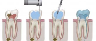
CT scan before implantation
Diagnostics using a computed tomograph before installing implants allows, first of all, to determine whether implantation is necessary at all. The image will give a complete picture, and the doctor will see where the teeth are missing, whether there are problem units, and whether they can be cured.
The 3D tomogram will show:
- hidden carious cavities;
- unerupted and “extra” teeth, which may interfere with the installation of artificial pins;
- properties of roots, canals - curved, narrow and long canals require a special approach, which should be taken into account before implantation;
- bone dimensions in height and width, on the basis of which the type and size of the implant is selected;
- condition of bone beams, partitions, voids in the jaw bone;
- the presence of inflammatory processes in the root area - cysts, granulomas, abscesses where implants are planned to be installed. All this needs to be treated or removed before surgery;
- inflammation in the paranasal sinuses and lacrimal ducts, which can become a temporary obstacle to implantation;
- density, size, inclination of the alveolar process, thickness of the cortical bone layer, taking into account which the optimal type of artificial pin is selected;
- the physiological structure of the maxillary sinuses, the mandibular canal to determine the angle of inclination of the implant rod;
- defects and anomalies in the structure of the dentofacial apparatus;
- quality of installation, strength of fixation of implants after surgery for their implantation;
- severity and nature of traumatic injuries in fractures.
Based on the results obtained, a virtual operation to install the rods is performed. The appropriate size of the titanium pin is selected, its inclination and the point of implantation are determined, bypassing the anatomical structures. Thus, the final outcome of implantation is modeled.
Next, the tomographic data is loaded into the computer, and the program creates a three-dimensional model. The patient’s personal surgical template is printed on a 3D printer - an overlay with guide holes for inserting rods.
During implantation, the template is placed tightly on the gums, and the placement of the pins is carried out with extreme precision.
Multispiral or cone beam?
A multispiral tomograph performs layer-by-layer scanning of an object along a spiral trajectory caused by the continuous movement of the table and the X-ray tube relative to each other.
Most often, MSCT - multislice computed tomography - is used in maxillofacial surgery for facial injuries and pathologies of the temporomandibular joint.
In dental practice, especially when planning implantation, this method is not widely used due to insufficient data accuracy. Since the patient lies down during the examination, the jaw connection is distorted.
In addition, the radiation level of MSCT can reach 1000 μSv, which is unacceptable, since implantological treatment involves more than one procedure over several months.
Cone beam CBCT is a more modern, accurate and safe method compared to MSCT. Its radiation exposure is less, about 25-50 μSv, which makes it possible to carry out the procedure several times a year.
Carrying out dental tomography
No preparation is required for a dental CT scan. The study is not carried out during pregnancy.
The research procedure itself does not cause any difficulties for patients. Dental computed tomography is performed in a standing position. It is necessary to place the chin on a special stand. After which the protection is put on. The picture is taken by the rotating part of the device in one revolution. Do not swallow during the examination.
You can get a dental computed tomography scan in Moscow at the Dental Center of Polyclinic No. 5 of JSC “Family Doctor”.
Sign up for diagnostics Do not self-medicate. Contact our specialists who will correctly diagnose and prescribe treatment.
3D CT or panoramic image?
An orthopantomogram allows the doctor to assess the condition of the teeth, root canals, and soft tissues. With its help, hidden inflammations, abscesses and abnormally located teeth are revealed.
However, a panoramic photo does not give a 100% accurate picture; the error is about 20%. Even a slight shift causes the focal spot to shift, and the image is compressed or stretched.
Due to the difference in the refraction of X-rays by tissues of different densities, it is impossible to assess the properties of the cancellous bone layer, since it is simply not visible behind the denser periodontium.
A two-dimensional orthopantomogram is, in fact, an auxiliary technique that gives a general idea of the condition of the oral cavity and identifies mainly obvious pathologies. It does not show the configuration and structure of the alveolar process at the desired level.
The advantage of a three-dimensional 3D tomogram is that it produces not one flat photo, but several consecutive images from different angles.
The doctor sees and evaluates all necessary objects located at any depth, from all sides and at different angles.
Contraindications for 3D dental imaging
The radiation exposure of CT scans ranges from 0.045 to 0.06 mSv. This is relatively little, given the recommendations of SanPiN, which indicate the upper possible threshold of radiation exposure for research purposes equal to 1 mSv per year. However, if you compare a CT scan with a dental x-ray, where radiation occurs in the range of 0.002 - 0.003 mSv, a 3D photograph of teeth no longer seems so harmless. In this regard, there are a number of contraindications to CT scanning. If we are talking about simple tomography, then, as a rule, it is not done during pregnancy, especially in the first trimester. The doctor will always consider the need for radiation exposure for a pregnant patient based on the balance of benefit to the mother and risk to the fetus. If tomography with contrast is necessary (for better visualization of soft tissues and blood vessels, it is rarely used for CT of the jaw), then the number of contraindications expands. It should not be given to pregnant women, nursing mothers, or to persons suffering from thyroid disease, severe diabetes mellitus, renal failure, or an allergy to iodine. If you have any allergies or have ever experienced drug intolerance, be sure to tell your doctor.
How to make a 3D tomogram
Usually the procedure is performed standing, the patient bites a small flat plate with his teeth and stands without moving for 15 to 30 seconds. The device makes several rotations around the head, managing to take about two hundred pictures in various projections.
In 10-15 minutes, the information is processed and transferred to electronic media.
We invite you to make a three-dimensional tomogram in our clinic using the latest generation dental tomograph. Sign up for the procedure online or by phone at a time convenient for you.
How is the research going?
The examination is carried out in 1-3 minutes:
- The doctor takes the patient to the CT scanner and helps fix the head in the desired position. Depending on the design of the tomograph, the examination takes place while sitting or standing.
- A small plate covered with a disposable cover is placed in the mouth, which fixes the position of the jaws.
- The scanning arc of the tomograph makes one revolution around the patient's head. The exposure time to X-ray radiation does not exceed 20 seconds.
The specialist writes the obtained data onto a CD and, if necessary, prints it onto film.