Safety of X-ray diagnostics
The standard of safe radiation exposure is regulated by the federal law “On Radiation Safety of the Population”. Article 9 of this law “State regulation in the field of radiation safety” states that the average effective dose for the population per year is 0.001 sievert (1 millisievert). In this case, the dose received during one cone-beam computed tomography is 0.09 millisieverts. Accordingly, at least 10 CT scans of teeth can be safely performed per year.
For comparison, during air travel the radiation dose is on average 0.08 millisieverts. The dose of so-called natural radiation is 2.4 mSv. It consists of the effects of cosmic and solar rays, the influence of atmospheric radioactive atoms (radon), radiation from soil and building materials, and food radionuclides. Thus, performing a dental CT scan is absolutely safe for your health.
Advantages
CBCT has a number of advantages over other diagnostic methods:
- Quick examination of the pathological area. A single rotation of the frame reduces the radiation dose by up to 15 times. Radiation exposure – up to 50 µSv.
- Scanning step – 0.125 mm.
- There are no prerequisites for the patient's position.
- The design is mobile and open.
- Creation of a layer-by-layer 3D model.
- A single planar sensor is used to increase image resolution. For comparison, with spiral CT there are a thousand point detectors.
- The image quality is 5 times higher than in MSCT.
- The 3D model is used to obtain a sample for implantation. A copy is created on a 3D printer.
- Special software that allows you to select the angle and scanning mode.
The procedure for performing CT scans of teeth in Moscow at Dental Guru clinics
Computed tomography of the jaw, Moscow. Any Dental Guru clinic has a full range of X-ray diagnostics, including a computer tomography machine. No special preparation required. Before the examination, all metal jewelry and items of clothing (earrings, chains, piercings in the dentofacial area, hairpins) are removed. A lead vest or apron is worn to protect against X-ray radiation. To fix the head, the chin rests on the stand and the forehead on the bracket. During the examination, the sensor scans the dentofacial area. At this time, you should refrain from any movements, including swallowing. The patient sits or stands and the examination takes 30 seconds. At Dental Guru clinics, cone beam computed tomography is performed using a multifunctional Rayscan device.
The essence of the procedure
During cone beam tomography, the machine moves around the head. On one side, the device is equipped with an X-ray source, on the other, with an X-ray receiver. The receiver processes the received data.
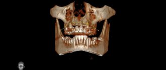
The tomograph is connected to a computer, where a special program creates a 3D image of the jaw. As a result of the study, the tomograph takes many pictures - on average 500. This allows the doctor to see a specific area of interest from different angles and projections.
Thanks to high accuracy and computer processing, the image provides the specialist with the most reliable and complete information about the condition of the tissues.
Implant planning using CT
A CBCT scan is necessary before dental implantation. Three-dimensional examination reduces the risk of the procedure, significantly improves the quality of implant installation, and shortens the patient’s rehabilitation period. CT scan shows the condition of the bone tissue, possible inflammatory processes, the location of the maxillary sinuses, nerves and blood vessels, helps to create a model of the implant, calculate the depth of its immersion in the bone and the angle of inclination, and the optimal physiological load on the implant.
Why is 3D examination important before implant placement? An artificial root will stand long and firmly if it is surrounded on all sides by bone tissue. And here an individual approach is needed. The structure of bone tissue varies in shape, width, height, density, and possible defects. For longevity, it is important to choose a site where bone volume and density are sufficient. You need to choose the angle of inclination for acceptable aesthetics. In order to determine the required installation depth, you need to know the location of the nerves and blood vessels. Thus, it is possible to obtain a surgical navigation template for implantation for the most accurate virtual planning.
Performing a CT scan with a radiopaque tray when planning a surgical template
Most patients who come for implants already have fillings, crowns, veneers or other dental restorations. All these materials can produce additional glare during CT scanning, so-called artifacts. To minimize them and ensure the accuracy of the CT examination, this procedure is performed using special radiopaque trays. They can be standard (if there are supporting teeth) or individual (when there are very few or no teeth and guidelines are needed for further work).
In these cases, special radiopaque trays are placed in the oral cavity, a CBCT (cone beam computed tomography of the jaws) is performed, then the trays are removed and sent to a dental laboratory. It is thanks to this type of research that it is possible to achieve accurate results and guarantee that the implant is installed in the area in which its location will be best. If implantation using surgical templates is planned, a CT scan with a radiopaque tray must be done. Before implantation with immediate loading, this study allows the fabrication of a temporary prosthesis to be worn before surgery.
Contraindications and prohibitions
When compared with traditional radiography, the quantitative exposure of the body to X-rays during multispiral immersion increases by 20 times. If you exceed the permissible number of studies, there is a risk of developing oncopathological processes. This is especially true for young patients, whose cellular structures are in a stage of constant growth and development.
The use of radiographic diagnostic methods during pregnancy in women is absolutely prohibited. The decision to prescribe this type of screening can only be made in the case of a life-threatening anomaly, when the risk of death is higher than the risk of harm from hardware diagnostics. The conditional threshold that removes this contraindication is the minimum age of the patient, which must exceed 14 years. Regarding the frequency of access to multispiral scanning, medical experts recommend not to exceed the schedule, which includes a one-time examination per year. An additional condition is monitoring of excess of permissible exposure over the past five-year period. During vascular tomography, color enhancement in the form of contrast is often used. The difference between MSCT and CT without contrast, the enhancing effect is achieved by intravenous administration of iodine solution. A contraindication is immune rejection to iodine-containing drugs. A true allergic reaction to the product is extremely rare. Associated unpleasant consequences of intravenous administration of the solution are:
- pressure drop;
- increased heart rate;
- fainting state;
- urge to vomit.
With minor manifestations of the reaction, these symptoms are considered to be the norm. The drug is administered under the careful supervision of a medical specialist. If negative consequences occur, urgent restorative manipulations are carried out.
If you do not do a CT scan before implantation, there may be:
- Loss of sensitivity and pain in the teeth and jaw.
- Injury to the gums due to incorrect calculation of the permissible load.
- Bite defects.
- Loosening and loss of the implant.
- Increased costs to correct problems, including installing a new structure.
This cannot happen in Dental Guru clinics. We care about the interests of our patients. All clinic specialists have extensive experience and high qualifications, confirmed by diplomas and certificates. CT scans of the upper and lower jaw are equally important.
Use of CBCT in dentistry
Such an accurate, fast and safe research method is widely used in surgery, dentistry and otolaryngology. In particular, CBCT in dentistry is used for:
- assessing the condition of root canals and pathological processes around them;
- studies of changes in periodontal tissue;
- assessment of traumatic injuries to teeth (except root fractures);
- diagnostics of internal destruction of the tooth root structure;
- recording changes in facial structure and bite after wearing braces and prosthetics;
- examination of the condition of the jaws before surgical treatment and implantation;
- diagnosis of other congenital and acquired anomalies of the maxillofacial region.
The role of full diagnostics and modeling capabilities - especially in orthodontics and implantology - is difficult to overestimate. A three-dimensional image without dimensional distortion will allow the doctor to determine the presence of inflammatory processes and seals that may interfere with the implantation of implants, and to accurately calculate the configuration of the supporting structure.
Accurate diagnosis is especially important during immediate dental implantation, which guarantees rapid healing and the ability to load the jaw almost immediately after installation of the implants.
In Dr. Kizim’s clinic, at an affordable price, you can do cone tomography of a segment (3-4 teeth), one or both jaws, as well as the temporal bone and middle ear (the results of the study are recorded on a CD).
Professional dentists who regularly improve their skills in the field of research methods will conduct a full diagnosis of the condition of the jaw and obtain a 3D model of the segment under study.
Computed tomography of the upper jaw
To diagnose the condition of the maxillary sinuses before implantation, it is important to do a CT scan of the upper jaw. After tooth extraction, thickening of the mucous membrane in the maxillary sinuses often occurs; there may also be manifestations of sinusitis, a tumor process, the presence of a cyst or polyp. A CT scan of the jaw will show whether there is perforation of the maxillary sinus due to the protrusion of the root or crown of the tooth. The term “odontogenic sinusitis” means that the cause of the disease is in the tooth. The bony septum between the oral cavity and the maxillary sinus is quite thin, so infection from dental tissue can cause sinusitis. Therefore, for implantation to be successful, it is necessary to promptly identify and treat possible diseases and eliminate the inflammatory process.
When is MSCT indicated?
Computer diagnostics is recommended in the following cases:
- stenosis of vascular canals;
- narrowing of patency;
- calcifications on the walls;
- pathological expansion (aneurysm);
- congenital developmental anomalies;
- presence of thrombosing objects;
- inflammatory processes;
- confusion and tortuosity of blood vessels;
- violation of the integrity of the walls, internal hemorrhages;
- aortic dissection;
- pulmonary hypertension;
- occlusal disorders;
- assessment of the degree of blood supply to oncological and benign tumors.
Computed tomography of the lower jaw
A CT scan of the lower jaw, as well as the upper jaw, shows the condition of the teeth and bone tissue: possible diseases, inflammatory processes, granulomas and cysts. A CT scan of the jaw is important to identify hidden processes and timely initiation of treatment. A CT scan of the upper and lower jaw is usually done at the same time, but some clinics can do a CT scan of the upper and lower jaw teeth separately. The lower jaw bears the main load when chewing and speaking; chronic subluxation of the lower jaw is quite common. CBCT of the temporomandibular joint helps to make a diagnosis and distinguish subluxation from dislocation and fracture.
Types and results of examination
CT is recognized as an indispensable diagnostic tool in the treatment of pathologies in various segments of dentistry:
- In therapy - diagnostics of soft tissues surrounding the teeth, examination of passages, determination of the degree of root defect and treatment results.
- In surgery - identifying the source of inflammation, determining its size and location for bone collection for implantation. The doctor determines the presence of tooth remains after extraction and other surgical procedures.
- Orthopedics – objective assessment of supporting structures and ancillary units. The doctor can localize complications and select a treatment plan.
- Orthodontics – determination of circumstances that impede the quality installation of the structure.
There are a number of restrictions on CT scanning:
- Pregnancy and lactation.
- Mental disorders.
- Kidney failure.
- The patient's inability to remain still during the procedure.
- Lack of vital necessity for children under five years of age.
During the procedure, the patient must independently hold his head still.
CBCT is recommended if:
- Injuries and mechanical damage to teeth and jaws.
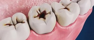
- Carious cavities hidden from other examination methods.
- Pathologies in the development and location of dental units.
- Indications for implantation and surgical interventions.
- Disorders of the maxillary sinuses.
- Complications after intracanal treatment.
It is advisable to use a diagnostic method to determine further tactics of dental treatment and its results.
Diagnosis of pathology of tissues surrounding teeth
In the presence of pathological changes in the periapical tissues, LCCT allows one to accurately diagnose the site of inflammation and identify a foreign body.
The examination is performed to detect cortical abnormalities and visualize the lingual and labial side.
Traumatic tooth decay
The doctor receives a 3D model when examining mechanical injuries:
- local defect or complete destruction of the root;
- mobility of units due to injury (luxation);
- tooth shortening;
- fracture of the alveolar process of the upper dental jaw.
The device is not capable of detecting root fractures in horizontal and vertical planes.
Dental examination of root damage
Root damage can be internal or external. External defects are diagnosed visually. Destruction of the unit from the inside is revealed by CT scan. The equipment builds a three-dimensional picture with changes in the transparency of the bone material.
CT algorithm
There is a method for a step-by-step description of targeted and panoramic dental 3D CT obtained as a result of the study. The detailed development of the algorithm provided dentists with the practical opportunity to study the image in depth and consistently.
Step-by-step algorithm for CT scanning:
- Study of a radiograph taking into account sharpness, distortion, contrast, and capture width.
- Assessment of bone tissue structure:
- condition of interdental septa;
- degree of change in the intraosseous structure;
- identification of impacted units;
- temporomandibular joint;
- paranasal and maxillary sinuses;
- mandibular canal.
- Determining the feasibility of additional diagnostic measures: targeted, extraoral radiographs.
- Study of dental units:
- identification of caries, fillings and their integrity;
- condition of roots;
- examination of root passages;
- periodontal fissure;
- examination of permanent orthodontic and orthopedic devices.
- Diagnosis of pathologies. The dentist evaluates the pathologically changed areas, their dimensions, location and nature.
Panoramic photo, CT scan of teeth in Moscow, price
For a CT scan of the jaw, the price in Moscow starts from 3,500 rubles. If you search online for “computed tomography of teeth price Moscow” or “computed tomography of jaw price”, then the average price of this examination in the search will be approximately 4,500 rubles. Dental Guru is currently running a unique promotion - a dental CT scan costs only 1,900 rubles. If the question of where to get a CT scan of a tooth is relevant to you, come to Dental Guru. When referred by a doctor, we guarantee non-interference in the patient’s treatment.
The doctor may also order two-dimensional examinations, such as spot and/or panoramic images. The targeted photograph shows the condition of 3-4 adjacent teeth. An OPTG (orthopantomogram) shows the condition of both jaws, including soft and hard tissues, malocclusions, impacted teeth and tumors, but you need to remember that this is a two-dimensional (single-view) image.
Execution steps
- The patient puts on a protective apron and takes a comfortable position - sitting or standing.
- The patient's head is securely fixed in one position using immobilization means. For maximum image clarity, the patient must remain motionless during the entire procedure.
- The specialist launches the program. The cone tomograph makes rotational movements around the axis of the head.
- The information received is sent from the tomograph to a special computer program.
- A three-dimensional 3D image of the jaw is formed.
- The entire examination takes 30-60 seconds
Commentary by the chief physician of the Dental Guru clinics
To understand the clinical picture, it is necessary to make an accurate diagnosis. Dental Guru dental clinics have all the necessary diagnostic equipment for x-ray diagnostics. These are multifunctional devices with the ability to take pictures of the temporomandibular joint, cephalometry, and a digital radiovisiograph for targeted imaging. We take a panoramic photo of the teeth, CT scan of the upper and lower jaw, the price is now unprecedentedly low. We are always glad to see you in our clinics. Call and make an appointment with specialists!