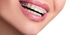
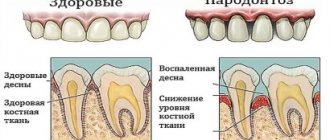
A deep bite is a developmental anomaly of the oral cavity in which the upper incisors almost completely overlap the lower ones. It not only distorts the proportions of the face, but can also cause speech disorders, increased tooth wear and trauma to soft tissues. Pathology can be detected both in early childhood and in adulthood. Treatment depends on the severity, but most often involves braces, plates, or orthodontic surgery.
Manifestations of distal bite in the mouth
Closing of lateral teeth during distal occlusion.
Distal occlusion is manifested in the mouth by the closure of the upper and lower teeth according to the “second class” (violation of the relationship between the projections of the antagonist teeth of the upper and lower jaw).
In the frontal segment (at the level of the front teeth) there are two closure options (the so-called first or second subclasses).
In the first case, a “sagittal gap” is observed, which reflects the discrepancy between the dentition in the sagittal plane (anterior-posterior direction). And the upper incisors are in protrusion (inclined forward). In the second option, the upper front teeth are tilted back (into retrusion). With the formation of a deep bite (when the lower teeth are not visible from under the upper ones).
Sagittal chain with distal occlusion. 2 class 1 subclass.
Distal occlusion. 2nd class 2nd subclass.
Thread lifting – tightening the skin of the neck
This effective treatment will appeal to those who are embarrassed by double chins and sagging skin. Thread lifting is performed by introducing polymer threads with notches that are well retained in the tissue. After placing the threads, the doctor lifts them up, cutting off the ends that are too long. Anesthesia allows you to make the procedure as comfortable as possible.
Thread lifting
Subsequently, the threads will be “embedded” in the tissue, stimulating tissue renewal and the growth of collagen protein. Rejuvenating and restorative processes will begin. The results of thread lifting last for 2–3 years.
Causes of distal bite (distal occlusion)
They are almost always (in the vast majority of cases) skeletal...
- Posterior position of the lower jaw. This is the most common cause of distal occlusion
- Small (short) lower jaw.
Many people write that the cause of a distal bite may be an overdeveloped (excessively large) upper jaw. They say that this is why there is a “gap” between the front upper and lower teeth (sagittal gap)... Many years of observations and our experience have shown that this is not so. With any anomaly of occlusion, there can be no talk of any OVERdevelopment. Everything, on the contrary, is UNDERdeveloped.
The lower jaw ends up in a posterior position (the first reason) only because the lower teeth bump into the upper teeth while the upper jaw is shortened and narrowed (generally underdeveloped). Or, when the upper incisors are “wrapped” back, into retrusion (second subclass). In addition, with distal occlusion, the upper jaw is always narrower than the lower jaw. Which, by the way, is another reason for blocking the lower jaw in the posterior position. And this same “blocking” does not allow the lower jaw, which is caught in the trap, to develop normally. Here is the second reason for distal occlusion - underdevelopment of the lower jaw.
There is also a “dental” reason for distal occlusion. Or more precisely, the dental cause of closure is “second class”. This is when mesial (front) displacement of the upper lateral teeth occurs. And then, even with a normal relationship of the jaws in the sagittal plane, the closure of the lateral teeth (molars) will also be in the second class. True, this is not a true distal occlusion...
A “dental” cause of the second class may complement or may camouflage true distal occlusion. Therefore, it will not be possible to figure out what is the reason for the closure in class 2 visually (by eye). Diagnostics required.
Introduction of lipolytics – removing excess fat, creating an ideal jaw line
This procedure is performed to reduce the amount of fat under the chin. Each session lasts 15-20 minutes and includes a series of injections. The active ingredient of the administered drugs is deoxycholic acid, which has the property of destroying adipose tissue.
After administration, the lipolytic immediately begins to destroy fat. The result is a noticeable reduction in fullness under the chin and a more defined jawline.
The number of injections required varies from person to person, although most patients require three to six treatments for best results. Improvements will gradually increase over another 12 weeks and will last for a very long time.
Diagnostics
The main goal of any diagnosis is to identify the cause of the problem. And make a diagnosis. Likewise, the diagnosis of distal occlusion is designed to identify the cause of closure in the second class. In order to separate the flies from the cutlets during treatment, so to speak. That is, dental causes of closure are in the second class from non-dental (maxillary, skeletal).
The main diagnostic methods are:
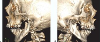
- Analysis of TRG (teleradiogram) of the skull in a lateral projection. It allows you to clearly determine all the skeletal (jaw) nuances of the problem. Both with the position of the jaws and with their sizes.
- Analysis of plaster models of jaws (for this, casts are taken). It allows you to clarify the “dental” nuances: the size of the teeth, the length and width of the dentition, the features of the relationship (including closure) of the upper and lower teeth
Analysis of TRG (teleradiogram) of the skull in a lateral projection.
Analysis of plaster models of jaws.
It is not recommended to fall below this diagnostic “bar”. Below - just by eye. But this, as we understand, is not the best option.
Diagnostics is the first and key step in the treatment of distal occlusion. Do you want to know why it’s impossible to do without diagnostics?
Evolution of tooth size
Our distant ancestors, who lived about 100,000 years ago, had very large teeth - about twice as large as those of modern humans. This was a vital necessity. With the help of teeth it was necessary to gnaw animal bones and tear raw meat. But with the development of the brain and changes in diet, they gradually decreased. Scientists have calculated that every thousand years, 1% of their surface “gone” (although it is possible that over the past centuries this process has accelerated significantly).
The changes affected not only the overall size of the teeth, but also their purpose. The largest tooth in modern humans is the first upper molar: in the process of evolution, the main functional role has passed to it from the second upper molar. The fangs, which were no longer so important for holding food, were transformed the most.
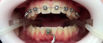
Errors in the treatment of distal occlusion
The first, main mistake is that many try to treat distal bite with one device. For example, only with braces (this is the most popular option). But braces, although a cool device, are not for skeletal problems. The level of hardware competence of the braces system does not extend further than “to place the teeth on the jaw.” And distal occlusion, as we have already said, is a purely skeletal problem. Jaws and posture are to blame. And braces are not capable of solving problems with posture or jaw problems. After all, braces cannot change the position or size of the jaw. No matter what the would-be doctors promise...
And therefore, such videos are just an advertising gimmick.
Or rather, a convenient “hook” that catches gullible patients. Convenient because it coincides with the desire to do everything quickly and efficiently... But it doesn’t coincide with reality.
And braces cannot be the main device in the treatment of distal occlusion.
Braces are the wrong choice when treating distal malocclusions. In any case, you definitely shouldn’t start with them. Braces, as mentioned and shown above, can be effectively used only at the final stage of treatment of distal occlusion. All major “feats” must be accomplished before working on the teeth. Before using braces. And they are accomplished at the level of the jaws and skeleton (posture). Otherwise, there will simply be nothing to “clog” (close) these teeth (read with braces). See “Fundamentals of the therapeutic concept of “Ortho-Arteli”.
The second common mistake in correcting distal occlusion is treatment with the removal of upper premolars (4 or 5 teeth).
And this mistake is much more serious. Because it can have the most dire consequences. See "Orthodontic treatment with tooth extraction."
After all, as we said above, the main causes of distal occlusion are skeletal. And most often the lower jaw is to blame. And if so, then a reasonable question arises: if the lower jaw is to blame, what does the upper teeth have to do with it (their removal). Where is the logic?
In general, all errors in the treatment of distal occlusion in an adult are usually associated with insufficient diagnosis. Which entails a misunderstanding of the reasons for the occurrence of distal occlusion in each specific case.
For example, the “dental” cause of distal occlusion is very often confused with the “skeletal” (jaw) cause. We remind you that in the first case you need to move the lateral teeth back. And in the second - to push the lower jaw forward and develop the upper one. And if you get confused, if you don’t figure it out... Start distalizing the upper teeth, or worse, remove them when it is necessary to advance the lower jaw... Or vice versa. It is clear that nothing good will come of it.
The third mistake is when, without understanding what’s what, what exactly is the cause of distal occlusion, but realizing, however, that the problem cannot be corrected with braces, they try to solve the problem surgically. Using orthognathic surgery. Through osteotomy of the lower and upper jaws.
This is where I would like to dwell in more detail. And explain something. Why do orthodontists send to a surgeon to “treat” a distal bite? They have already said: they understand that braces will not cope. But they don’t understand how to treat it without braces. They don’t understand because in dentistry in general, and orthodontics in particular, there are a lot of prejudices regarding the possibilities of moving the lower jaw forward and developing the upper jaw. And these, as we saw above, are the main (“key”) points in the treatment of distal occlusion.
Most orthodontists simply do not believe in this (that it is possible in principle). Because someone (even a “great one”) said two hundred years ago that this cannot be done. Time has passed. Everyone has already forgotten why it is “impossible”. But no one even bothered to check why, in fact, it was impossible...
And therefore, those doctors who blindly believe in these moss-covered “dogmas” simply do not see an alternative. And for them there are only two extreme poles in the treatment of distal occlusion: braces and surgery. That's all.
During a direct examination by a specialist, you will be able to find out your exact diagnosis, as well as receive a referral for diagnosis or a treatment plan.
And in our clinic we treat distal occlusion between these two poles. Using methods of maxillofacial orthopedics and craniodontics. Here are the ones: changing the position of the lower jaw and changing the size of the upper. And we get good results, without regard to the fact that someone once said something... We must always double-check what was said. We checked. And everything turned out to be somewhat different. Or rather, not at all the way they “frightened” us. You can also push it out. It can be developed. You just need to know how, because there are some “secrets” here. And we, at the Orto-Artel clinic, knowing these “secrets”, take advantage of these opportunities, successfully eliminating distal occlusion of any complexity.
By the way, if we are talking about surgical treatment of distal occlusion, then we need to determine when and in what cases surgery is generally appropriate. But the indication is basically the same - a small lower jaw, the so-called lower micrognathia, and the micrognathia is very pronounced.
We indicated this reason above as one of the skeletal reasons. I will add: the lower jaw should be VERY short. Literally ugly. Only then can this be resolved surgically. In all other cases, you can do without surgery. Proven and tested many times.
By the way... In our clinic, a good half of the patients have formal indications for surgical treatment of distal occlusion. But only a few got to the surgeon. Because formalities are formalities, but compromise solutions were found that satisfied everyone: both the patients and us. This is because we approach treatment not formally, but in essence. And we are guided by logic and common sense.
Well, as a special case of a short lower jaw, which cannot be corrected except by surgery, this is a small chin. If it is small and spoils the face, then even by pushing the lower jaw forward we will not get the aesthetics we want. Simply because orthodontists do not have the means and instruments (devices) to selectively influence the chin. It is impossible to stimulate the chin to grow. Only surgery.
Inferior micrognathia.
This is where the indications (and there is essentially one) for surgical intervention for distal occlusion end.
Going with a knife into the upper jaw - God forbid! Even if the upper jaw is indirectly to blame for the distal occlusion. More precisely, the upper jaw also, as a rule, suffers (dimensionally) with distal occlusion. The upper jaw is directly connected to the base of the skull. And such interventions can then backfire. Literally. And the most important thing is that there is no need to go there surgically. The upper jaw can be developed!!!
Massage – the hands of a massage therapist are able to create a new neck line
Various types of massage give good results, especially with the use of warming components that activate blood and lymph circulation in the cervical area. The massage therapist’s hands will break up fat deposits and strengthen the subcutaneous muscle.
Massage
An additional positive effect will be created by herbal and other natural ingredients included in the massage products. They will improve the condition of the skin and help remove wrinkles.
The number of procedures performed is selected individually. Improvements will be observed for another two weeks after the last massage session. In the future, you will need to periodically contact specialists to keep your neck muscles in good shape.
Prevention of the development of micrognathia and macrognathia
Let's talk about possible measures to prevent the development of maxillofacial anomalies. Since the influence of negative factors on the health of the child’s future dental system is especially active during the mother’s pregnancy and in the first months of the baby’s life, preventive measures must be followed even before the birth of the child and in the first year of life . To promptly prevent the development of dentofacial deformities, you should adhere to the following:
- carefully monitor the course of pregnancy, try, if possible, to avoid the risk of infectious diseases of the expectant mother;
- try to avoid long stays of a pregnant woman, mother and baby in an unfavorable ecological environment;
- eat a nutritious and varied diet and lead a healthy lifestyle during pregnancy;
- give up bad habits in the process of planning pregnancy, during pregnancy and breastfeeding;
- adhere to proper feeding of the baby (use an anatomically correct nipple and discard it in a timely manner; when artificially feeding, make sure that the bottle with the mixture does not put pressure on the alveolar processes);
- if possible, stop bad habits (sucking and biting fingers and cheeks, tongue, various objects) that provoke deformations of the dentition, disturbances in the growth of the jaw bones;
- observe the timing and sequence of teething, monitor the development of speech;
- promptly identify and eliminate congenital pathologies in the form of a shortened frenulum, cleft lip, palate, etc.;
- wean the child from sucking fingers, objects, etc.;
- conduct myogymnastics with the child to prevent disorders of respiratory, swallowing, and speech functions; ensure that the child does not breathe through his mouth;
- timely treatment of baby teeth and periodontal diseases;
- adhere to preventive measures and promptly treat common diseases (rickets, diseases of the ENT organs, endocrine pathologies, etc.)
Thus, in the prevention and timely identification of risk factors that contribute to the occurrence of various dental anomalies, the most important thing is the active and careful participation of parents at all stages of the child’s growth and development.