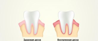
This is the name given to a pathological process manifested by excessive growth of gingival tissue, which often leads to the closure of a significant area of the tooth crown. Often, gum hyperplasia is not a consequence of an inflammatory reaction, and the pathological process does not involve any other tissues of the oral cavity (tongue, cheeks, etc.).
This pathological process is often accompanied by hyperemia, in which excess blood enters the tissues, leading to bleeding at the time of consumption of rough food, as well as pain reactions.
If gum hyperplasia is not eliminated in a timely manner, it can stop the process of replacing baby teeth with permanent ones, cause osteoporosis, and contribute to the disruption of the integrity of the partitions between the teeth. Among other things, this anomaly negates oral hygiene and provokes the appearance of various oral diseases.
Symptoms of gum hyperplasia
Gum growth in the initial stages may resemble swelling or tumor formation. Specific symptoms gradually appear:
- seals appear on the surface of the mucous membrane or under it, which gradually increase in size and may become more voluminous;
- The volume of the gums increases: they become wider, the gingival margin rises higher. At the same time, the tissues remain dense, there are no signs of edema;
- the interdental spaces are filled with gum tissue;
- the edges of the gums rise, covering the surface of the crowns;
- when chewing or brushing teeth, overgrown areas are injured and can become inflamed, after which their surface becomes compacted;
- the color of the mucous gradually becomes bright pink.
What symptoms can be used to identify hypertrophy?
You can see the increased size of the gums visually. Hyperplasia is a non-inflammatory disease that can occur in both adults and children.
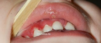
The first and most important symptom is the growth of gums, which seem to be on the tooth. This can be observed in the area of one tooth, between dental units, or throughout the entire jaw. In this case, the mucous membrane may not change its color, but may become bright red, and even turn a little blue. Patients with hypertrophy speak not only about the aesthetic disadvantage of the periodontium, but also about the occurrence of difficulties when chewing food, and pain when pressing on the gums.
Pathology can occur not only in humans, but also in cats and dogs. However, hyperplasia in animals is not contagious and is caused by other reasons.
Types of gum hyperplasia
Periodontal tissues can grow in different ways, involving different areas of the gums. There are several types of hyperplasia:
- localized: the growth of new cells occurs in a limited area, next to one tooth or group of teeth (only on one or both sides);
- generalized: the gums grow over the entire surface on one or both jaws;
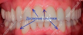
- papillary: new cells are formed only in the area of the gingival papilla, the pathology does not affect other gingival tissues;
- marginal: the gingival papilla and the area of the gingival margin grow;
- diffuse: growth of both marginal and attached gums occurs (an increase in periodontal tissue to the entire height from the gingival margin to the mucogingival border).
Hyperplasia may be limited. The increase in tissue in this case is similar to a tumor, strictly isolated, and has a wide base or pedicle. It looks like a growth or bump.
Reasons for appearance
Gum overgrowth can be a consequence of taking certain medications. There are a number of studies that have proven that periodontal hyperplasia is a consequence of taking antibiotics and other medications. In all the cases studied, either the dosage of the medications was violated, or the patient self-medicated, that is, he prescribed medications for himself, without consulting a specialist.
Other reasons include:
- genetics: in this case, the pathology manifests itself already at a young age, as a rule, at the time of the eruption of milk and then molars;
- hormonal changes in the body during pregnancy;
- blood diseases;
- incorrect structure of the maxillofacial apparatus.
Causes of hypertrophic gingivitis
Hypertrophic gingivitis can act as a separate pathology or be one of the complications of catarrhal gingivitis.
At the same time, factors that increase the risk of its occurrence and development are:
- occlusion disorders;
- twisting or crowding of teeth, their supernumerary;
- presence of dental plaque;
- improper fastening of the bridle;
- poor hygiene practices;
- leukemia;
- use of inappropriately selected prosthetic structures;
- injury to gums due to incorrectly installed fillings;
- hormonal imbalance (during menopause, during gestation or puberty);
- endocrine disruptions;
- hypovitaminosis;
- taking certain medications (immunosuppressants, antiepileptic drugs, oral contraceptives and others);
- lack of a competent approach to the treatment of dental diseases.
Diagnostics
Gum hyperplasia is diagnosed by a periodontist or dentist. Initially, the patient is offered to undergo an X-ray examination. This allows you to diagnose the disease more accurately. In addition, a sample of the enlarged gum tissue is sent for histological analysis.
- Cytological examination of a smear (scraping) from the vaginal dome (in the absence of the cervix)
Diagnosis of the disease
More research is being done to differentiate generalized hyperplasia from other diseases, such as periodontitis or gingivitis. These include differential diagnosis of gingival cysts. After the doctor makes a diagnosis, he prescribes treatment for the patient.
Treatment
The first thing a doctor does is diagnose the pathology. The disease is easily confused with other gum diseases such as gingivitis and periodontitis.
If drug-induced hyperplasia is diagnosed, they try to eliminate the disease by discontinuing medications. If this is not enough, surgery is performed to remove excess gum.
That is, if therapeutic methods are not enough, the pathology is eliminated using gingivectomy. The essence of the operation is to excise the overhanging edge of the gum. After gingivectomy, when the gums have healed, damaged teeth are treated. Plaque is removed using professional oral hygiene, and the enamel is strengthened with fluoride-containing products.
The sooner treatment begins, the easier it will be to eliminate the pathology and return the gums to a healthy and attractive appearance.
Forms of the disease and stages of development
There are two forms of pathology: generalized, that is, affecting one or even both jaws, and limited, which appears in a small specific area.
In the generalized form of the disease, there may initially be several lesions in different places in the oral cavity, which later merge into one. A limited form of hyperplasia may not manifest itself at all for a long time.
Stages of disease development:
Stage 1 – the gingival papillae increase in size, the periodontium is slightly enlarged and extends onto the tooth. Stage 2 – the tissue grows over half of the crown part of the tooth. Stage 3 – most of the tooth is hidden under the gum. In this case, the mucous membrane is regularly injured, which causes bleeding, swelling and general inflammation of the oral cavity.
Also, periodontal hypertrophy is divided into fibrous and medicinal forms. Fibrous is extremely rare. Due to similar symptoms - the appearance of periodontal pockets that are filled with plaque, the pathology is often confused with periodontitis. The most common form of the disease is the drug form.
Gum diseases in adults
Gingivitis
Inflammation of the mucous membrane is localized around the diseased tooth; the pathological process can be acute or chronic.
Types of Gingivitis:
- catarrhal,
- atrophic,
- hypertrophic,
- ulcerative-necrotic.
If you start treatment on time, you can defeat the disease very quickly and without consequences in the form of tooth loss. If the situation is neglected, the disease can develop into a more dangerous form - periodontitis.
Periodontitis: what is it?
Periodontitis is a gum disease that destroys the supporting structure of the affected tooth, as well as the ligament between the bone and the root of the tooth. As a result of the inflammatory process, the gum partially peels off from the tooth, forming a gum pocket in which food debris accumulates, which is a breeding ground for pathogenic microorganisms. It is not possible to remove these accumulations with a toothbrush.
Without proper treatment, bleeding and swelling of the gums quickly develops, and pus can form in periodontal pockets. Teeth become loose and eventually fall out. The acute phase is characterized by general malaise, weakness and severe pain in the affected area.
Periodontal disease: photo
Periodontal disease, like periodontitis, affects the supporting part of the tooth, but inflammation does not occur. There is a disruption in the blood supply to soft tissues, their dystrophy develops, the necks of the teeth are exposed, and the gums are slowly destroyed. Periodontal disease does not remain local - over time it affects both jaws, so it is very difficult to fight this disease. For periodontal disease, physiotherapy, local treatment and splinting of loose teeth are effective.
Complications in patients with hypertrophy
In addition to aesthetic problems, the disease leads to other negative consequences.
The patient experiences pain when chewing food, and the mucous membrane becomes sensitive to food irritants.
But the most important complication is the difficulty of maintaining high-quality hygiene. The accumulation of plaque under the gum leads to the formation of tartar, the mucous membrane becomes inflamed, periodontal bleeding and bad breath appear. All this can lead to the development of caries, pulpitis, and periodontitis.
In addition, periodontitis, an inflammatory gum disease, may develop. The consequence of periodontitis can be the destruction of the ligamentous apparatus with which the tooth is held in the socket.
In childhood, periodontal hypertrophy can lead to abnormal tooth growth, malocclusion, and, without proper treatment, problems with the maxillofacial apparatus. Therefore, you cannot delay treatment, and when the first signs of the disease appear, you should immediately contact a specialist.
Development of gingival hyperplasia
Regardless of the fact that the pathogenesis of gingival hyperplasia is not completely clear, it has been found that quite often this pathology occurs because the movement of calcium through the fibroblast membranes of the gums has been disrupted. And this is fraught with an imbalance of homeostasis in the cells from which the gums are built, as well as the production of collagenase, leading to gingival growth.
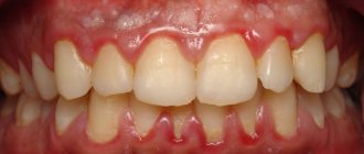
Gum hyperplasia develops slowly. Initially, formations of dense consistency, resembling a roller, appear in the gingival areas (mainly in the area of the front teeth). In their shade they do not differ in any way from other areas of the gums, and when pressed they do not give a painful reaction. At this stage, dental crowns are covered with gum tissue by about one third.
Then progression of gingival hyperplasia is observed, as a result of which the papillae are deformed, and the overgrown gum covers half of the dental crown.
And the extreme degree in which gingival hyperplasia manifests itself can be considered one in which the edge of the gum grows so much that it covers two-thirds of the crowns, reaching the chewing surface or the cutting edge. The enlarged papillae are covered with bleeding granules of different sizes.
Treatment methods
The treatment regimen largely depends on the cause of the disease. If gum growth is caused by taking medications, they are replaced in consultation with the attending physician. In other cases, sanitation of the oral cavity and surgery are indicated - overgrown tissues are removed with a laser, and the mucous membrane is sutured.
To prevent repeated cases of gum overgrowth, it is necessary to eliminate the provoking factors - restore hormonal levels, undergo orthodontic treatment, cure diseases of the teeth and gums.
Diagnosis and treatment of gingival hyperplasia
The main task facing a physician diagnosing a pathology is not to confuse it with other diseases. For this reason, in addition to external examination, X-ray examination and histological analysis are necessary.
Treatment for gum hyperplasia today is carried out exclusively through surgical intervention. It is performed by removing excess gum tissue. In parallel with this, complete sanitation of the oral cavity is carried out.
If the pathology occurs after taking special medications, then they are changed to other medications. In this case, refusal of the drug leads to the return of the gums to a physiological state. If this does not happen, the excess tissue is removed surgically. If a person returns to taking medications that caused gum hyperplasia, then a relapse of the disease is inevitable.
Enamel hyperplasia
Dental hyperplasia is an increase in dental tissue cells, resulting in the formation of small oval seals on the surface of the tooth enamel. They can be located on the surface of the tooth root, on the neck of the tooth, and also on the surface of the dental crown. Depending on the location of the enamel seal, molar, cervical and coronal hyperplasia of teeth is distinguished.
Typically, tooth enamel hyperplasia does not require treatment, since it does not annoy the patient and does not cause pain. The exception is cervical hyperplasia, in which enamel seals are located close to the gum and constantly injure it. In this case, the doctor will polish off the seal and prescribe the patient phosphate-containing drugs for further treatment.
Types of disease
Doctors call two types of gum hyperplasia:
- Focal, which is practically not registered. It looks like single gingival growths observed in the area of the lingual gingival zone or in the area of the tubercle of the upper dentition. Moreover, single growths can cover all gums, or just one;
- Generalized gingival hyperplasia. It is registered quite often. It is visualized as granular growths, which eventually grow together into one, subsequently covering a significant area of the tooth surface.