V.F. Privorotsky, N.E. Luppova, T.A. Gerasimova, A.V. Orlov, F.P. Romanyuk, E.A. Antonova
Medical Academy of Postgraduate Education
Consultative and diagnostic center for children
Children's City Hospital of St. Olga, St. Petersburg
The last decade has been characterized by an increase in the pathology of the gastrointestinal tract (GIT) in both adults and children. In the structure of these diseases, the main place in the frequency and diversity of damage to organs and systems belongs to acid-dependent diseases, which usually include gastroesophageal reflux disease (GERD), chronic gastritis, chronic gastroduodenitis (CGD), peptic ulcer, as well as chronic pancreatitis.
If the frequency of gastritis, gastroduodenitis and peptic ulcers in the pediatric population has remained relatively stable over the past 5–7 years, then with regard to GERD there is a clear tendency towards an increase in the frequency of this disease. It is these trends that gave the WHO expert group the basis to figuratively call GERD “the disease of the 21st century.”
Gastroesophageal reflux (GER) is one of the most common motor disorders of the upper gastrointestinal tract. Its frequency, according to various authors, ranges from 18–25% in the population of children suffering from digestive disorders [2,3,4].
GER itself as a mechanism, its clinical manifestations, as well as possible complications that significantly reduce the patient’s quality of life, constitute a relatively new nosological form - GERD.
Among the numerous existing definitions of this disease, we think the most successful is the following:
“GERD is a chronic relapsing disease characterized by certain esophageal and extraesophageal clinical manifestations and various morphological changes in the mucous membrane (SM) of the esophagus due to retrograde reflux of gastric or gastrointestinal contents into it” [3].
From a theoretical point of view, GERD represents that relatively rare “model” of the disease, within which it is difficult to separate etiology and pathogenesis. Among the many factors that explain the occurrence and development of GERD, the main one is a violation of the “obturator” mechanism of the cardia, which can be absolute or relative.
In addition, the mechanism of development of this disease implies insufficient efficiency of esophageal cleansing (clearance), as well as a change in the resistance of esophageal mucus. An increase in the acid-forming function of the stomach also plays a certain negative role, as a result of which refluxate (components of gastric contents) acquires special aggressive properties. Approximately according to these laws, the so-called “acid” GER is implemented.
At the same time, some children experience reflux of alkaline components (bile and duodenal juice) into the esophagus, which is commonly called “alkaline” GER. According to the literature, true “alkaline” GER is very rare, not exceeding 5% in frequency in the population of children with motor disorders of the upper gastrointestinal tract [3,6].
In addition to these factors, an uncompensated increase in intragastric or intra-abdominal pressure plays a significant role in the genesis of GERD.
Summarizing the above, we can say that GERD is a disease with a complex pathogenesis and the prevalence of one or more factors determines a wide range of clinical and morphological variants of this nosological form.
Esophagitis symptoms and treatment
The clinical picture of GERD is polymorphic and often depends on the nature of the underlying gastrointestinal pathology. However, it is quite specific and is represented by esophageal (esophageal) and extraesophageal (extraesophageal) symptoms.
Esophageal symptoms include complaints such as heartburn, belching, regurgitation, bitterness in the mouth, “wet spot” symptom, dysphagia, odynophagia (pain in the sternum while eating). The last two complaints are quite rare in childhood and, as a rule, indicate significant motor and/or structural disorders of the esophagus. It should be noted that the severity of clinical signs of the disease in children does not always correlate with the intensity of reflux and the quality of the refluxate.
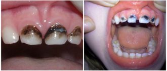
Extraesophageal symptoms of GERD are also very diverse. Currently, the following extraesophageal “systemic” manifestations of this disease are distinguished: 1) bronchopulmonary (cough, shortness of breath, difficulty breathing, asthma attacks), 2) otorhinolaryngological (hoarseness and loss of voice, recurrent pain in the throat and/or ears), 3) cardiological (heart pain, rhythm disturbances), 4) dental [3,4,8].
that GER - associated bronchopulmonary disorders are in first place on this list. They are the most studied, better known to practitioners and have acquired a certain evidence base in recent years.
The history of studying the relationship between the pathology of the esophagus and the bronchopulmonary tree begins from the end
19th century. Thus, back in 1892, W. Osler noted the need to avoid excessive eating and was the first to describe attacks of suffocation associated with aspiration of gastric contents. In 1934, G. Bray pointed out the connection between the digestive tract and bronchial asthma (BA). Further study of this problem was continued by the works of Mendelson (1946) and Frieland (1966). They also introduced the term “reflux-induced BA” [1].
In subsequent years, Mansfield and Stein identified decreased airway patency associated with heartburn. Serious pediatric studies on this topic were first conducted in the 60–70s of the last century, and by now there is sufficient factual material. A peculiar reflection of this experience was the classification of BA edited by I.M. Vorontsova (1996), in which “gastroesophageal reflux asthma” is highlighted as a separate item.
GER - associated respiratory disorders are traditionally divided into 2 groups: “upper” (apnea, stridor, laryngitis) and “lower” (bronchial obstruction syndrome, bronchial asthma). The symptoms inherent in these conditions are characterized in the English literature by a special term - RARS (reflux-associated respiratory syndrome).
The connection between the esophagus and the bronchial tree is explained by their common origin from the primary digestive tube and common innervation by branches of the vagus nerve [9].
GER can cause respiratory diseases in two ways: 1) direct, with the development of mechanical occlusion of the lumen of the tracheobronchial tree by aspiration material and 2) indirect (neural) with the development of discrinia, edema and bronchospasm [3,5].
What to do during diarrhea without fever
Often, if you eliminate the cause of diarrhea in a two-year-old child, then this illness, provided that the baby does not have a fever, goes away.
With diarrhea, the baby does not eat well. But once you get rid of the disease, the child will start eating with pleasure.
diarrhea in a child
The appearance of intestinal infections often affects those children who go to kindergartens. To avoid illness, you should teach your child to wash their hands thoroughly after using the toilet. The child should know that dirty hands after using the toilet are a source of diarrhea.
If diarrhea continues for quite a long time, it is recommended to find the root cause to avoid serious consequences. If a child is not treated for diarrhea, it can cause depletion of the child's body.
The most important task for parents when a child has diarrhea is to prevent dehydration of the child’s body. To prevent dehydration, the loss of fluid and salts in the baby’s body should be compensated. Here it is suitable to use special solutions that treat dehydration, which are sold in pharmacies (Rehydron, Oralite). Add a tablespoon of sugar to 1 liter of the resulting solution.
It is important that during diarrhea the child drinks plenty of liquid. Large intake of fluid compensates for its loss in the body during diarrhea.
You can also make the following solution that will help save your child from dehydration: a teaspoon of baking soda, three-quarters of a teaspoon of salt, and eight small spoons of sugar should be diluted in a cup of orange juice and mixed with a liter of water. The child should be given a small spoonful of the solution to drink every fifteen minutes for two hours. If after this time the child does not begin to feel sick, then you can double the dosage of the solution. As soon as the baby begins to feel improvement, then gradually you need to return to the child’s usual diet. But if symptoms of dehydration appear more and more, it is recommended to immediately call an ambulance.
You should call an ambulance if diarrhea is accompanied by certain symptoms, such as:
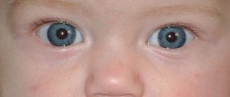
- sunken eyes;
- lethargy and drowsiness;
- darkening of the color of urine, as well as a decrease in its quantity;
- dry mouth;
- the appearance of dryness and roughness of the skin;
- lack of tears while crying.
Reflux gastritis
The direct path of development of respiratory disorders in children suffering from GERD is primarily due to macroaspiration of gastric contents with the development of mechanical bronchial obstruction and (less often) pneumonia. Macroaspiration of acidic material (pH <2.5) can cause reflex closure of the airway, decreased surfactant, epithelial damage, and in severe cases, pulmonary edema and bleeding.
In children, such a dramatic development of events rarely occurs. Typically, a pediatrician is faced with clinical manifestations of laryngitis or bronchial obstruction of varying severity, which can often become recurrent. The leading mechanism in this case is often microaspiration, which provokes the development of laryngo- or bronchospasm in a reflex way with the formation of such pathologies as chronic bronchitis, repeated pneumonia, pulmonary fibrosis, apnea.
Protection against bronchopulmonary aspiration involves coordinating the swallowing reflex and closing the glottis during swallowing. Based on this, it can be assumed that in some cases the condition of the epiglottis as such acquires special significance. The latter circumstance rarely becomes a subject of thought for ENT doctors and endoscopists, despite the fact that it is visually possible to determine the features of the anatomical structure of the epiglottis, as well as the features of its functioning in a particular patient. In addition, the development of microaspiration in GER is facilitated by the condition of the upper esophageal sphincter and esophageal peristalsis.
The indirect (neural) path of development of GER-dependent respiratory manifestations is realized through the afferent fibers of the vagus with the development of a system of bronchoconstrictor reflexes and, as a consequence, bronchospasm. There is a point of view according to which a necessary attribute for the development of bronchospasm in patients with asthma is esophagitis, which irritates afferent vagal receptors.
The schematically described mechanism looks like this: GER – esophagitis – irritation of afferent vagal receptors – increased reactivity of the tracheo-bronchial tree – increased lability of the bronchial muscles – bronchospasm [5,7].
A number of authors suggest the presence of specific receptors for damage to the esophageal mucus, the so-called nociceptors. It is assumed that the latter react only when the esophageal mucosa is changed, and in the absence of damage they do not function. This hypothesis may explain why physiological GER does not lead to coughing and choking [3,7].
There is information in the literature about the effect of some neuropeptides on changes in bronchial conductivity, especially in the case of damage to the esophageal mucus. Influencing the tone of smooth muscle fibers of the bronchi and blood vessels, stimulating the release of histamine, leukotrienes and other mediators, they change the reactivity of the tracheobronchial tree [9].
Considering the problem under study “from the opposite direction”, it is necessary to note the provoking influence of respiratory pathology on the development of GER. Any respiratory disorder or symptom can trigger GER if it alters any aspect of the antireflux barrier.
The main mechanisms by which this pathological effect is realized can be presented as follows: 1) an increase in the pressure gradient - an increase in negative intrathoracic and positive intra-abdominal pressure, 2) a decrease in pressure in the LES area, 3) an increase in acid production, 4) impaired food evacuation from the stomach, etc. [1,6].
To illustrate the above, some well-known examples can be given: intra-abdominal pressure increases, in particular, with forced exhalation during coughing or sneezing in asthma, cystic fibrosis (CF), bronchopulmonary dysplasia, and respiratory infections. At the same time, the frequency of GER also increases, especially in children with cardia insufficiency or sliding hiatal hernia. Negative intrathoracic pressure increases, for example, with stridor or hiccups.
In addition, one should remember the possible side effects of theophyllines and glucocorticosteroids (GCS), widely used in the treatment of asthma. These drugs reduce the tone of the LES, thereby provoking a breakthrough of the antireflux barrier. There is evidence that oral administration of theophyllines and b2-adrenomimetics reduces the tone of the LES and stimulates the secretion of hydrochloric acid, while inhalation of systemic corticosteroids and theophyllines does not change the tone of the LES. It is also known that when using inhaled corticosteroids without the use of a spacer, 80% of the inhaled dose enters the stomach, which adversely affects both the tone of the LES and gastric motility [1,9].
One of the most important respiratory complications of GER is asthma. According to various authors, pathological GER is detected in 20–80% of children suffering from asthma (according to our data - 65%). The number of refluxes often correlates with the severity of respiratory symptoms, and individual episodes of GER directly coincide in time with attacks of suffocation.
There are indications in the literature that with adequate therapy for GER, the frequency of bronchospasm symptoms decreases. The most characteristic sign of GER-dependent bronchial obstruction is a night cough due to prolonged acidification of the esophagus in a horizontal position, decreased salivation and clearance of the esophagus, as well as the development of esophagitis.
GER-dependent bronchial obstruction may be suspected in children who have:
1) attacks of coughing and/or choking, mainly at night; after a heavy meal;
2) a proven combination of respiratory and “upper” dyspeptic symptoms (belching, heartburn, regurgitation, etc.);
3) positive effect of antireflux therapy ex juvantibus;
4) signs of torpidity to adequate basic therapy;
5) non-atopic variants of BA [1,3,6,8,9].
The system of evidence in such cases is built on the basis of a reliable diagnosis of GER using all available methods (both purely gastroenterological, and pulmonological and allergological).
In our opinion, the optimal method is to use the following specialized methods for examining children:
1) FGDS with targeted biopsy, histological examination of biopsy samples, helpel test (a type of urease test to determine Helicobacter pylori infection), contrast fluoroscopy of the esophagus and stomach, daily pH monitoring, acid test (Bernstein test in any modification).
2) Spirometry; bronchoprovocation tests with physical activity, saline, histamine, methacholine; impulse oscillometry; X-ray of the lungs (according to indications); allergy examination (general and specific: IgE, RIST and RAST).
Currently, there are numerous endoscopic classifications of GER in adults (Savary–Miller, Los Angeles, etc. classification). At the same time, all of them are not sufficiently adapted for children. The modified version of the classification given below by G. Tytgat (1990, 1996) takes into account the severity of both morphological and motor disorders in GER.
Prevention
Inflammation of the paranasal sinuses is a disease that is very often diagnosed in children of any age. Usually occurs against the background of reduced immunity. Taking this into account, maximum attention should be paid to measures that will restore and strengthen the body's defenses. Doctors advise starting to take care of this from childhood. It is very important to provide a balanced diet rich in vitamins and minerals.
Children's multivitamin complexes "Vitrum" and "Azbuka" have proven themselves very well. If the child is not allergic to the components of the drugs, use immunomodulators - pharmaceutical and natural (ginseng, echinacea, lemongrass, etc.).
Always try to properly treat seasonal infectious diseases - colds, flu and other diseases - until complete recovery. One possible cause of rhinosinusitis is dental disease. Particular attention should be paid to the treatment of the teeth of the upper jaw. Living a full life, daily physical activity and proper nutrition will help your child stay fit and get sick less often. Sinusitis is dangerous due to severe complications. Constant headaches and prolonged runny nose - such symptoms should cause vigilance in parents. You should not self-medicate. The best option is to consult a doctor immediately. Only he will be able to determine the cause of the illness and select therapy taking into account the individual and age characteristics of the child.
Non-traditional treatment for reflux
System of endoscopic signs of GER in children (according to G. Tytgat as modified by V.F. Privorotsky et al., 2002)
Morphological changes
0 degree. There are no signs of damage to the esophageal mucosa.
I degree. Moderate focal erythema and (or) friability of the mucous membrane of the abdominal esophagus.
II degree. The same + total hyperemia of the abdominal esophagus with focal fibrinous plaque and the possible appearance of single superficial erosions, often linear in shape, located at the tops of the folds of the mucosa.
III degree. The same + spread of inflammation to the thoracic esophagus. Multiple (sometimes merging) erosions located in a circular pattern. Increased contact vulnerability of the mucous membrane is possible.
IV degree. Esophageal ulcer, Barrett's syndrome, esophageal stenosis.
Motor disorders
A. Moderately expressed motor disturbances in the area of the LES (raising of the Z-line up to 1 cm), short-term provoked subtotal (along one of the walls) prolapse to a height of 1–2 cm, decreased tone of the LES.
B. Clear endoscopic signs of NKJ, total or subtotal provoked prolapse to a height of more than 3 cm with possible partial fixation in the esophagus.
B. The same + pronounced spontaneous or provoked prolapse above the crura of the diaphragm with possible partial fixation.
Histological examination of biopsy specimens of the esophagus and (or) stomach, as well as x-ray examinations of the upper gastrointestinal tract are carried out according to standard methods and are described in the relevant manuals.
The “gold standard” for determining pathological GER is daily pH monitoring, which allows not only to record the fact of reflux, but also to determine its nature (physiological or pathological). The study is carried out using Gastroscan-24 (Russia) or Sinectics-medical AB (Sweden) devices. To date, there are no uniform standards that allow us to definitely establish the connection between GER and bronchial obstruction. To document this fact, equipment is needed that allows one to simultaneously assess respiratory function and register GER. Therefore, various indices are currently being developed to judge the presence of such a relationship [9].
The principle of the acid perfusion test (Bernstein test) is to assess the child’s subjective sensations during artificial acidification of the lower third of the esophagus with a 0.1% solution of hydrochloric acid (or pure lemon juice, the pH of which is known). The test is assessed as positive in cases where the child experiences chest pain or heartburn in the first 3 minutes of the test.
The basic method for studying the functional state of the lungs is spirometry (study of external respiration function). The study is carried out using a computer spirograph, in which air flow is measured using flow meters, and the volume is calculated by integrated flow.
Bronchoprovocation tests allow us to determine the reactivity of the tracheobronchial tree. Tests with methacholine and histamine are carried out only during remission. These irritants directly affect the bronchi, which have smooth muscle fibers. This leads to a reduction in the latter, stimulation of cholinergic activity and an increase in vascular permeability. In low concentrations, these drugs do not have side effects and do not cause long-term broncho-obstructive reactions.
Pulse oscillometry allows you to determine the components of total respiratory resistance (friction and reactance in a certain frequency range).
In the above diagnostic algorithm there is no mention of scintigraphy of the esophagus (to confirm the fact of reflux) and lungs (to ascertain aspiration of gastric contents into the lungs) using food labeled with a technetium isotope. The limited data available in the literature indicate the low sensitivity of this method [3,6,8,9].
One of the key questions of the “relationship” of GER and BA is the following: does GER play the role of the only triggering point of the bronchial obstruction mechanism or does it act as an integral part of some combined mechanism?
In the latter case, there may be a triple component of the triggering of bronchial obstruction: atopy, infectious dependence and GER. What place does each factor occupy on this kind of “pedestal of honor”? This is a question, the answer to which would significantly facilitate the development of adequate treatment programs and allow us to avoid the formation of forms of AD that are torpid to the basic treatment.
Unfortunately, there is no exact answer to this question yet, and we have to come to terms with the fact that in real life we have only two situations that allow us to associate GER with bronchial obstruction syndrome or BA: 1) “pure” GER-dependent variant; 2) a variant in which GER plays a certain negative role in the genesis of AD along with other factors.
CF, unlike AD, is not a model of a “classical” GER-dependent disease, and the relationship between GER and CF is less clear. However, the disorders that occur with CF, such as increased intra-abdominal pressure during coughing, slower gastric emptying, impaired motor function, increased production of hydrochloric acid, are “fertile ground” for the occurrence of GER and the development of GERD.
Differences between diarrhea and loose stools
Any mother should clearly know what distinguishes diarrhea in a 1-2 year old child without body temperature from loose stool. Loose stools may appear periodically. All children, without exception, face this. Loose stools can be caused by drinking a large amount of juice, as well as a consequence of a viral infection or some other disease.
Diarrhea is very loose stool. With diarrhea, the child goes to the toilet up to five times a day. This malaise poses a danger to the child - it provokes a lack of water in the body.
Treatment of GERD
Treatment of gastroesophageal reflux disease is aimed at improving the motility of the upper gastrointestinal tract, suppressing the acidity of gastric juice, as well as eliminating factors leading to increased intra-abdominal pressure or decreased tone of the lower esophageal sphincter.
Much attention is paid to the lifestyle and nutrition of patients. The diet of patients suffering from GERD excludes foods that irritate the mucous membrane (spicy sauces, seasonings, onions, garlic, peppers, ketchup, sour fruit juices), reduce the tone of the lower esophageal sphincter and/or slow down gastric emptying (alcohol, coffee, chocolate, cakes, pastries, fatty foods, lard, margarine), as well as products that increase gas formation, leading to an increase in intra-abdominal pressure. It is necessary to explain that overeating, as well as eating in a hurry, before bed or at night will lead to increased symptoms. Fractional meals have a positive effect. Food should be thoroughly chewed, the temperature of the food should not be very high or very low, but should be around 37–38°C. The last meal should be at least 2 hours before bedtime. The head end of the bed is raised 15 cm using stands. This helps improve esophageal clearance due to the effect of gravity on the esophagus and stomach. It is important to note that elevating only the head (for example, using a high pillow) is not allowed, as this increases intra-abdominal pressure, which aggravates reflux. Exclude physical exercise after eating, tight belts, tight clothing - all this leads to an increase in intra-abdominal pressure and contributes to the reflux of stomach contents into the esophagus.
The main groups of drugs used in the treatment of GERD are prokinetics, acid suppressants and antacids.
Prokinetics include dopamine receptor blockers - metoclopramide, domperidone and cisapride. Cisapride has now been discontinued due to the risk of cardiac arrhythmia. Metoclopramide penetrates the blood-brain barrier and blocks dopaminergic receptors in the thalamus, hypothalamus and brain stem. The antiemetic effect is associated with inhibition of the vomiting center, as well as with increased propulsive gastric peristalsis associated with blocking peripheral dopamine receptors. The use of metoclopramide in children is undesirable, since the development of side effects such as extrapyramidal disorders, drowsiness, fatigue, and anxiety associated with the penetration of metoclopramide through the blood-brain barrier is possible. Hyperprolactinemia may develop.
Another drug that blocks dopamine receptors is domperidone. It is devoid of the side effects of metoclopramide and is currently the drug of choice in the treatment of children with GERD. Domperidone blocks only peripheral dopamine receptors of the stomach and duodenum, which leads to increased tone and peristalsis of the upper gastrointestinal tract. Anthro-duodenal coordination improves, gastric contractility increases and thereby accelerates gastric emptying. In addition to tablet forms, the drug exists in the form of syrup, which facilitates its use in pediatric practice. Domperidone is prescribed at a dose of 2.5 mg per 10 kg 3 times a day for 1–2 months. Side effects are rare (0.5–1.8% of patients). These include headaches and general fatigue. Extrapyramidal disorders and hyperprolactinemia are extremely rare.
Proton pump inhibitors (PPIs) or H2 blockers are used to reduce gastric acidity. However, the results of numerous studies have shown that the use of proton pump blockers in the treatment of gastroesophageal reflux disease is significantly more effective. Inhibition of the proton (acid) pump is achieved through inhibition of H+, K+–ATPase of parietal cells. The antisecretory effect in this case is realized not by blocking any receptors (H2-histamine, M-cholinergic) involved in the regulation of gastric secretion, but by a direct effect on the synthesis of hydrochloric acid. Proton pump inhibitors are substituted benzimidazole derivatives.
Being chemically weak bases, they accumulate in the tubules of parietal cells, where the pH values are the lowest (1.0–0.8). In the tubules of parietal cells, benzimidazole derivatives are converted into tetracyclic sulfenamide. Sulfenamide binds covalently via disulfide bonds to the cysteine groups of the proton pump, which leads to inhibition of the enzyme and inhibition of acid secretion. The resulting sulfenamide does not pass through membranes well because it is a cation. This ensures selective accumulation of the active form of the proton pump inhibitor in the secretory tubules of parietal cells. By acting on this stage, proton pump inhibitors cause maximum inhibition of acid formation. In children, omeprazole and rabeprazole are used in a dose of 10 to 20 mg once before dinner. The dose is selected and adjusted individually under the control of pH-metry.
We monitored the effectiveness of the use of the proton pump inhibitor rabeprazole. 30 children aged 7–15 years with gastroesophageal reflux disease were examined. 17 children received rabeprazole at a dose of 20 mg once a day, 13 children received rabeprazole at a dose of 10 mg once a day. Control was carried out by studying intragastric acidity using the Gastroscan 5-M and Gastroscan-24 devices (NPO Istok-Sistema, Russia). pH-metry was carried out on days 7–14 of taking the drug and after 1–3 months.
Data on the clinical effectiveness of the drugs are presented in the table.
The pronounced positive dynamics on the first day of therapy while taking rabeprazole is due to the peculiarity of its metabolism.
When conducting daily pH-metry, the following indicators were assessed: from 9 a.m. to 9 p.m., basal acidity, the buffering effect of food, and the presence of GER were assessed. At 21:00 the patient was given the drug and the latent period and period of action were determined. The criterion for patient resistance to PPIs is the absence of an increase in pH in the body of the stomach above 4 units. The following data was received.
A plateau of level 4 and higher in the body of the stomach during the first single dose was obtained in 4 patients; in the rest there was a periodic increase in pH in the body of the stomach, but a stable level was not obtained. The latent period averaged 5 hours. The duration of action of the drug did not exceed 5 hours.
Short-term pH-metry was carried out 7–14 days from the start of taking the drug and 2–6 months after the end of treatment. At the same time, the effectiveness of the drug was assessed.
In children who received PPIs for 7–14 days, a body pH of 4 or more was noted in 8 people. In 15, the pH in the body above 4 rose intermittently. In 10 of them, the pH in the body remained at a level of up to 1.5 throughout the day, while the rest showed normal acidity.
Control after 2–6 months revealed the absence of a lasting effect in 16 patients. 2 maintained body pH at 4–6 for 2 months. In 18 children, normacidity was noted (body pH 1.5–2). However, only 7 people made complaints.
Thus, despite the proven effectiveness of PPIs in relieving clinical symptoms within 1–3 days from the start of taking the drug, there is an individual sensitivity of the patient’s body to PPIs. It can only be assessed by pH measurement. Carrying out daily pH-metry allows you to select the dose and drug in each specific case.
Gel-based antacids are used as an enveloping agent. By covering the gastric contents, the antacid prevents trauma to the mucous membrane during reflux into the esophagus, forming a protective film on its surface. Antacids for gastroesophageal reflux disease are used immediately after meals 3-4 times a day.
We observed 180 children with chronic nonspecific lung diseases (CNPD). The first group consisted of 120 children with asthma (d–74, m–46); the second – 36 children with CF (d–12, m–24); the third – 24 children with recurrent bronchitis (d–16, m–8). The age of the subjects ranged from 6 to 18 years. The vast majority of children of all groups (85%) had clinical and endoscopic signs of CGD.
Among patients with BA, 24 children (20%) were diagnosed with a severe course of the disease (in all cases, BA was atopic, infection-related), 80 children (66%) had a moderate course (68 had a mixed variant, 12 had an atopic variant) , 16 children (14%) had a mild course (13 had a mixed course, 3 had an atopic variant).
A severe course of CF was observed in 11 (30%) children (all had a mixed form), a moderate course in 14 (40%) people (9 had a mixed form, 5 had a pulmonary form), a mild course – in 11 (30%) ) children (5 have a mixed form, 3 have a pulmonary form, 3 have an intestinal form).
Identification of variants of the course of recurrent bronchitis is not accepted in modern pulmonology. All children with recurrent bronchitis were examined at the height of exacerbation.
Among patients with asthma, complaints typical of GER were noted in 58 children (49%). Intragroup analysis revealed such complaints in 19 children (78%) with severe disease, in 34 children (43%) with moderate asthma, and in 5 children (31%) with mild asthma.
In patients with CF, similar complaints in various combinations occur in 31 children (86%), including all children with a severe form of the disease, 12 children (86%) with moderate CF and 8 children (73%) with mild .
12 children with recurrent bronchitis (50%) had complaints characteristic of GER.
According to the results of instrumental examination, almost 2/3 (65%) of the examined children with BA (regardless of the severity) were diagnosed with GER (the vast majority of them had an endoscopically positive variant and only 4% had an endoscopically negative variant).
Among patients with CF, GER was diagnosed in 31 children (88%), and all of them had an endoscopically positive variant.
An endoscopically positive variant of GER was found in 9 children (41%) with recurrent bronchitis.
According to the results of intragroup analysis, GER of varying severity was detected in 68% of children with severe asthma (20% of them had GER grades 2–3), in 30% of children with moderate severity (17% of children had GER grades 2–3) , and in 50% of children with mild - (5% of them have GER grade 2-3). According to the results of FGDS and fluoroscopic examination, a sliding hiatal hernia was detected in every sixth child with severe asthma and in every tenth child with a milder course of the disease.
The majority of patients with CF (88%) had esophagitis of varying severity. At the same time, endoscopically positive GER was found in all patients with severe CF, in 54% of patients with moderate CF, and in 45% of children with mild disease.
Of particular interest, in our opinion, are the following data: in 90% of children with asthma who have a sliding hiatal hernia, pulse oscillometry revealed significant changes in reactance and frictional resistance, which indicates heterogeneous ventilation of different parts of the lung tissue. In all patients in this group, based on the results of bronchoprovocation tests, the III threshold of hyperreactivity was determined.
Carrying out a correlation analysis based on spirometry data in patients from the compared groups was difficult due to objective reasons: the children were examined during different periods of the disease. At the same time, spirometry, of course, refers to screening methods for examining children with COLD, and its implementation is necessary to assess the severity of the disease and develop optimal treatment programs.
How is diarrhea not accompanied by fever treated?
Diarrhea should be treated by drinking plenty of fluids, as well as introducing a large amount of vegetables and fruits into the child’s diet. But it is recommended to remove complex carbohydrates and carbonated water from the diet.
After the specialist determines the cause of diarrhea, some additions will be made to the baby’s diet. For example, a child may be prohibited from consuming whole milk, fiber and fats. With all the diet, the baby will have to be given medications prescribed by the doctor.
To prevent diarrhea in a two-year-old baby , parents should carefully monitor the child’s diet, as well as the cleanliness of his hands before and after eating, as well as after visiting the toilet. Children need to be taught hygiene skills from an early age.
Respiratory infections in children
According to bronchoprovocation tests (with histamine and methacholine), the third threshold of hyperreactivity was detected in all children with severe BA, in 20% of children with a moderate course of the disease and in 2% of children with a mild form (the differences between the severe form and the other two are significant).
In patients with severe CF, the maximum (third) threshold of hyperreactivity was diagnosed in 66% of children.
In the group of children with recurrent bronchitis III, the hyperreactivity threshold was not diagnosed.
Based on the above, the following conclusions can be drawn:
1) in 65% of children with BA, in 88% of children with CF and 50% of children with recurrent bronchitis, GER of varying severity was detected;
2) a relationship was noted between the severity of GER and the severity of COLD (in severe forms of BA and CF, high-grade GER was detected significantly more often (p<0.05) than in patients with milder forms of these diseases and recurrent bronchitis);
3) in the vast majority of children with COLD who have GER, an endoscopically positive version of the latter has been proven;
4) the most unfavorable is the combination of BA or CF (regardless of the severity) with a sliding hiatal hernia and reflux esophagitis; in these children, significantly more often (p<0.05) than in children with recurrent bronchitis, positive tests with histamine and methacholine with a high threshold of hyperreactivity are detected, and serious disorders are determined according to pulse oscillometry;
5) there was no connection between HP infection and the degree of esophageal damage in children with COLD;
6) all cases of GER-associated BA were identified in children with atopic and/or infectious-dependent forms of this disease;
7) all children with CF should be prescribed a special instrumental examination for early detection of esophageal damage.
Despite significant advances in recent years, there are still many “blank spots” in the topic of respiratory manifestations of GERD in children. In particular, the role of alkaline GER and other non-acidic stimuli in the genesis of respiratory disorders is not fully understood, and not all aspects of the reflex mechanism of reflux-dependent laryngo- and broncho-obstruction have been revealed. The question remains to be answered why only 18–20% of children with pathological GER develop respiratory manifestations to one degree or another, while the opposite situation occurs with a 4-fold prevalence. Answers to these questions may be the topic of future publications.