A scary story about terrible killer bacteria is one of the most effective ways to influence people's consciousness and try to make them more careful about maintaining hygiene rules, as well as draw attention to vaccination issues. But without visual material to show, dry data on microbiology research, interspersed with scientific terms, most often does not gain even a tenth of the necessary attention. Often such information remains unclaimed. To convince the listener or reader that bacteria are indeed literally everywhere and can play a decisive role in human destiny, these bacteria need to be shown and this display must be made as colorful as possible.
Observing bacteria through a microscope. Photo
The structure of bacteria is much simpler and more uniform than the structure of protozoa, and there is not such a wealth of forms as in ciliates.
However, this uniformity and simplicity of structure make bacteria a very good model for many experiments. Viruses are even simpler in structure, and therefore even better as a model. But about them - later, in a special chapter. To look at living bacteria, you and I will have to look for stronger and more complex microscopes than those with which we can examine ciliates. You can't do without magnification of 600-800 times.
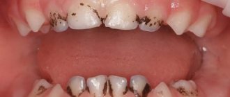
But the source, in which you can always find a wide variety of bacteria, is always available. This is your own mouth. Scrape off plaque and mix it with a drop of water or saliva on a slide. This will be enough for you to get acquainted with the main forms of bacteria.
If you look at them through an ordinary microscope used in medical and biological laboratories, you will probably be disappointed. Grayish, with unclear contours, very small sticks, balls, and threads will be visible. Can they be compared with ciliates, as fancy as tropical fish?
With a so-called phase contrast microscope, you can see more. The difference between this microscope and a conventional one comes down to the fact that particles that are equally transparent to light rays, but with different densities, look different here: denser ones are darker, less dense ones are lighter.
It is interesting to observe living bacteria using a so-called dark-field microscope. The light rays here do not go through the object of observation into the microscope lens, but from the side. You've probably seen how brightly dust particles glow in a ray of sunlight breaking through curtains or shutters in a dark room.
Bacteria look roughly the same way in a dark-field microscope - like light dots on a jet-black or brownish background. Their general outlines are a little blurred, but the movement of the bacteria is clearly visible. And the nature of the movement makes it possible to recognize the causative agents of certain diseases.
Photo: Saroj Regmi
Photo: US Geological Survey
Photo: Umberto Salvagnin
Other bacteria do not have flagella necessary for movement. But this does not mean that they will be motionless in the field of view of the microscope. No, it will seem to you that the bacteria are moving, all at once, like ants in a torn up anthill. However, this is not independent, active movement of the microbe, but the so-called Brownian movement.
Brownian motion of any small particles floating in a liquid (not just microbes) is a consequence of the random thermal movement of the molecules of this liquid. Molecules press on the particle from all sides, and it, so to speak, “marks time.”
But if you look at mobile bacteria under a microscope, you will see how quickly they cross the field of view, freeze in place, and then rush on again. It is especially interesting to observe spirochetes, which look like an animated spiral from an electric stove. They are so thin that under a regular microscope it is difficult to see a living spirochete.
They are visible much better in a dark-field microscope. You'll probably find them in plaque; just take a good look - it's best to look for spirochetes while they are moving. They either float, wriggling like snakes, or twitch in place and even fold in half.
It is not as convenient to examine living bacteria through a microscope as dead and colored ones. The details of the structure of these organisms were studied specifically on colored preparations. To stain bacteria, you need to apply them to glass (as they say, make a smear), dry it, heat it on a burner flame (so that the cells subsequently tint better) and drop a drop of special paint onto the smear.
If you go to a microbiological laboratory, then, of course, there will be a set of various paints. One of the most common is methylene blue. Since it is part of the ink for a fountain pen, for lack of anything better, you can splash a drop of ink onto the smear. After 6-8 minutes, the paint should be washed off with water and the smear should be dried.
Depending on what type of bacteria was stained, you will see balls or rods under the microscope - straight, curved or comma-shaped. Chains can be formed from sticks and balls.
Microbes that live under your nails
The area under the nails contains all types of bacteria that live on the hands. But the concentration of such microbes is hundreds of times higher. This is due to the inaccessibility of the space under the nails for treatment with disinfectants and anti-inflammatory agents.
Interestingly, there are many times more such microbes under artificial false nails than under natural ones.
Permanent residents of the body inhabiting this zone:
- fungal bacteria;
- staphylococci,
- streptococci.
The difficulty of disinfecting fingers is that the nail plate reliably protects this part of the body from the action of the most effective antibacterial agents.
Choosing the right model
Many novice researchers are interested in which device to choose in order to examine fermented milk, as well as other common categories of bacteria.
The budget segment of microscopes demonstrating 640x magnification will not give the same effect that can be appreciated in a video made with a more powerful microscope. Bacteria in urine, for example, can only be seen under equipment lenses that magnify 1000x or more.
The phase-contrast type of device works by detecting different particle densities. This microscope, which allows observation and magnification of bacteria, colors the elements in a light gray or dark gray shade. In this video you can see a multiple increase in bacteria in the urine.
A dark-field microscope allows you to see lactic acid bacteria (you can also see what they look like in the photo). Its advantage is the scattering of light coming not directly through the lens, but from the side. The device also allows you to understand the actual movement patterns of bacteria.
Development of the disease
Nail mycoses, or onychomycosis, affect every tenth representative of the globe. The main pathogens are dermatophytes, such as T. rubrum. But there are forms caused by mold or yeast-like fungi. Factors that provoke onychomycosis: wearing uncomfortable, tight shoes and neglecting hygienic care of hands and feet.
Onychomycosis occurs in several stages:
- Spots of a yellowish or gray-white hue appear on the platinum.
- The plate becomes brittle, flakes, crumbles, and some areas thicken.
- The bed takes on a brownish tint, and the nail plate comes off.
Pathological changes can be observed both in a local area and on the entire surface. The fungus gradually spreads to neighboring fingers.
Studying Cipollino
A microscope will help your child learn that all living things are made up of cells. Under a microscope you can see not only a cell, but also examine its structure. To do this, together with your child, prepare a simple and visual preparation from ordinary onions. Why onions? This plant has very large cells, and they are clearly visible under relatively low magnification. So, cut the onion into several parts and separate one juicy layer. Cut a small piece from it, and then use tweezers to separate the thin film from the concave side of the piece. Drop distilled water onto a glass slide, place a film in it and carefully straighten it with a needle. Then add a couple of drops of an aqueous solution of methylene blue or an aqueous solution of iodine. This should be done so that the colorless cells become colored and become better visible. If you can find a red-violet onion, you don’t need to add any dye. Cover the resulting “beauty” with a cover glass and blot away any escaping liquid. Try to view the drug first at low and then at high magnification. Tell your child that both plants and animals are made up of tiny cells. These are the ones that are visible through a microscope, like little bricks. Why were they called cells? This name was invented by the English botanist R. Hooke. Examining a section of the cork under a microscope, he noticed that it consisted of “many boxes.” He also called these “boxes” chambers and... cells. After all, it really looks like someone drew the onion film into squares.
At high magnification, the cell wall, nucleus, and vacuole are clearly visible. Explain to your child that a cell wall is a partition, a wall between cells. It protects the cell and helps maintain the desired shape. Thanks to the nucleus, the cell grows and reproduces. And inside the vacuole there is cell sap. The one that splashes in different directions and brings tears to our eyes when we cut onions.
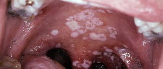
Fungi on the skin
On the body, fungal infections can develop anywhere: on the head, feet, buttocks or palms.
The most common forms are:
- Dermatophytosis: damage to the deep layers of the skin, which is caused by both yeast and mold fungi.
The most common fungal disease. This infection triggers an inflammatory process and leads to the formation of pink or red spots or plaques. The size of such plaques is very diverse. Fungi that cause dermatophytosis are permanent residents of hair and skin. They are able to perfectly absorb and assimilate keratin from hair and nails. - Keratomycosis: a disease that affects the top layer of skin. Symptoms of the disease are small, hard nodules on the skin that are filled with pus. The forms of the disease vary and include pityriasis versicolor, axillary trichomycosis and erythrasma.
- Deep mycosis: the disease affects the subcutaneous tissue, muscles, joints and bones, mucous membrane, tissues of internal organs and parts of the nervous system. With such a lesion, warts and deep fistulas appear on the skin.
- Candidiasis: The infection is caused by yeast. The most common sites of lesions on the body are in the groin area, in the armpits and under the female breast.
The manifestation of the disease depends on the location of the lesion and the type of fungus.
Candida and dermatophytes cause itching, discoloration of the skin to red-bluish, peeling of infected areas, dandruff and dry hair, and discoloration of nails.
With mycosis of the foot, in addition to peeling of the skin, blisters up to 2 mm in diameter appear, filled with liquid. These bubbles collect between the toes. The smell that appears when the fungus decays also causes trouble for the patient. Often a person gets such diseases in a sauna or in the summer, when wearing tight shoes for many hours.
When fungi get on the skin of the face, they first attack dead epithelial cells. If treatment is not carried out, the skin begins to peel, become rough, and red or yellow spots appear. During the transition to the severe stage, purulent foci and ulcers appear. Fungal infections of internal organs begin.
On the head, fungal infections lead to hair loss, flaking, dandruff, and the formation of bald patches.
Mobile and immobile organisms
The reason is not independent movement, as in those with additional elements that allow them to move, but Brownian movement (random, thermal type). Sticks and threads can:
- cross the field
- freeze,
- fold in half
- form a spiral.
Having a microscope on hand to observe various bacteria, you can examine your everyday environment and physiological fluids - microorganisms in urine and saliva. Interesting things are nearby, but seeing life hidden from prying eyes is not easy. On the one hand, there are various categories of videos and photos available, but it is much more effective to conduct the experiment yourself.
Tags: what, microbe, need, increase, see, to
Use an antibacterial spray
Despite the fact that the main ingredient in an antibacterial gel or spray is ethyl alcohol, it effectively kills many bacteria, disinfecting the skin.
Stock up on this product when you are traveling on public transport or going on a trip. Even if you want to have a snack on the way, you don’t have to worry about poisoning. Just apply a little gel to the surface of your hands, rub it in and wait until it dries completely. Do the same when you visit a public restroom. While at home, treat frequently used surfaces (faucets, door handles, sinks) with an antiseptic that contains bleach or alcohol.
Found a violation? Report content
Astronomy and microscopy
Paramecium caudatum) lives in fresh waters. The unicellular organism received its name for the elongated cilia on the back half of the body. Between the cilia, of which there are more than ten thousand throughout the body, there are trichocysts or small fusiform bodies. They are organelles (organs in multicellular organisms) of attack and defense, which are thrown out with force and pierced into the enemy body or victim. On the side of the body of the ciliate there is a pre-oral recess that passes into the mouth. The ciliate digests food by forming special digestive vacuoles, separated from the pharynx, which pass through the entire body, carried away by the flow of cytoplasm. Under favorable temperature conditions and an abundance of food, vacuoles are formed every minute. The secretion function is performed by two contractile vacuoles. Ciliates feed on other protozoa, single-celled algae, and themselves serve as food for larvae of fish and amphibians. That is why protozoa of the genus Paramecium are intensively grown in fisheries, as well as in aquarium farming.
Now we can begin to examine the ciliates under a microscope. It doesn’t matter if a ready-made microslide is not at hand. Any aquarist will share with you a couple of secrets of breeding slipper ciliates or the individuals themselves, along with water from the aquarium. You can also obtain protozoa in any stagnant body of water and, in order to obtain a critical mass sufficient for research, create the most favorable conditions for the reproduction of slippers. These protozoa are easily bred at home on dried banana peels or an infusion of hay dust.
We will share with you the simplest, but no less effective, method of breeding ciliates on a piece of carrot. A soaked piece of carrot (gram per liter) does not decompose by bacteria for a long time, and the water remains clear. The container is placed in a dark place with a temperature slightly above room temperature. After a few days, you can see with the naked eye a whitish suspension surrounding the carrots, which is a cluster of ciliates-slippers floating chaotically in the water column.
The slipper ciliate reproduces once or twice a day, initially in an asexual way, that is, by dividing the cell in half along the equator. After several such divisions, the cell is ready to reproduce sexually - a complex exchange of particles of a small nucleus. Moreover, during sexual reproduction, the number of individuals remains the same and does not increase, but the cell gains an improved ability to adapt to environmental conditions.
Next, place a drop of water between the slide and cover glass. Living ciliates under a microscope, even at 80x magnification, are a never-ending accumulation of cells 0.2-0.3 mm long. Therefore, the structure of an animal cell under a microscope can only be studied on a protozoan dying from desiccation. Drying ciliates under a microscope look more puffy and practically do not move. Changing the lens, we set the magnification to 200 times: the picture is the same, but larger, the internal structure of the protozoa is visible.
The two-dimensional image of the protozoan does not correspond to what you will see in the lens. A cell under a microscope does not at all look like the notorious lady's slipper or spindle, as animal artists depict ciliates. The body shape of a single-celled organism has a “ridge” and in cross section is not an oval, but a rhombus. Apparently, the protrusion enhances hydrodynamics and improves the maneuverability of the ciliate. The body of the protozoan takes on an oval shape only when it dries out.
Although the slipper ciliate under a microscope looks somewhat different than in the illustration from a school textbook, nevertheless, at eight hundred times magnification you can see the basic elements of the structure of an animal cell. Under a microscope, the nucleus, cytoplasm and other shaped elements of an animal cell are visible. The cell membrane, consisting of polysaccharides and proteins, is not visible under a microscope (light). The lucky owners of an electron microscope will be able to study its structure.
We are sure that now you will spend whole hours with the Altami microscope, observing the life of a by no means primitive protozoan with the complex Latin name Paramecium caudatum or slipper ciliate. The photos you take with the Altami video eyepiece will remind you that nature is perfect.
Author of the article Gorelikova Snezhana
Pasteurization of milk
This is also an interesting experiment that can be done at home, only aimed at destroying bacteria.
The world owes the appearance of shelf-stable milk (pasteurized) to the Frenchman Louis Pasteur. This scientist developed a process to kill microorganisms found in liquids. True, Pasteur processed wine and beer, not milk.
In a regular kitchen you can easily pasteurize milk. To do this, place the container with milk in a steam bath (in a pan with hot water) and, with constant stirring, bring it to a temperature of 63 - 65⁰C. After half an hour, the container with milk is transferred to cold water to quickly reduce the temperature.
Conclusion:
Typically, dental plaque has nothing to do with the pathology of the gastrointestinal tract.
I had separate observations that plaques (not like Priestley’s plaque in appearance) decreased in parallel with the treatment of reflux in children with specialized complaints, but global conclusions cannot be drawn from this.
If the dentist, when trying to clarify the cause of plaque that forms too quickly, reveals dyspeptic complaints and some suspicious symptoms (sour breath, thick coating on the tongue), a referral to a gastroenterologist seems quite logical.
6, total, today
Great and terrible
Well, the most beautiful objects for children's research are, undoubtedly, insects. Where to take samples for examination is up to you. But I think you shouldn’t catch and kill insects on purpose. Even for science. There is no need to make this approach the norm for the baby. Exceptions may include “harmful” insects: flies, mosquitoes, cockroaches, and Colorado potato beetles. These “annoyances” can always be found in abundance. It is very interesting to look at a fly under a microscope (especially binocular)
Pay the baby's attention to the structure of her eyes, legs, and wings. Look at both sides of the wing
From above you can clearly see its structure, and from below you will see a very beautiful picture: iridescent brocade tints. In a mosquito, pay attention to the “biting” device – the proboscis.
Look for a butterfly wing in the meadow. Under a microscope, pollen is visible on it. Examine the web. You can always find dead small insects there. It is simply amazing how complex such tiny, inconspicuous creatures are. Read the book by Y. Larry “The Extraordinary Adventures of Karik and Valya” with your child. Probably, Karik and Valya saw insects almost the same - huge and terrifying.
What can you consider?
Everyone can now see photos and videos of all bacteria known to science in the public domain. Fermented milk is cocci and rods, bacteria in the urine are regular shaped balls (staphylococci), straight rods, threads (proteus). They are especially clearly visible under the electronic device in the photo.
The material being studied must be fixed using a special method to avoid rapid disintegration and reduce the level of toxicity (the second is relevant for the study of microorganisms in urine that are not always safe).
You can see bacteria in an electron microscope after preheating the glass on which the sample is applied for examination. It is not necessary to buy a burner - household fire sources and standard tweezers will allow you to do this. Methyl alcohol or acetone can be used for the same purposes.
Chemical fixation requires caution (it's best to review the video first). Next, the sample is stained and then magnified under a microscope (the most common paint is methylene blue)
Nobody cares about hygiene
An American microbiologist conducted research and found out that every fifth person does not wash their hands with soap after using the toilet, especially a public one. This means that even if these four people from the statistics thoroughly clean their hands, apply antiseptic gel and dry the surface of the skin, they can still “pick up” pathogenic microorganisms.
Know your enemy by sight - bacteria, photos of which you have never seen before
Microorganisms called bacteria surround us everywhere. Sources for learning about these simple but interesting organisms can be found literally everywhere. Even on the hands, in the mouth, in the urine and saliva of a person, millions of interesting samples live. By placing bacteria under a microscope, you can see their structure, features, and understand by what characteristics they are classified.
You can watch videos demonstrating magnification of these organisms under a microscope. These are modern devices that allow you to see particles invisible to the human eye. They make it possible to find out quite accurately how the world of unicellular organisms works, as well as what a bacterium is, in as much detail as possible.
This bacterium loves to live in our intestines, but it shouldn't be given a warm welcome. According to US health data, every year it causes illness in 73 thousand people, of whom just over 60 die. In third world countries the statistics are even worse.
But how beautiful this E. coli looks! Like mushrooms in the forest. I don’t even want to talk about all the diarrhea that it causes. The drawing of the bacterium was made using three-dimensional graphics, since photographs taken using an electron microscope are always black and white.
Bacilli are gram-positive microorganisms that resemble rods in their shape.
The bacteria shown in the picture require oxygen to reproduce and live a normal life cycle, but they have various tricks and mechanisms to help them survive even in a very dangerous environment without access to oxygen.
Here is a video that shows how bacteria multiply, everything is clear even without translation.
Look how beautiful the green bacteria are, you can admire this 3D graphic for a long time.
But it’s better to never encounter this microorganism in real life - all kinds of salmonellosis and even typhoid fever - that’s what they cause.
Most often, such bacteria are found in the intestines of people and animals; in a healthy person, the immune system and own microflora inhibit their reproduction.
twisted bacterium
Campylobacter, which means “twisted bacterium” in Latin, is a genus of Gram-negative bacteria that usually has a spiral shape. They are very mobile - with the help of one or several flagella they can develop a decent speed for their size.
A modest member of this genus, Campylobacter jejuni is now considered the leading culprit of poisoning in adults and children.
When you tell others: “I won’t go to work today, I’m probably poisoned by something,” most likely this “beauty” has entered your digestive tract.
But if we translate it into Russian, we get a terrible phrase - Plague stick. It was these microorganisms that reduced the population of Europe in the Middle Ages.
Scientists analyzed the remains of people who died during the worst epidemics and found the DNA of this bacterium in them.
And this is Staphylococcus aureus, the most dangerous bacterium, a photo of which every person should see at least once. I have already written about what diseases these “bunches of grapes” cause – from pneumonia to various sepsis.
General information about microbes
These tiny organisms get their name from the abbreviation “microscopic object.” It was proposed back in 1878 by the French Emile Littre and Charles Sedilot.
Billions of such small organisms inhabit the air, the earth's surface and water everywhere. They are also found in human, animal and plant organisms. The first time microbes were seen was by the Dutch optician Antonio Leeuwenhoek, looking at a drop of ordinary water through a magnifying glass.
Microbes are the most ancient inhabitants of the planet. Their earthly history is more than 3.5 billion years. And for the first billion years they were the only living organisms on Earth. In the human body, the ratio of microbes and our own cells is 50/50. Most microorganisms benefit the body.
The 2 largest groups of microbes are nuclear-free prokaryotes and eukaryotes, which contain a nucleus in their cells.
The total number of microbes inhabiting the Earth and their ubiquitous distribution make them the most important link in maintaining the balance of the earth's biosphere.
Microbes vary in shape:
| Microbes | Form |
| Staphylococcus or streptococcus | Round cocci |
| Spirochetes | Spiral-shaped organisms |
| bacilli | Similar to sticks |
| Bifidobacteria | They live in fermented milk products and look like a two-toothed saw. |
There are also multifaceted microbes in the shape of stars, triangles and polyhedra. Some microbes are immobile, but mostly these microorganisms have flagella and are capable of movement.
The following classification:
- bacteria;
- the simplest single-celled organisms, for example amoebas;
- microscopic fungi.
Setting up the microscope
First of all, you need to set up the lighting. To do this, rotate the mirror under the stage so that the light from the table lamp is reflected from it and passes through the diaphragm hole. While viewing through the eyepiece, rotate the mirror until the entire field of view (i.e., what you see through the eyepiece) is evenly illuminated. Now place your specimen on the stage and secure it with special holders. Set the lens to the lowest magnification. While looking through the eyepiece, use the adjustment screws to slowly raise or lower the microscope tube until an image of the specimen appears in the field of view.
While focusing, you can carefully move the preparation. This will make it easier for you to position it correctly.
Once you have found the image, turn the screws even more slowly so that the object you are examining is as sharp as possible. Then, if necessary, set the magnification to a higher magnification. Everything can be considered!
If your microscope comes with a built-in illuminator, you won't need a mirror. There is also no need to adjust it if you are going to view objects in reflected light. In this case, simply place the object on the stage, which should be as illuminated as possible, and adjust the focus.
Wear gloves
In some professions, this is not just a desire or request, but a requirement. For example, dentists must put on clean, disposable, sterile gloves before beginning a procedure. The same goes for chefs who serve food in a public place.
Try this rule in your family, it will help prevent the spread of germs. Surgical gloves are not such an expensive product that an ordinary person cannot buy them. However, this is how you can stop Campylobacter from getting into your food - a bacteria that can cause serious poisoning.
Wear surgical or latex gloves when handling raw poultry and discard them immediately after you have handled the product and placed it in the pan or pan. This reduces the risk of spreading the virus to the kitchen faucet when you go to wash your hands. It is also possible that when working with raw meat you can contaminate other food products.