Darkening of the tooth near the gum can be caused by various reasons. Not all of them are associated with dental diseases and pathological processes in the oral cavity. For example, sometimes a tooth darkens due to taking medications containing iron gluconate, or excessive consumption of grape or tomato juice.
However, in most cases, the causes of darkening of the tooth near the root and gums are pathological processes. This is a reason to immediately contact a dentist. The following diseases lead to this symptom:
- Cervical caries;
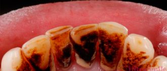
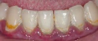
- Tartar;
- Fluorosis.
Let's talk about these diseases and methods of treating them in more detail.
Gingivitis: my gums hurt
The process, which is characterized by gum inflammation, swelling, redness and bleeding, is called gingivitis and is one of the most common periodontal diseases in both children and adults. Only 3% of people can boast of absolutely healthy gums. How to get into such a small percentage of lucky ones? The answer is simple - follow the necessary measures to prevent gum inflammation, regularly visit the doctor and not let even seemingly harmless signs of an incipient disease take their course.
Gingivitis is the last of all diseases in periodontology in which the inflammatory process can still be stopped. Next comes periodontitis, a disease in which inflammation spreads to other periodontal tissues. From this point on, treatment is based only on bringing the disease into remission and attempts to relieve symptoms as much as possible at the time of relapses, as well as in the future when it is necessary to resort to tooth extraction. Therefore, gingivitis in children and adults requires increased attention in order to avoid serious periodontal problems.
How to care for your gums after implantation
After dental implantation, it is recommended to completely abstain from eating and drinking for two hours. Next, for 2-3 weeks, nutrition should be gentle, without consuming solid, excessively hot or cold foods.
One of the most important rules is to regularly rinse your mouth with prescribed antiseptic solutions, chamomile or other ingredients recommended by your doctor.
It is preferable to chew on the side opposite the suture, however, this recommendation will be individual for each patient. During the first 24 hours after the installation of dental implants, it is not recommended to brush your teeth, but in more detail about how to care for your gums after implantation, the doctors of the Apex dental clinic will tell you at your consultation.
Types of gingivitis
Gingivitis differs in the nature of its course:
- Acute gingivitis is a disease whose symptoms appear suddenly and progress quite quickly.
- Chronic gingivitis is a sluggish process, the symptoms of which increase gradually.
- Aggravated gingivitis (recurrent stage of a chronic process) is an increase in the symptoms of a chronic disease.
- Gingivitis in remission is the moment of complete relief of all symptoms.
The form is:
- catarrhal gingivitis, which is manifested by swelling and redness;
- ulcerative (ulcerative-necrotic) gingivitis, with necrotic (dead) areas of the gums;
- hypertrophic gingivitis, in which there is a significant increase in the volume of gum tissue and its bleeding;
- atrophic gingivitis, on the contrary, is characterized by a decrease in the volume of gingival tissue;
- desquamative (geographic) gingivitis, which is manifested by intense redness and abundant desquamation of the epithelium of the mucous membrane.
According to its distribution in the oral cavity, gingivitis can also be local (affects some areas of the teeth) and generalized (the process affects the gums of the entire jaw or both jaws). And according to severity - mild, moderate and severe gingivitis.
Why can baby teeth turn black?
Baby teeth are much more susceptible to caries than molars, since they do not have such a dense structure. Enamel can change color due to the development of various pathological processes that can be provoked in a child:
- fragility of tooth enamel;
- calcium deficiency;
- improper oral care;
- chips and cracks in the enamel;
- vitamin deficiency;
- endemic fluorosis;
- chronic gastrointestinal diseases;
- genetics.
Sometimes the cause of darkening of the teeth of very young patients is artificial feeding of the baby at night. Consuming formula or milk at night reduces saliva production. The acid that accumulates on the teeth during feeding is not washed away by saliva and the process of destruction of tooth enamel begins.
Causes of gingivitis
Most often, gingivitis develops as an independent disease, but sometimes the causes of its occurrence are acute and chronic diseases of the gastrointestinal tract, cardiovascular system, hematopoietic organs, infectious diseases, and changes in hormonal levels. In this case, gingivitis is one of the symptoms of the underlying pathology. The causes of gingivitis can be internal or external.
Internal reasons include:
- tooth growth that injures the gums - the eruption of wisdom teeth;
- vitamin deficiency, hypovitaminosis (most often lack of vitamin C and zinc);
- weakened immune system;
- metabolic disease;
- allergic diseases;
- diabetes;
- stress, mental illness;
- anomalies and various deformations of the gums;
- diseases of the gastrointestinal tract.
External reasons are a number of factors:
- physical (injuries, burns);
- chemical (the influence of aggressive substances);
- medical (incorrectly applied fillings, incorrectly installed veneers, traumatic wearing of braces);
- bad habits (smoking, mouth breathing);
- biological (infectious process);
- hygienic (insufficiently thorough hygienic procedures).
Toxins from microorganisms that enter the oral cavity with food and water, as well as those that live there permanently, form dental plaque (plaques) due to insufficient hygiene measures. They are the most common cause of the inflammatory process.
Inflammation can develop differently depending on the cause. Chronic catarrhal gingivitis occurs most often due to unsatisfactory hygiene measures or as a result of gum injury or burns. Hypertrophic gingivitis is caused by crowded teeth, incorrectly installed fillings or dental crowns, as well as changes in hormonal levels, for example, during pregnancy (pregnant gingivitis) or puberty (adolescent or juvenile gingivitis). Necrotizing ulcerative gingivitis (Vincent gingivitis) is usually caused by an infectious process. It occurs due to the activation of two microorganisms (Vincent spirochete and spindle bacillus) against a background of weakened immunity, hypothermia, stress or malnutrition.
Fluorosis
Fluorosis is a dangerous dental disease that is caused by consuming extremely large doses of fluoride. Usually this substance enters the human body along with tap water. In addition, the source of fluoride can be the work environment or even the wrong toothpaste.
Fluorosis begins with the appearance of white, dull spots on the surface of the teeth. They are not always visible to the naked eye. Over time, the spots change color - they first turn yellow, then turn brown. If you don’t consult a dentist at this point, fluorosis can turn into erosive or destructive forms - when the disease begins to destroy the enamel.
Treatment of fluorosis is almost always conservative. The dentist remineralizes the enamel, restoring the balance of calcium, phosphorus and fluoride. Darkened areas are bleached. In addition, the patient is recommended to change his lifestyle to reduce the amount of fluoride consumed - for example, install a special water filter at home or change his place of work.
Fluorosis is often “epidemic” in nature. If it is caused by an increased content of fluoride in tap water, it develops in the whole family. If the reason was a violation of industrial safety - for the entire team. In the Ryazan region, fluorosis is often detected in residents of the Rybnovsky, Chuchkovsky and Shatsky districts, where the water contains an increased amount of fluoride.
Forms of gingivitis and symptoms
Signs of gingivitis directly depend on the nature of the disease and its form. Let's look at each form of gingivitis separately. So, complaints and visual inspection.
Catarrhal gingivitis
This form of the disease usually occurs without obvious pain. Its immediate symptom is bleeding gums when brushing teeth, eating solid foods and other mechanical effects on the dental system.
Ulcerative-necrotizing gingivitis
This is one of the most unpleasant forms of gingivitis, which is characterized by a feeling of itching of the gingival papillae, severe pain, copious flow of saliva, fever, inflammation of the lymph nodes and the formation of necrotic areas of the gums.
Hypertrophic gingivitis
Patients suffering from this form of gingivitis complain of severe pain, constant bleeding of the gums and a significant increase in the volume of the gums, which can partially cover the crowns of the teeth from the outside (not from the tongue). At the same time, the patient’s gum remains quite hard and under it, on the teeth, tartar forms, which creates favorable conditions for the proliferation of microorganisms. With hypertrophic gingivitis, teeth may move slightly.
Atrophic gingivitis
The last and most advanced stage of gingivitis, often leading to periodontitis, is atrophic gingivitis. With it, the gum tissue becomes thinner, decreases in size, the necks of the teeth, and sometimes their roots, are exposed. Teeth become more sensitive to temperature changes (cold or hot drinks, frosty air), to sour or sweet foods, to the mechanical impact of a toothbrush.
Desquamative (geographic) gingivitis
The symptoms of this form of gingivitis differ from others by pronounced red spots on the gums, desquamation of the upper layer of the epithelium, the appearance of blisters on the gums and the formation of mouth ulcers and erosions.
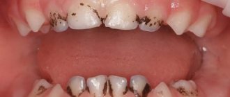
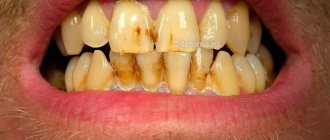
What is Priestley plaque on teeth?
This is a dental problem that occurs in children. In this condition, deposits are found on the enamel of the teeth. Dark plaque on a child’s teeth can be black or brown, sometimes with a light or pearlescent tint. As a rule, it is located as a thin border along the lower edge of the teeth closer to the gums. But plaque can also cover a large area of the tooth or look like a small spot on the enamel. Most often, dark deposits appear on the inside of the teeth, less often in the interdental space or on the outer surface of the teeth.
Photo courtesy of InWhite Medical Clinic
Diagnostic tests
- Schiller-Pisarev test
This test is based on determining the level of glycogen in the gum. Its amount increases significantly during inflammation, while healthy gums do not contain glycogen. Lubricating the inflamed gums with Schiller-Pisarev solution gives a color change reaction from light brown to brown. This research method is used to make diagnoses of both periodontitis and gingivitis.
- Assessment of oral hygiene level
A solution (2 g of potassium iodide, 1 g of crystalline iodine, 40 ml of distilled water) is applied to the outer surface of the six lower front teeth.
The assessment is carried out using a five-point system and each tooth is assessed separately:
- 5 points – complete staining of the entire tooth surface;
- 4 points – staining of ¾ of the tooth surface;
- 3 points – staining of half the tooth surface;
- 2 points - staining of a quarter of the tooth surface;
- 1 point - absence of any staining of the tooth surface.
Then the scores of all examined teeth are summed up and divided by their number (usually the test is carried out on six teeth). This is how the hygiene index is obtained.
As a result, the quality of hygiene is assessed:
- 1.1-1.5 points – good hygiene index;
- 1.6—2.0—satisfactory hygiene index;
- 2.1—2.5—unsatisfactory hygiene index;
- 2.6—3.4—poor hygiene index;
- 3.5-5.0 - very poor hygiene index.
- Vacuum test according to Kulazhenko
Using a Kulazhenko vacuum apparatus, it is possible to determine the time of hematoma formation when a vacuum is applied to the gum area. Typically, the test is carried out in the incisor area by placing a tube of the device on the gum. The formation of a hematoma in 60 seconds indicates the normal condition of the gums, while the appearance of a hematoma in 29-30 seconds signals an inflammatory process.
- Oxygen tension in gum tissue
The sensor of the device is applied to the gum, and the device records the level of tissue hypoxia. Reduced oxygen tension indicates a prolonged inflammatory process.
After implantation, the gums turned blue: other symptoms and diagnosis of peri-implantitis
Regardless of the reasons that caused peri-implantitis, this disease can be characterized not only by changes in the color of the gums, but also by other symptoms. These include bleeding gums in the area of the dental implant, deformation of the gums in this area, mobility of the crown, as well as painful or uncomfortable sensations in this localization.
Only a dentist can diagnose peri-implantitis when conducting an objective examination of the patient’s oral cavity. As a rule, this complication is characterized by all signs of inflammation and may involve swelling and redness, a local increase in temperature in the soft tissues of the oral cavity, etc.
When contacting the Apex dental clinic, already at the initial appointment, the doctor will accurately determine the cause of darkening of the gums after installation of the implant,, if necessary, prescribe additional instrumental research methods (radiography), and also select an individual treatment regimen.
By choosing the Apex dental clinic, you are making a choice in favor of high-quality dentistry and a quick solution to all dental problems that arise!
Differential diagnosis of gingivitis
It is based on the complaints presented to the patient, a visual examination of the patient, the results of functional tests and laboratory tests. The goal of differential diagnosis is to distinguish gingivitis from other periodontal diseases, such as periodontitis and periodontal disease.
The main feature that distinguishes gingivitis from other periodontal diseases is that the inflammatory process affects only the gum tissue, the remaining structures (muscle ligaments that hold the tooth in the jaw and bone tissue) remain unchanged.
Along with this symptom, gingivitis is not characterized by periodontal pockets, exposure of the necks of teeth, or their mobility. And the x-ray shows no signs of bone resorption.
Identifying gingivitis in a timely manner, determining its form and prescribing the correct treatment is the task of a periodontist. But not to forget about prevention and regularly visit the dental clinic is the maximum program for the patient. This is the only way to avoid a more serious periodontal disease – periodontitis.
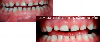
Treatment of a darkened tooth
Darkening of the tooth near the gum requires immediate consultation with a doctor, regardless of the cause. If you leave the disease to its own devices, you can face more dangerous and serious consequences.
The doctors at our regional dental clinic are ready to help. We use modern equipment, progressive therapeutic practices and always put the patient's comfort as a priority. Therefore, treatment of a darkened tooth will be quick and effective. Don’t let the disease lead to dangerous consequences - make an appointment with the dentist!
How to detect dark plaque in time
- regularly examine the child’s oral cavity independently;
- do not postpone scheduled visits to the dentist.
It must be taken into account that Priestley plaque on the teeth often accumulates slowly, which is why the child gets used to the stains on the enamel and does not tell his parents about them. Darkening of teeth can occur quickly, literally overnight. Reactive appearance most often occurs during illness, accompanied by intoxication, dehydration and increased body temperature. This is due to the fact that the child’s body reacts sharply to various changes in diet, the functioning of internal organs and in external conditions.
Deposits most often occur at the age of 2-3 years. However, they can also appear on the very first milk teeth of a one-year-old child. Or it could affect the baby teeth of a younger teenager who haven’t had time to change. The darkening may also spread to permanent teeth.
Content:
- Why isn't every spot a black tooth decay?
- When there is black caries, but it is still not treated
- How to deal with little black caries
- When black caries still needs to be filled?
Sooner or later, small dots or dark-colored depressions appear on the teeth of any person. They are usually perceived as caries. But, when visiting a dentist, the patient may be told that he does not need to treat his teeth. Why is this happening? Is it true that not every black spot on the crown of a tooth is a cavity?
Causes of a black spot on the gum as a result of necrosis
A black spot on the gum due to necrosis often appears in patients who pay insufficient attention and neglect to the health of their teeth and oral cavity. Untimely removal of chips, cracks and treatment of caries risks leading to the most unpleasant consequences. Without receiving sufficient nutrition, the cells of the mucous membrane age and begin to die.
Over time, a small black dot on the gum will spread and turn into a very noticeable dark spot that is easy to see even with the naked eye. This is the main sign of necrosis of gingival tissue. In order to prescribe effective treatment and slow down the process of gum death, it is important to correctly determine the cause of the pathology:
- improper adherence to hygiene procedures with the use of low-quality pastes and rinses;
- exacerbation of pulpitis, accompanied by the accumulation of pus, which decomposes all nearby tissues;
- wearing an incorrectly fitted prosthesis;
- disruptions in the functioning of the endocrine system (detected when additional examination is ordered if preliminary diagnostic methods have not yielded results);
- complications of radiation therapy.
Dentists often have to deal with necrosis of gingival tissue in patients who have undergone dental treatment using outdated techniques, in particular, using arsenic. The substance is dangerous; it is deeply absorbed into cells, contributing to their destruction.