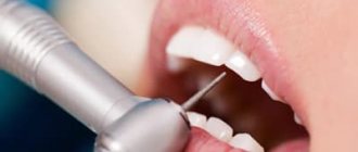
Closed curettage of periodontal pockets
Closed curettage or curettage is one of the few methods of treating periodontitis that allows you to eliminate granulations and various types of dental plaque from periodontal pockets. However, due to the lack of visual control, the doctor is not always able to completely eliminate these problems, which reduces the effectiveness of this method. That is why it is used for shallow periodontal pockets, which is typical for the earliest stages of the disease. With a more extensive lesion, the technique can only temporarily improve the patient’s health, but sooner or later periodontitis will begin to recur, which will require re-treatment.
Among the advantages of this type of surgical treatment are:
- Good aesthetic result.
- Rapid gum recovery.
- Minor blood loss.
Due to the features described above, closed curettage is not always used.
Where to contact?
Modern treatment methods used in the CELT clinic can significantly slow down the development of the disease, and often stop it for a long period. Our experts recommend regular dental examinations, which allow you to identify the disease at an early stage, but even if the disease is advanced, we will do everything to stop it.
Surgical methods for the treatment of periodontitis are selected by our specialists based on diagnostic studies and examination of the patient. Our specialist will explain the current situation to you and tell you about the treatment methods that will be optimal in your case. He will provide full information about their advantages and disadvantages, calculate the cost of treatment and answer any questions you may have.
Make an appointment through the application or by calling +7 +7 We work every day:
- Monday—Friday: 8.00—20.00
- Saturday: 8.00–18.00
- Sunday is a day off
The nearest metro and MCC stations to the clinic:
- Highway of Enthusiasts or Perovo
- Partisan
- Enthusiast Highway
Driving directions
Open curettage of periodontal pockets
This type of operation does not have the disadvantages of the previous method. A good overview of the problem area allows the doctor to remove all granulation tissue and thereby prevent further progression of the disease. Open curettage is performed under local anesthesia and takes place in several stages:
- The surgeon makes an incision in the gum in the projection of the neck of the tooth.
- A certain area of the mucous membrane peels off.
- Granulations and deposits in periodontal pockets are eliminated.
- If necessary, plastic surgery of bone defects is performed.
- The flap is sutured, the surgical field is treated and protected with an aseptic dressing.
Due to the high traumatic nature of soft tissues, a limited area of gum can be treated in one operation, usually up to 8 teeth. After the wound has healed, repeated interventions are performed.
Preparation and stages of treatment
Based on an assessment of the results of preliminary diagnostics, a treatment plan is formed that takes into account the need to comply with standards that prevent the spread of infection. The surgical site is anesthetized for the entire period of the operation, which lasts no more than 1.5-2 hours. Upon completion of the procedure, slight discomfort is allowed, caused by a radical change in the structure of the tissues, the signs of which persist for a certain period of time.
The standard protocol for periodontal surgical treatment includes:
- Carrying out sanitation and professional cleaning;
- The use of medications that limit and relieve inflammation;
- Fixation of an orthopedic device or medical splint on movable units;
- Prescribing antibiotics that stimulate the patient’s immunity.
Tissue restoration is accompanied by the application of a soft bandage. If necessary, the doctor also prescribes a course of medications that eliminate the possibility of infection and stimulate regeneration.
Why is curettage needed?
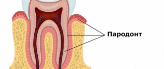
The process of development of periodontitis is slow and occurs in a certain sequence. First, due to poor oral hygiene, the active proliferation of microorganisms begins, which in the process of their vital activity release a large number of toxins. These toxins have a negative effect on the gums and cause inflammation.
If the patient does not seek medical help at this stage, the inflammation gradually leads to the detachment of the gum from the tooth and the formation of periodontal pockets. These pockets contain food debris and soft dental deposits, which over time mineralize and become hard. Ideal conditions are created for the proliferation of microorganisms that affect not only the gums, but also the bone structures. As a result, granulation tissue is formed, which contributes to the rapid destruction of bone and loosening of teeth. If it is not completely removed, periodontitis will progress further, leading to more severe consequences. In order to effectively clean periodontal pockets from dental plaque, hard deposits and granulations, curettage is used.
Symptoms and signs of periodontitis
Mild periods of periodontitis are usually asymptomatic. The patient may experience moderate discomfort, slight gum bleeding and redness. These symptoms often go unnoticed and are therefore not a reason to seek medical help.
In later stages, the signs of periodontitis become more pronounced. It can be:
- severe bleeding gums;
- pain when pressed or when eating;
- bad breath, which is difficult to get rid of with home remedies;
- increasing spaces between teeth;
- noticeable tooth mobility;
- discharge of pus from gum pockets;
- increased sensitivity to hot and cold foods;
- visual lengthening of teeth due to exposure of the neck.
These signs may be combined with general symptoms, which include fever, weakness, fatigue, and poor appetite.
You should contact your dentist if you experience any symptoms from your gums and teeth. The initial stages of periodontitis respond well to treatment using conservative methods, while advanced forms require long-term, complex and expensive treatment.
Flap surgery for periodontitis
Flap surgery is indicated for patients with pronounced periodontal changes: pocket depths of more than 5 mm, active inflammation, etc. This technique allows not only to eliminate various types of deposits and granulations, but also to restore interdental defects and bone tissue defects. To form a flap, precise cuts are made in a specific direction:
- Horizontal cuts are always made. They can run either inside the gingival sulcus or parallel to the gum edge.
- Vertical incisions allow you to expand the surgical field, but lead to the formation of scar changes in the gums, so they are performed only when necessary.
Among the disadvantages of flap surgery, there is a possibility of flap displacement and exposure of the neck of the tooth. This complication worsens the aesthetics of the oral cavity, contributes to increased tooth sensitivity and the development of tooth root caries.
Bone grafting
In severe periodontitis, there is a loss of bone tissue, which contributes to the loosening of teeth. In order to stop this process, bone grafting is used. The patient's own tissue, as well as artificial substitutes, can be used as a graft. In the first case, a section of bone can be taken from the chin, upper palate, or lower jaw. Since the patient’s own tissues are absolutely biocompatible, this method is the most effective, but requires two operations: tissue collection and transplantation.
Special bone substitutes do not have this disadvantage, so bone grafting in such cases is carried out in one stage. However, this method is characterized by a lower tissue survival rate, so it is not always possible to achieve the desired result with its help.
Plastic surgery of lip cords and frenulums
In the presence of pronounced strands of the mucous membrane, the risk of periodontal disease increases significantly. Massive folds that are fixed to the gingival papillae, when stretched, can displace them and lead to various problems. In order to prevent such conditions, band plasty is performed. Typically, the technique consists of excision of these folds and subsequent suturing of the resulting defect.
Lip frenuloplasty can be performed in the following cases:
- Pronounced tooth gap between the front incisors.
- Preparation for orthodontic treatment.
- Presence of gum recession.
- Before installing removable dentures.
- Incorrect sound pronunciation.
Surgical treatment can be performed in various ways. If the frenulum is very narrow, then it is cut transversely. If there is a wide frenulum, its excision is performed. In addition, individual sections can be moved to the desired position.
Among modern techniques, laser frenuloplasty is noted. The procedure is very quick, does not require stitches, has a short recovery period and rarely leads to complications.
After plastic surgery of the cords and labial frenulum, a period of rehabilitation begins. Typically, the patient is required to follow simple rules, which include careful oral hygiene and a certain diet. Typically, recovery takes about a week.
Closing gum recessions
Closing gum recessions is carried out only by surgical methods, which involve moving donor flaps to the recession zone and plastic surgery of the existing defect. You can receive a transplant in several ways:
- Pedicled flap. In this case, the donor tissue is a nearby area of the mucous membrane. The technique is used in the presence of localized defects.
- Gingival flap. It is taken from the gum area, provided that the volume of tissue in the selected area is sufficient.
- A scrap from the sky. In this case, the donor fragment is taken from a specific area of the hard palate
To close recession zones, the method of directed tissue regeneration can be used. In this case, special membranes are used as donor tissue, which can be absorbable or non-absorbable. These membranes promote the migration of cells from natural tissues and subsequent regeneration of the damaged area.
This method does not require the formation of a donor wound, but is not suitable for all patients. To obtain a successful result, the membrane must be covered with a layer of mucous membrane, which will provide the necessary nutrition, which is not always possible. In addition, due to the long process of formation of new tissue, it is necessary to observe certain rules of oral hygiene, therefore this method requires the active and conscientious participation of the patient in the treatment process.
Surgical methods for treating periodontal diseases
Inflammatory periodontal diseases are currently the most common diseases in the world. Any restoration, from a small filling to a complex implant bridge, can only be performed when the supporting periodontal structures are healthy and free of inflammation[1].
It is advisable to treat periodontal diseases in a comprehensive manner using general and local therapy. The surgical method in the complex treatment of the vast majority of periodontal diseases is a priority. It is carried out after conservative therapy and is aimed at eliminating local causes that support inflammation: removing dental plaque, including subgingival plaque. Curettage of granulations and deepitalization of the gingival pocket.
All periodontal interventions can be divided into two groups. The first group includes interventions aimed at eliminating periodontal pockets:
- Closed curettage of periodontal pocket.
- Open curettage of periodontal pocket.
- Gingivectomy.
- Flap operations.
- Apically displaced flap.
- Guided regeneration of periodontal tissues.
The second group includes interventions aimed at eliminating structural disorders of the soft tissues of the vestibule of the oral cavity, which not only aggravate the course of the inflammatory process in the periodontium, but in some cases are themselves the causes of its specific lesions. This:
- Plastic surgery of frenulum and cords (frenulotomy and frenulectomy).
- Vestibuloplasty.
- Operations to eliminate recessions. [2]
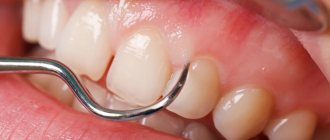
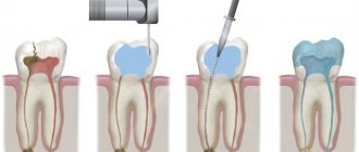
Curettage.
The purpose of curettage is to remove granulations, vegetative epithelium, decayed tissue, tartar, and damaged cement from the periodontal pocket. Scraping of the contents of the periodontal pocket is carried out with special instruments[3].
As a result of curettage, a clean wound surface should be obtained. Curettage is indicated in the presence of pockets no more than 4 mm deep, dense gums, and in the absence of bone pockets. For deeper pockets, preference is given to gingivotomy or gingivectomy, or one of the flap operations.
Curettage is contraindicated in the presence of acute inflammatory processes - abscesses, thinned and fibrously changed gums (regardless of the depth of the pocket), since the fibrously changed wall does not adhere well to the wall of the tooth. And also in the presence of bone pockets and tooth mobility of 3-4 degrees [4].
Gingivectomy.
Kinds:
- Simple.
- Radical.
- Partial.
Indications for simple gigivectomy are gingival pockets more than 4-5 mm deep with horizontal, uniform destruction of the alveolar bone; hypertrophic gingivitis; fibrous gum compaction; the need to lengthen the clinical crown of the tooth before orthopedic treatment; providing better support for the clamps of the rubber dam system, during patchwork operations, and with gingival fibromatosis.
Gingivectomy is contraindicated in the presence of deep bone pockets and a narrow zone of attached gum.
Flap operations.
Used to eliminate periodontal pockets, restore lost supporting tissue, i.e. formation of new connective tissue attachment and resumption of bone growth[5].
These surgical interventions are based on the Cieszynski-Widman-Neumann operation, which is performed when the pocket depth is more than 6 mm. The essence is to cut out and fold back the mucoperiosteal flap, followed by careful treatment of the roots of the teeth, bone pockets, and the inside of the flap. To date, several dozen modifications of this operation have been proposed. The advantage of these operations is the complete removal of pathologically altered tissues under visual control, ensuring longer-term stabilization of the periodontal process. The disadvantages are exposure of the necks of the teeth, some trauma, decreased alveolar process height, increased tooth mobility, dentin hypersthesia, and cosmetic defects[3].
Guided regeneration of periodontal tissues. (NRT)
The histological development of the root from the Hertwig epithelial sheath has been well studied. As in the case of embryonic development and healing of any other tissues, the process is regulated by genetically determined mediators. These include growth and differentiation factors, including bone morphogenetic proteins and special matrix proteins of the Hertwig epithelial sheath.
The classic NRT procedure involves installing a physical barrier (membrane), without the use of biological factors. A physical barrier placed in the desired location prevents apical epithelial proliferation or the formation of long junctional epithelium during defect healing.
All barrier materials are divided into the following groups:
- Synthetic non-resorbable.
- Synthetic resorbable.
- Naturally resorbable.
When using absorbable membranes, a second surgical intervention is not required. Therefore, such membranes are preferred in periodontics. There is a wide range of membranes on the market, none of which are universal and suitable for all cases. Directed regeneration requires sustainable materials[1].
An ideal membrane should have the following characteristics:
- Safety in terms of transmission of infections.
- Biocompatibility (lack of toxic and immunogenic properties).
- Easy adaptation to root and bone surfaces.
- Rigidity (the membrane should not sink into the bone defect).
- Permeable to some molecules, but not to cells.
- Immobility after integration into tissue.
- Long lasting stability to maintain tissue space.
- Controlled biological resorption.
- Additional antimicrobial and biostimulating properties.
Frenulotomy.
It is carried out with a shortened frenulum of the tongue by cutting it. It is advisable to perform this operation as early as possible - in infancy and childhood.
Frenulectomy.
It is performed when there is a short frenulum of the tongue or lip, as a result of which a diastema subsequently develops. Two semi-oval vertical incisions are made, excising the frenulum along with compact osteotomy. Having mobilized the mucous membrane along the edges of the wound, the latter is sutured tightly [6].
Vestibuloplasty.
It is carried out with a shallow vestibule of the oral cavity and involves moving the facial muscles attached to the crest of the alveolar process deep into the vestibule of the oral cavity.
Vestibuloplasty is used to create conditions for the anatomical retention of complete removable dentures, to restore the buffer function of the oral vestibule in case of periodontitis of the anterior teeth, in the case of a small vestibule of the oral cavity, as well as for endosseous implantation, when high muscle attachment to the alveolar process causes ischemia and inflammation of the gingival tissue. cuffs of functioning dental implants. Healing occurs by secondary intention or the wound is closed using free mucosal grafts[2].
Operations to eliminate recessions.
To date, the Muller classification is most often used: Class 1.
The recession does not reach the mucogingival border. 2nd Grade. The recession crosses the mucogingival boundary. 3rd Grade. Loss of attachment is also proximal (loss of gingival papillae) Class 4.
Proximal loss of attachment combined with tooth position abnormalities. Recessions 1 and 2 are eliminated surgically. For recessions of classes 3 and 4, only partial elimination is possible[7].
Before carrying out manipulations, it is necessary to eliminate factors that may be the causes of recessions. Currently, there are many modifications of surgical interventions to eliminate recessions:
- Coronal displacement flap.
- Laterally displaced flap technique.
- “Envelope” technique using a subepithelial palatal flap.
When performing interventions, it is desirable that the thickness of the mucoperiosteal flap in the intervention area be at least 1.5 mm.
The success of surgical treatment of periodontitis depends on a set of measures, including conservative therapy and adequate orthopedic treatment.
Literature:
- Wolf G.F., Rateitskhak E.M., Rateitshak K. “Periodontology”, 2008, “MED-press-inform”, Moscow
- Grudyanov A.I., Erokhin A.I. “Surgical methods for the treatment of periodontal diseases”, 2006, “Medical Information Agency”, Moscow
- Tsepov L.M., Nikolaev A.I. “Diagnostics and treatment of periodontal diseases”, 2002, “MED-press-inform”, Moscow
- Ivanov V.S. “Periodontal diseases”, 1998, “Medical Information Agency”, Moscow
- Borovsky E.V., Ivanov V.S., Vagner V.D. “Therapeutic Dentistry”, 2004, “Medical Information Agency”, Moscow
- Robustova T.G., Romacheva I.F., Afanasyev V.V., 1996, “Medicine”, Moscow
- Müller H.-P. “Parodontology”, 2004, “GalDent”, Lviv
Transplantation of a flap from the palate
Recession flaps are usually taken from the hard palate. In this case, the graft can consist only of connective tissue, or be epithelialized. The thickness of the flap depends on the tasks and the result that needs to be achieved. Direct closure of gingival recessions requires thick flaps, while restoration of visible gingival areas is carried out using thin and epithelialized fragments.
The graft is usually taken from the area of the hard palate, which borders the canines and premolars. In this case, it is necessary to retreat 2 mm from the edge of the gum. These rules allow you to avoid crossing the palatine artery and the development of bleeding. The thickness of the mucous membrane from the donor area should not be less than three millimeters, so probing is carried out first. After obtaining a flap, the defect is sutured