A fistula on the gum (also called a fistula) is an opening in the gum from which pus is released, often with blood. A fistula appears against the background of caries, periodontitis, and also due to poor-quality dental services. Over a long period, the fistula develops asymptomatically, but subsequently can lead to severe complications. Therefore, if a purulent formation appears on the gum, you should immediately contact your dentist.
Fistula on the gum: causes
A fistula on the gum is a hole that forms as a complication after inflammatory pathologies, including untreated ones. It can also occur after dental intervention (poorly provided services). Very often, a fistula is formed as a complication of long-term chronic periodontitis.
Also, the reasons for its appearance may be:
- damage to the tooth root;
- advanced caries;
- pulpitis;
- inflammatory processes of the cyst;
- problems with the growth of wisdom teeth;
- improper teething in children;
- granuloma - inflammatory processes in the tissues of the periosteum and the apical region of the tooth root (accompanied by an increase in temperature, enlarged lymph nodes and other signs).
Fistula in the soft periodontal tissues after tooth extraction
The formation of a hole after tooth extraction is normal. As a rule, the hole heals within one to two weeks. If the operation was performed on wisdom teeth, the tightening process may take several weeks. Impaired socket tightening can occur due to infection or in case of injury to the area where the tooth was removed.
In a situation where the hole has tightened and a gap has formed in the periodontium, it may indicate that the tooth was not completely removed. Small fragments of the tooth gradually begin to decompose, forming a fistula.
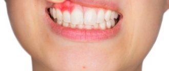
How a fistula is formed
The process of fistula formation looks like this. A small hole appears in the gum near the base of the tooth. Its color stands out a little against the background of healthy tissues - the color is rich pink or red. At the same time, the remaining teeth remain healthy and do not hurt.
At first, the fistula looks like a small swelling, then it grows and resembles a pimple or an abscess. The last stage - the seal opens, after which mucus or pus comes out of it. Then the wound becomes covered with a scar, but does not disappear. In the future, the development of the fistula can become chronic - it will open several times a year and exude pus. During exacerbations, mild pain will be felt.
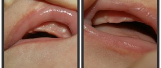
The appearance of a hole in the periodontium in a child
The formation of a hole in the gum near a tooth in a child can be due to reasons such as: periodontitis caused by complicated caries, or injury to the oral mucosa. With periodontitis, the lesion reaches the jaw bone. A cavity appears inside the periosteum, which is filled with pus. When there is an excess of pus, it breaks out through the fistulous tract, thereby bringing severe pain.
Frequent mechanical damage to the mucous membrane and gums causes infection to enter the oral cavity. Bacteria entering the periodontium cause inflammation and fistula formation.
Symptoms of a fistula: how dangerous is it?
Fistula manifests itself with a number of symptoms:
- unpleasant odor from the mouth;
- tooth mobility;
- feeling of foreign taste;
- mucous membrane is bluish or, conversely, pale in color;
- discharge of purulent mucus, often with blood;
- painful sensations during mechanical contact (chewing food, drinking hot drinks, brushing teeth).
The disease may also be accompanied by symptoms not directly related to the oral cavity. It is manifested by such signs as apathy, general weakness, and fever.
Despite the small area affected, the fistula poses a certain health hazard. Thus, purulent discharge can penetrate the lymph or blood, which will lead to diseases of the internal organs, pathological processes in facial tissues and even loss of teeth. Therefore, even at the first relatively mild symptoms, it is important to consult a doctor.
It is worth keeping in mind that the disease is asymptomatic for a very long time. Moreover, if treatment is delayed, this can lead to partial death of the periosteum. And then the patient will have to remove not one, but several teeth at once.
Prognosis and possible complications
If the fistula is detected and treated in a timely manner, the prognosis is favorable. If you follow all the specialist’s recommendations, within a week the resulting wound will completely heal and heal.
If proper treatment is not carried out, the following consequences are possible:
- abscess formation;
- phlegmon;
- osteomyelitis;
- tooth loss.
Preventive measures
- regular teeth cleaning;
- carrying out professional hygiene in the dental office;
- preventive examinations by a dentist every six months;
- daily consumption of fruits and vegetables.
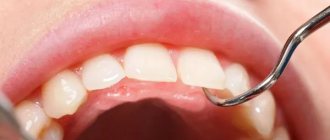
Diagnosis and treatment of fistula
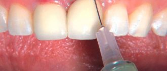
To carry out a diagnosis, you must contact your dentist. The doctor orders an x-ray and conducts a visual examination. Treatment is carried out in a clinical setting. Therapy depends on the cause of the fistula. For example, fillings are used to treat caries, and when an infection is detected, appropriate medications are used. The dentist can also perform treatment with ultrasonic waves or laser.
In some cases, a fistula appears due to poorly done filling of the canal. Then the patient is given an anesthetic, the filling is removed, the dental canal is cleaned and then a new high-quality filling is performed.
If the cause is a cyst, it is removed, which also requires the intervention of a dentist. For recovery, the use of antimicrobial agents, antibiotics, and antihistamines is indicated. If swelling occurs, the doctor may prescribe rinsing with a salt solution.
Pericoronitis: treatment
If you have inflammation of the gums near the wisdom tooth, the treatment most often consists of a dental surgeon removing the hood over the wisdom tooth. However, if severe purulent inflammation is observed, then complete excision of the hood is undesirable immediately, because this can lead to various inflammatory complications.
In case of severe purulent inflammation, the hood is first only dissected to facilitate the outflow of purulent discharge, and anti-inflammatory therapy is prescribed. And the doctor will prescribe you for its complete removal after the active inflammation has subsided. Also, in some cases, the doctor may recommend immediately removing the wisdom tooth (24stoma.ru).
Excision of the hood over the wisdom tooth –
Removing the hood of a wisdom tooth involves excision of the overhanging mucous membrane over the erupting eighth tooth. Excision of the hood over the wisdom tooth leads to the elimination of conditions for the proliferation of pathogenic bacteria. This minor surgical procedure is usually less traumatic, but in some cases a large amount of gum tissue must be excised.
Excision of the hood over the wisdom tooth is performed by a dental surgeon under local anesthesia. The procedure is completely painless if you see a good specialist, if the anesthesia is administered correctly and a good anesthetic is used, and not something like novocaine. Pain will appear only after anesthesia has passed (after 30 minutes), so it is worth taking an analgesic even before the pain appears.
- Hood removal: price for 2022 in an economy class clinic in Moscow, a similar service costs about 2,500 rubles. In the regions, the cost of the procedure may be 2 times lower. By the way, in the clinic at your place of residence (if you have an insurance policy and a passport), you should undergo this intervention completely free of charge.
Stages of excision of the hood –
- Conducting local anesthesia,
- Using a scalpel and surgical scissors (less commonly, a surgical laser), the dental surgeon excises the gum overhanging the tooth.
- Treating the wound with antiseptics.
- An iodoform turunda is usually placed in place of the excised hood.
- The doctor gives recommendations and schedules a re-examination.
Removing a wisdom tooth hood: video
Please note that both hood excision operations are performed with a surgical laser and not with a scalpel. Using a laser avoids bleeding, swelling and severe pain. In Russian dental clinics, lasers are practically not used (due to their absence), and only a few clinics can boast of their presence.
After the intervention, the following are prescribed:
- Antiseptic baths with chlorhexidine solution 0.05% (3-4 times a day);
- Antibiotics are not prescribed in every case;
- for pain - good tablet analgesics.
Usually this is enough for you to completely forget what wisdom tooth pericoronitis is after 4-5 days.
However, if the doctor performed the operation traumatically, the pain may last for 7-10 days. If you want to remove inflammation as quickly as possible, then after antiseptic rinses, you can additionally apply CholisalGel to the hood 2 times a day in the morning and evening (it has a pronounced analgesic and anti-inflammatory effect). Remember that if you put gauze soaked in iodoform on the wound surface, you need to remove it yourself no later than the next day. Then it itself will become a breeding ground for infection. After you take out this turunda, it may cover a little. Then it is advisable to treat the wound with a gauze swab soaked in 3% hydrogen peroxide.
Important: in some cases, the hood may form again, in which case either a repeat operation may be required, or the issue of tooth extraction will be decided. Often these teeth are in an incorrect position. An experienced doctor can quickly determine the chances of the eighth tooth taking the correct position using an x-ray and external examination of the tooth.
In what cases is it better to immediately remove a tooth with a hood -
If your gums near your wisdom tooth are inflamed, the most radical treatment method will be the removal of the 8th tooth, above which the ill-fated hood appears.
This will solve the problem permanently, but you must be prepared for the fact that the eighth teeth may have curved roots (this can be checked by taking a photo) and then removal may be difficult. Situations where deletion is the best solution to the problem -
- Firstly , when the lower jaw is insufficiently long, which means there is not enough space for the eruption of a wisdom tooth. Removal in this case will prevent the remaining teeth from being displaced by the erupting tooth, and will prevent the development of crowding of teeth in the anterior part of the lower jaw.
- Secondly , if the 8th tooth has a strong inclination towards the cheek or the seventh tooth, then it will still have to be removed sooner or later, because it will injure either the buccal mucosa or the root of the 7th tooth, respectively.
For more information about the difficult eruption of wisdom teeth, read the article: → “Features of the growth and eruption of eighth teeth”
Is treatment possible at home?
A fistula can only be treated in a clinic. But if it is impossible to urgently visit the dentist, for example, the pain occurred at night, it is recommended to rinse the mouth with the following means:
- infusion of chamomile;
- infusion of oak bark (dry raw materials can be purchased at a pharmacy);
- if there is purulent discharge, the mouth should be rinsed with a solution of salt and soda or antibacterial agents.
The described measures help relieve pain, but do not eliminate the cause of the disease. Therefore, in any case, you need to see a dentist.
Diagnostic methods
Typically, parents or a dentist detect fistulas at a very early stage. They are localized on the outer or inner side of the gums and reveal themselves by noticeable swelling and the appearance of a whitish lump.
It is very important not to self-treat when a problem is discovered. If your baby has baby teeth, improper treatment can pose a risk to the formation of the dentition. When the teeth are molars, there will also be a lot of problems, and there is a risk of needing extraction.
At the dentist's appointment, the doctor will carefully examine the child and take an x-ray. The location of the inflammation, its nature, and estimated depth are taken into account. Already at the first visit, the doctor begins to develop treatment methods to try to solve the problem without surgical intervention.
Prevention of fistulas
The main measure for preventing fistulas is maintaining oral hygiene. It is necessary to undergo an annual examination by a dentist and, if necessary, begin treatment for caries and periodontitis in order to prevent the development of chronic diseases.
It is also advisable to be treated by experienced, qualified dentists, since poor-quality services can also lead to the formation of a fistula. After the intervention, you need to monitor your well-being to prevent the development of a fistula.
Symptoms
Signs of fistula formation include:
- aching pain that increases in the evening;
- burning and itching of gums;
- swelling in the affected area;
- gum hyperemia;
- increased body temperature;
- general weakness, chills;
- increased irritability;
- apathy.
The appearance of symptoms of pathology is individual for everyone. Some may experience very severe pain and discomfort in the mouth, while for others the symptoms may be almost invisible.
What reasons can lead to its appearance?
The main cause of epulis on the gums is considered to be constant mechanical impact (rubbing) of soft tissues.
Injury to one area can cause gum swelling:
- poorly installed seal;
- uneven edges of the opposite tooth, which is destroyed or chipped;
- tartar;
- poor quality or incorrectly installed prosthesis;
- agonist tooth for malocclusion;
- adjacent teeth when teeth “pile up.”
Supragingival tissue occurs and, as a consequence, injury to epithelial tissue as a result of a burn, bruise or constant ingestion of irritating foods. In pregnant women, tumors occur due to hormonal imbalances. Epulis on a child’s gums can form due to general causes or disturbances in the eruption of permanent teeth.
What is epulis
A tumor on the gum is characterized by a benign nature. It is classified as a group of periodontal diseases. Epulis is also called “giant cell granuloma” or “supragingival”. Epulis looks like a lump that is attached to the gum in the area of the interdental space on a stalk. A formation is formed from the epithelium of the alveolar process.
In diameter, an epithelial tumor of the gums can reach a size of more than 3 cm. The shade of a giant cell granuloma can be different: bluish, brown, red-brown, or the color of the gums. Ulcers may appear in the affected area.
If the epulis on the gum has a diameter of up to 2 cm, does not cause pronounced symptoms and progresses slowly, then the pathology is classified as a benign form. With rapid growth of the supragingival tissue, severe pain, inflammation and swelling of the gums, there is a risk of developing a malignant tumor.
According to statistics, the pathology is most often diagnosed in pregnant women, but supragingival formation also occurs in women who are not pregnant. Less commonly, the disease occurs in men and young children when replacing milk teeth with permanent ones.
What to do with it: remove or treat
Epulis on the gums is considered a serious pathology that requires specialist help as early as possible. Delaying a visit to the doctor can cause complications. What treatment will be carried out depends on the type of disease and clinical picture.
Treatment always begins with identifying and eliminating the provoking factor. If the cause lies in internal processes, then therapy is carried out simultaneously with the treatment of epulis. When a tumor is caused by orthopedic or orthodontic structures, tooth damage, poor-quality restoration, etc., the cause is first eliminated. Professional teeth cleaning using a gentle method is mandatory. The dentist may prescribe the use of local antiseptics, hormonal drugs, antibiotics and other types of medications depending on the cause of the pathology.
In most cases, angiomatous and fibromatous epulis of the gums can be cured conservatively. After the causes of the pathology are eliminated, the tumor may begin to decrease in size and even disappear completely. Giant cell epulis is an indication for surgical treatment.