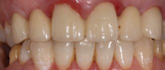
High-quality complete dental prosthetics with crowns in Moscow implies not only the restoration of their chewing function. Teeth are the most important component of a beautiful, attractive smile that evokes the sympathy and trust of others. Therefore, both affordable metal-ceramic crowns and more expensive aesthetic crowns should fit perfectly into the dentition and serve for a long time without causing undesirable consequences. Modern medicine has all the materials and technologies necessary for this.
Filling or crown: which option to choose?
The crown of a tooth refers to both its part located above the gum mucosa and an artificial structure in the form of a case, put on the natural crown destroyed by caries. The need for such tooth restoration arises after treatment of deep caries and pulpitis with the passage of root canals, and sometimes after mechanical trauma to the jaw and tooth.
Many patients are afraid of having a crown installed and beg the dentist to limit themselves to a filling. But if you fill a pulpless tooth, of which less than 50% of its own tissue remains, then it will most likely have to be removed very soon. After all, hard tissues after removing the pulp become very fragile and easily crack under load. Prosthetics after removal will become inevitable, but will cost much more.
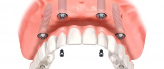
Fixing crowns on implants
In case of complete loss of a tooth or the impossibility of classical prosthetics, the best solution would be implantation, thanks to which the functionality of the dental unit can be completely restored. The implant replaces the root of the tooth, and the prosthesis replaces the crown part. An abutment is a kind of adapter between these two components. As a result, the patient receives a solid structure, all elements of which are securely connected to each other. There are two methods of fixing implant-supported crowns.
- Cement fixation.
The essence of the method is similar to installing a crown on a natural tooth, only an abutment is used as a support. - Screw vixation.
The crown is connected to the abutment using a special screw that passes through the entire body of the crown. The resulting hole is subsequently closed with a composite material.
Each of the two methods has its fans and opponents among clinicians, however, both cement and screw fixation on implants are actively used in modern practice.
Pros and cons of cement fixation of crowns on implants:
- Lower cost.
- Ease of implementation.
- Better occlusion for prosthetics of chewing teeth.
- Better suited for bridges.
- Lack of possibility of prompt repair and prevention.
- There is a risk of cement resorption and decementation of the crown, which leads to a violation of the tightness.
- Risk of peri-implantitis when cement is removed beyond the working area.
Pros and cons of screw fixation of crowns on implants:
- A more modern type of fixation.
- A screw-retained crown is considered the best solution for single prosthetics.
- Integrity of structure and absence of cement.
- Good maintainability.
- Higher cost and manufacturing complexity compared to cement fixation.
- Risk of screw breakage or loosening.
- Many experts consider this method less universal.
The choice of the type of fixation is made by the doctor after analyzing the diagnostic results, assessing the clinical case and the anatomical features of the patient’s dental system.
Why do they grind down a tooth for a crown?
The dentist must remove part of the tissue to form a stump of a certain shape:
- a ledge is formed at the edge of the gum, on which the crown will be placed in the future;
- The edges of the tooth should remain almost parallel, and not cone towards the chewing edge.
The service life of the artificial crown largely depends on the quality of preparation and compliance with these two conditions. If there is no smooth and even ledge, its edge will not adhere to the tooth, but will hang over the gum, forming a gap. Soft plaque will accumulate around, and colonies of pathogenic microflora developing in it will cause caries and gum inflammation.
If the tooth stump takes a cone-shaped shape after filing, it will be difficult for the dental technician to produce a functional structure that can withstand the chewing load. This is fraught with depressurization (or decementing) of the crown. Residues of food and soft plaque with microflora will penetrate under it, which will eventually lead to its loss.
Preparation for the crown of a living, non-pulpless tooth should be carried out with plenty of cooling. After all, overheating of the tissues will cause inflammation of the pulp, which may appear after the installation of the crown. Due to the need to treat pulpitis, it will have to be removed and redone after treatment of the canals.
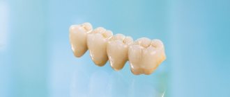
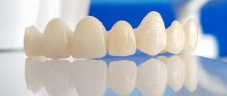
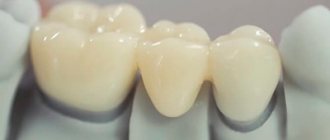
Fitting of various bridge designs.
The application and fitting of a solid bridge frame includes the following steps.
1) External assessment of workmanship on models in the articulator.
2) Fitting, paying special attention to: the accuracy of the frame fit in the cervical area (marginal fit); no gap between the edge of the crown and the tooth stump; correspondence of the contour of the edge of the supporting crown to the contours of the gingival edge; the degree of immersion of the crown edge into the gingival sulcus; approximal contacts, occlusal contacts with antagonist teeth; space under the intermediate part.
3) If necessary, correction of occlusal relationships is carried out.
4) Before permanent fixation of the bridge, electroodontodiagnosis of the supporting teeth is carried out to exclude inflammatory processes in the dental pulp. If there are signs of pulp damage, the issue of endodontic treatment is decided.
5) Instructing the patient on the rules of care and use of the prosthesis and instructions on the need for thorough brushing of the teeth with a brush and toothpaste twice a day and regular visits to the doctor once every 6 months.
The fitting stage of the bridge begins with the cast frame being carefully examined on the model, paying attention to the quality of the casting and processing of its outer surface. Here they also check the accuracy of the fit to the plaster model. After this, its position in relation to the antagonists and adjacent teeth is assessed, based on the thickness of the future ceramic coating. Its thickness ranges from 0.5 to 1.7-2.0 mm. On plaster models of the jaws fixed in the articulator, the space between the frame and the surrounding teeth - adjacent and antagonists - is determined. In cases where the gap between the frame and neighboring teeth, including antagonists, is clearly insufficient for applying a ceramic coating, it is necessary to find out the reason. It may consist, firstly, in insufficient precision in the preparation of the supporting teeth, when the layer of tissue removed does not correspond to the thickness of the walls of the metal-ceramic crown. Secondly, a thick frame can also take up part of the space intended for applying ceramics. Thirdly, the space for veneering is significantly reduced by inaccurate fitting of the cast metal frame on the plaster stump of the tooth. If any of these reasons are detected, the question of how to eliminate the defect is decided. The framework that meets the requirements is disinfected and tested on the supporting teeth in the oral cavity.
The cast frame is not always applied immediately, without preliminary fitting, exactly to the prepared teeth; in these cases, one has to resort to a painstaking procedure of sequential fitting. To do this, use a special occlusive spray. Having received prints of areas of the inner surface that prevent overlap, they are ground off with diamond heads (cylindrical or truncated cone-shaped). The manipulation is repeated several times until the cast frame is accurately installed on the supporting teeth. It is necessary to check the accuracy of its fit to the cervical part of the tooth. After fitting, the frame is returned to the laboratory for coating.
A working model with a metal-ceramic prosthesis is transferred to the clinic for testing in the oral cavity. Assessing the quality of a manufactured prosthesis begins with examining it on a plaster model. First of all, attention is paid to the accuracy of restoration of the anatomical shape, the presence of interdental contact points and the nature of closure with antagonist teeth. It is useful to once again evaluate the fit of the edge of the crowns to the gingival part of the tooth.
The disinfected metal-ceramic prosthesis is placed on the teeth. Pay attention to the accuracy of the application. After checking the metal frame, only ceramic mass can prevent the application of a bridge if there is an excess of it on the contact surfaces facing adjacent teeth, or on the edge of the metal frame adjacent to the ledge or neck of the teeth. In the first case, areas of excess ceramic are identified using carbon paper placed in the interdental spaces and the ink layer facing the ceramic. In the second case, ceramics found on the edge of the cap can be detected when examining this area of the crown or when checking the tightness of the fit to the cervical part of the tooth using an articulation spray or a layer of silicone fluid mass. Regardless of the reason, the excess ceramic material is ground off with shaped diamond bits until the denture fits perfectly into place. After this, occlusal contacts with antagonist teeth are carefully verified, both in central and other types of occlusion.
Having achieved an accurate installation of the prosthesis, pay attention to its similarity with symmetrically located teeth. If necessary, appropriate corrections are made. To do this, use diamond shaped heads to remove part of the ceramic coating or transfer it to a dental laboratory, where an additional layer of ceramic is applied. Particular attention is paid to matching the color of the veneer and natural teeth in dim daylight. In some, most complex cases, with an unusual color scheme of natural teeth, dyes are used to correct the color. After this, the prosthesis is sent to the laboratory for glazing and thorough polishing.
After glazing, the prosthesis is placed on the supporting teeth. If the patient is satisfied with the aesthetic qualities of the prosthesis and does not experience any inconvenience when closing the dentition, it is advisable to temporarily strengthen the prosthesis on the supporting teeth for 1-2 months. Temporary fixation of a metal-ceramic prosthesis allows, in case of any complications, to eliminate them without compromising the integrity of the prosthesis. Such complications include traumatic pulpitis, apical periodontitis, the appearance of areas of increased pressure under the body of the prosthesis, early chipping of the ceramic veneer, etc. For temporary fixation of metal-ceramic prostheses, you can use Tempbond cement, Repin (Slovakia). "Provicol" and others.
If no complications arise during the period of temporary fixation and the patient does not complain, the prosthesis is removed from the supporting teeth and, in the absence of signs of pathology, is strengthened on the teeth with permanent cement.
Scheme: “Evaluation of the quality of manufacturing a bridge prosthesis”
| Assessing the quality of a bridge: | 1. Correct modeling of the intermediate part. 2. The quality of the casting of the bridge. 3. Quality of cladding. 4. Quality of processing and polishing of the prosthesis. | |||||
| Criteria for assessing the quality of a manufactured bridge: | The prosthesis should fit easily onto the supporting teeth. | Abutment crowns should tightly cover the necks of the abutment teeth. | The prosthesis should not interfere with the closure of teeth in all types of occlusion. | |||
Fixation of a bridge with cement .
Before fixing the finished prosthesis in the oral cavity, remove the scale that formed as a result of firing on the inner surface of the crowns. For this purpose, a sandblasting machine with glass beads is used as the working medium.
To fix metal-ceramic prostheses, special cements should be used - uniface, aquameron or other ionomer class cements.
When strengthening a bridge, you need to thoroughly dry the crowns and supporting teeth with alcohol, ether, and warm air. The cement is mixed to a creamy consistency and filled into the crowns. The supporting teeth are covered with cotton rolls and changed from time to time, keeping the teeth dry until the prosthesis is applied and the cement hardens. The prosthesis is fixed on the supporting teeth, after the cement has hardened, excess cement is removed with a probe or a smoother, from the gum to the crown, so as not to injure the gum.
There are features that are unique to the fixation of bridges. They are due to the fact that the bridge is fixed. prostheses, it is necessary to simultaneously strengthen two or three, and sometimes more crowns located at a certain distance from each other. Therefore, it takes longer to degrease and dry teeth. The most common complication is increased bite and decementation of supporting structures. An increase in bite occurs due to the fact that excessively hardened cement is not fully squeezed out from under the crown, and the crowns become uncemented because saliva gets into them.
Thus, good isolation of the supporting teeth from saliva and the rapid application of bridges are the main conditions for the successful implementation of this manipulation.
If after fixation of the bridge prosthesis, bite separation is detected outside the bridge prosthesis, it is necessary to immediately remove the prosthesis and carry out the above-described manipulations again.
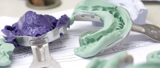
How to take impressions correctly?
If the mucous membrane was damaged when grinding the native crown, the procedure for taking impressions should be postponed until it is restored.
- In some patients, the injured gum can give recession - move away from the neck of the tooth and expose its root.
- Due to the presence of blood, the materials used to obtain impressions may not clearly depict the formed ledge.
As a result, the edge of the manufactured and installed crown will stick out above the gum, which will significantly reduce the aesthetics of prosthetic treatment of the front teeth.