Causes of a crack in the root of a tooth
Not all patients understand why a root crack occurs in a living tooth. The reason lies in the uneven distribution of the load. Moreover, the load can be strong and sudden, or it can accumulate over years and lead to the appearance of a crack after some time.
Typically, the cause of a crack is caused by the following factors:
- domestic trauma
- blow, fall, sudden load while chewing; - improper treatment
- strong pressure when cleaning canals or filling the root can injure the walls and in the future, with the slightest excessive load, cause a crack in the root; - error during prosthetics
- strong pressure when installing a pin or the manufacture of inappropriate dentures that unevenly distribute the load during chewing, leading to injury; - Bruxism
- unconscious clenching of the jaws, for example, during sleep, leads to a load that the patient does not notice and cannot control.
Why do cracks appear in enamel?
There are several reasons why chips and cracks appear. The most common of them:
- Mechanical
. The habit of chewing pens and pencils, husking seeds, and opening bottles with teeth lead to microtraumas that grow into large ones. - Chemical
. Sour and sweet foods, carbonated drinks, and frequent whitening destroy tooth enamel. - Temperature
. Hot food washed down with ice-cold drinks is a direct path to enamel cracks. - Physical
. It is better to use abrasive toothpastes periodically, and then give the enamel a chance to “rest.” - Traumatic
. This can be a one-time injury, for example, falling from a bicycle, or regular microtrauma, as during sports. - Age
. With age, teeth become more fragile and restoration mechanisms slow down.
Do you have small cracks on your teeth? This already indicates that you need to reconsider your habits, make changes to your diet and be sure to visit the dentist.
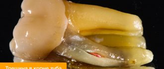
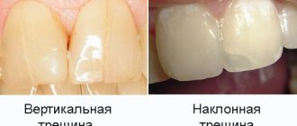
Types of tooth root crack
Pressure can be applied both vertically and perpendicularly to the crown. The following types of cracks are distinguished:
- oblique crack of a tooth
- a diagonal fracture, usually the healthy part is much larger, and the doctor can save the tooth by removing the affected part; - transverse crack of a tooth
- part of the root is completely broken off and deprived of nutrition; the closer the crack is to the coronal part, the higher the risk of complications; - longitudinal crack in the root of a tooth
- the fracture runs vertically, along the axis of the root, often accompanied by a fracture of the crown; - comminuted tooth crack
- multiple root fractures intersecting with each other.
Removing part of a tooth, treating gums with a laser, treating tooth canals with a microscope
First, the chipped wall of the tooth was removed.
After this, the gum was trimmed with a laser to free the edge of the chipped tooth from under the gum.
As a result of the split, the nerve in the tooth was damaged. The damaged nerve had to be removed.
Removing nerves from a tooth is a complex procedure that requires the highly qualified doctor who performs the procedure and the availability of the necessary equipment, including an operating microscope. At Dial-Dent, such procedures, called tooth depulpation, are always carried out using microscopes by doctors who have at least 10 years of experience. Yu.A. Borisova, a dentist-endodontist at Dial-Dent, a specialist in the treatment of dental canals with extensive experience, knows about all the advantages of dental treatment with a microscope.
Dial-Dent expert in dental canal treatment with a microscope Yu.A. Borisova removed the nerve from the tooth and filled the canals.
The photo shows tooth canals treated with a microscope:
The interval between visits is 2 weeks, since on the first visit an antimicrobial medicine was placed in the tooth and the tooth was closed with a temporary filling. Under no circumstances should you fill the canals right away or rush. The infection could enter the bone through a crack, and if the canals are filled in one visit, there is a high probability of complications, the tooth will continue to hurt, or a cyst will appear near the root of the tooth over time.
During the second visit to the endodontist, the tooth canals were thoroughly washed and hermetically sealed with hot gutta-percha.
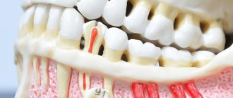
Root crack diagnosis
Only a doctor can diagnose an injury. First, the specialist makes a preliminary diagnosis based on the patient’s symptoms and complaints. More detailed research is required to clarify.
- Instrumental examination.
By tapping the tooth, the doctor determines whether there are irregularities, fractures or bleeding in the root of the tooth. - X-ray.
The image clearly shows the tooth root crack, its location and the severity of the injury. - Electrical exposure
helps determine the condition of the pulp.
Strengthening a tooth with a pin insert and temporary prosthetics
After root canal treatment by endodontist Yu.A. Borisova, the patient switched to another doctor - orthopedic dentist A.S. Ivankov for further prosthetics. An orthopedist is a specialist in restoring damaged teeth and prosthetics.
This tooth, significantly damaged by a crack, had to be restored with a pin (pin stump insert) and a ceramic crown. Our chief physician did not want any metal ceramics. He insisted on using the best, most innovative and modern materials and technologies. The restoration of the tooth by the orthopedist also took place in several stages. A pin insert (pin) is installed in the tooth.
A tooth with a pin inlay is prepared for a crown:
Orthopedic dentist A.S. Ivankov put a temporary crown on the tooth, which he was supposed to wear for six weeks.
Why was six weeks needed? It's all in the gum! The gum, damaged as a result of the chip, was bleeding. It was necessary, by recreating the shape of the tooth on a temporary crown, to “deceive the gum”, make it think that there are no problems, and the tooth is in place. The gums around the temporary tooth will heal and take on a normal appearance. After 6 weeks, the final ceramic restoration, a ceramic crown, will be made and will look like a natural tooth, tightly enclosed by the gum.
The photo shows the healed gum around the temporary crown after 2 months:
Crack in the root of a tooth: what to do
After diagnosis, patients are concerned with the question of whether it is possible to cure a crack in the root of a tooth. It all depends on the severity of the injury, the type of fracture and location. But there is no therapeutic treatment - tablets, ointments and injections; in any case, surgical intervention is necessary.
Ⅰ.
In case of a transverse fracture, the tooth can be saved, but the pulp is removed without exception. The doctor cleans and fills the root canals, and fastens the crack with a pin. Now the tooth needs rest and a minimum of chewing load - to reduce mobility, the teeth are splinted, that is, fixed in the correct position with a special splint-onlay.
Ⅱ.
Splintered, vertical and oblique cracks in most cases are only the initial stage of destruction of the root system, and over time the fault will grow. Therefore, depending on the severity of the injury, the doctor removes either part of the root with a crown or the entire tooth. A partially lost tooth can be restored using an inlay on a pin, or a complete tooth can be restored by installing a prosthesis or implant.
Treatment of a crack in the root of a tooth requires following the following recommendations:
- limit the chewing load - it is better to eat soups, cereals and other soft foods, avoid hard foods;
- monitor the temperature - all foods and drinks should be slightly warm, ice cream or hot tea have an undesirable effect on the injured root;
- eliminate pressure - do not open your mouth wide, do not close your jaw too tightly, brush your teeth carefully, avoid blows.
Treatment
The treatment method is selected according to the size and depth of the fracture. In one case, artistic restoration is sufficient, in another, prosthetics with a crown may be required.
- In case of microcracks, surface grinding of the enamel is performed, with remineralization, restoration with light-curing material and coating with dental varnish.
- Cracks in the front teeth worsen the aesthetics of the frontal area. Veneering will help restore the beauty of your smile - fixing thin plates of ceramic or composite onto the enamel. The enamel layer is prepared to a depth identical to the thickness of the plate, and a microprosthesis is attached to the prepared surface with dental glue.
- Deep horizontal and vertical splits are restored using dental crowns. Prosthetics with crowns are also carried out if the damage has affected the pulp. After depulping and filling the dental canals, the tooth is given the desired shape (grinded), and an artificial crown is fixed on it.
A tooth split into two halves with a damaged root must be removed followed by prosthetics. Endodontic treatment of cracks in a tooth is possible if the root remains intact. Deep damage to the pulp with extensive spread of the pathological process to the periodontium also leads to removal of the dental unit.
Complications and consequences of a root crack
In this situation, timely treatment is important: seeing a doctor at the initial stage will help avoid consequences. If this is not done, you may encounter the following complications:
- abscess.
A crack without treatment can provoke an inflammatory process, which in the future will turn into suppuration of soft tissues and lead to the formation of an abscess. Treatment of a purulent cavity is much longer, more expensive and unpleasant than treatment of a fissure; - phlegmon.
Suppuration, which, unlike an abscess, does not have clear boundaries and does not flow into the formation of a cavity. Cellulitis is a decay of cells that can spread to any tissue in the cavity; - periodontitis.
Inflammation of the periodontium, that is, the tissues surrounding the tooth, leads to their death and the inability to hold the tooth in the gum. The result is the complete loss of one or more teeth; pulpitis. The death of the pulp - the neurovascular bundle of tissues in the tooth through which it receives nutrition. Pulpitis is quite painful and can affect neighboring tissues; - tissue injury.
A root fragment can injure soft tissue. In this case, the doctor will have to extract a piece of the root and remove the infected tissue.
Saving the tooth of the chief physician of Dial-Dent S.V. Zukora
It so happens that even good dentists themselves need dental treatment. Our chief physician S.V. Zukor had a toothache. Or rather, this tooth had been hurting for a long time. Periodically, once or twice a week, while chewing, a short but very intense painful shooting occurred in the teeth of the upper jaw on the right. The pain was short and very intense. After a couple of seconds everything passed. Nothing was found during the examination. A thorough visual examination of all the teeth in this area, x-rays and computed tomography of the teeth revealed nothing. Our specialists could not find the source of the pain. The cause of pain in the teeth, that is, toothache, can be not only problems with the teeth. Often diseases of the ENT organs or neurological symptoms are disguised as toothache. That is why the Family Dental Clinic employs both a neurologist and an ENT doctor, who are involved in diagnosis in complex cases. Tsukor S.V. was examined by these doctors, who gave an expert opinion that everything was normal with the neurological and ENT systems.
So, after all, teeth! But nothing is visible. The tactics of observation and wait were chosen. Yes, painful impulses were repeated, but nothing could be done until the exact cause of the pain was identified. Monitoring was carried out throughout the year, periodically repeating examinations.
Prevention of root injuries
The best treatment is disease prevention. To prevent cracks at the root, try to follow a few recommendations:
- be careful and attentive, because half of the cases of root cracks are household injuries, which can be avoided if you take care of yourself and your health;
- carefully choose a clinic and a doctor - before visiting a dentist, check the reviews and ratings of the clinic, certificates and specialist diplomas, do not go to questionable dentists;
- strengthen your teeth - eat healthy foods, do not forget about the importance of calcium and fluoride in strengthening your teeth, choose suitable dental hygiene products.
If symptoms of a root crack occur, do not self-medicate and do not wait for the pain to go away on its own. See your doctor. If the pain is caused by a temporary phenomenon, you will be calm, and if a crack is discovered at the root, treatment at the initial stage will help maintain the health and beauty of your smile, avoid complications and unnecessary expenses.
Where to carry out preventive measures
Regular dental examinations (at least once a year) will help diagnose the problem at an early stage. When cracks appear, remineralization therapy aimed at strengthening the enamel will help stop further tissue destruction and prevent complications. The doctor will treat the teeth with a composition with a high concentration of fluoride, phosphorus and calcium. Next, the crack is sealed with varnish to strengthen and protect the enamel layer. You can carry out preventive remineralization of enamel in Moscow at the Ilatan clinic.
Varieties
In addition to those mentioned above, there are the following types of fractures:
- Transverse. This is the least problematic option, especially if the pulp is not damaged. The choice of treatment method is determined taking into account the degree of the fracture. If only the crown is damaged, it can be increased. If the pulp is damaged, in most cases it must be removed, after which the tooth is restored based on the existing root. If, as a result of the fracture, only the root remains, the tooth is built up with pins.
- Longitudinal or vertical. In this case, it is impossible to restore the tooth with a pin, since the remnants of the root are not capable of serving as the basis for a crown or pin. If an element of the dentition is broken in half, it is very difficult to identify the problem. People often waste time, and the condition of the tissue quickly deteriorates, as a result of which the tooth is usually removed.
- Splintered and oblique. In such situations, the development of events occurs approximately the same as in the case of longitudinal fractures.
Dental prosthetics with a ceramic crown - the result
We tried on the crown and it fit perfectly!
After fitting, the crown is fixed on the tooth.
During fixation, due to polishing the seam between the crown and the tooth, the gums were irritated and the photograph shows that the gums are bleeding a little. It will go away on its own in a few days.
Comment by the chief physician of the Dial-Dent clinic S.V. Tsukora: “ I experienced the work of the Dial-Dent team myself! Understanding the problems doctors face in cases like mine, I could only trust our specialists! The treatment was comfortable and went exactly according to schedule! I've never gone without a tooth. There was always a temporary crown that allowed me to chew and smile through all stages of treatment! I am grateful to all participants - doctors and dental technicians of Dial-Dent! And also to our assistants who monitored my comfort during the treatment process, and to the administrators who always reminded me about the visit! If we talk about the cause of the crack, it could have been boxing training, for which I forgot to wear a mouth guard. It always seems that training is not a fight, that there is no danger of injury, but this is not so. Take care of your teeth - always wear a mouth guard!
»
Monitoring of the treated tooth continues. Dial-Dent specialists, like the patient himself, are ready, if necessary, to remove this tooth and install a dental implant. Currently, the tooth has ceased to cause concern and is functioning perfectly.
Making a ceramic crown
The permanent dental crown was made in the dental laboratory of the Dial-Dent clinic by dental technician D.V. Ceramic wolf E.max. Photographs of teeth in different modes were used to give the technician maximum information to recreate the color.
The dental technician will match the color of the crown to make it indistinguishable from natural teeth in your mouth.
Having our own laboratory in our center simplifies many tasks related to dental prosthetics and makes all stages better and more predictable. All necessary corrections and fittings are made in the presence of a technician!
Permanent ceramic crown, material – E.max ceramics:
Alveolar ridge fractures
The appearance of such fractures occurs as a result of a direct blow, which affects several adjacent teeth at once. Since the upper jaw is in front, it is the one that suffers in most cases. This fracture can be determined by painful palpation, as well as the presence of damage to the mucous membranes. The process begins to move, and elements of the dentition may be broken or dislocated. Part of the jaw may be torn out or supported only by soft tissues.
Such a significant jaw injury is usually accompanied by dizziness, severe headaches, traumatic brain injury, and nausea. Using an x-ray, you can determine the location and direction of damage, as well as determine whether the broken part can be repaired. If it is viable, the specialist installs a splint that provides fixation of the bone for full fusion. In case of serious injuries, teeth that cannot be restored are removed, followed by fixation of the alveolar process.
If the injury is irreversible, the broken part is removed, since in the event of a rupture of the periosteum and adjacent soft tissues, the process usually cannot take root. In such situations, the damaged area is covered by the periosteum.
What is
Not everyone knows about a tooth fracture, but dentists encounter this problem quite often. Many fractures occur in children, boxers, stuntmen, and active sports enthusiasts. Another common option is accidents when a fracture is caused by accidental mechanical damage. It often occurs during an accident or fight.
As is the case with various parts of the body, a fracture negatively affects the anatomy and natural shape of the tooth.
A tooth fracture is a violation of the integrity of dental tissue (one or more of its layers) down to the tooth root. In most cases, the upper front teeth, in particular the central incisors, are affected. A fracture of the alveolar process, root or crown may also occur.
Common Causes
In addition to the factors mentioned above, it is also worth noting unqualified treatment. For example, when removing a lower jaw tooth with forceps, you can damage one of the upper teeth with the instrument. If a tooth is weakened by various diseases or is in poor condition, the likelihood of a fracture is very high. Of course, such cases are very rare, but they do happen. Another reason for a fracture is an error at the stage of selecting the size of the holes in the pins. It is important to note that the lack of treatment for caries also negatively affects the condition of the enamel, as a result of which sometimes eating solid foods can provoke a crown fracture. Do not forget about the anatomical features of the jaw. If its structure has certain defects, the pressure will be distributed incorrectly.
Signs
The most common and obvious sign of a tooth fracture is pain, which in some cases can be very severe. Pain sensations increase noticeably at the moment of closing and opening the jaw. If we are talking about a minimal fracture (the enamel is damaged, but the pulp and dentin layer are kept healthy), the pain will be less noticeable compared to a tooth root fracture.
Fractures can be complete or incomplete. During the first, the pulp is opened, as a result of which various irritants can make the pain more severe. Complete fractures are considered to be a fracture of the entire root or its apex, as well as a fracture of the neck of the tooth. In case of an incomplete fracture, damage to dentin and enamel may occur.
Additional symptoms of tooth fractures may include loosening of teeth, problems with speech, bleeding gum tissue, and difficulty opening or closing the mouth.