Removing a tooth is the last thing a dentist will suggest. Due to a number of subjective and objective reasons, it is impossible to maintain the dentition in ideal condition until old age.
Dental defects are the loss of one or more dental units, leading to a violation of the integrity of the dentition, deviation in the development of physiological occlusion, and an abnormal arrangement of individual teeth.
Reasons contributing to the development of dental defects
Dental defects arise due to:
- untimely treatment of caries;
- inflammatory processes in the oral cavity;
- presence of malocclusion;
- genetic conditioning;
- diseases leading to changes in metabolic processes;
- damage to the jaw apparatus;
- various neoplasms.
Classification of dentition defects
There are several types of classification. Dentists mainly use three of them:
- Kennedy classification:
- Group 1 - bilateral absence of chewing teeth;
- Group 2 - unilateral absence of a molar;
- Group 3 - unilateral absence of lateral teeth with preservation of distal support;
- Group 4 - defect of the anterior jaw.
- Beltman classification:
- Class 1 - the defect includes edentulous chewing teeth.
a) dentition with unilateral terminal defects;
b) dentition with bilateral terminal defects.
- Class 2 - there is one or more included defects (if the terminal units are present, there may be no teeth in other parts of the row).
a) the number of missing units does not exceed three;
b) the number of missing units is three or more.
- Classification according to the Gavrilov system:
- Group 1 - dentition with terminal one- and two-sided defects;
- Group 2 - dentition with anterior and lateral one- and two-sided defects;
- Group 3 - combined defects;
- Group 4 - single preserved dental units.
IROPD - index of destruction of the occlusal surface of the tooth
Author: Polilov D.A. — 2022
Many believe that today, with the advent of modern high-strength, low-shrinkage composites, IROPZ has become obsolete, and the indications for direct restorations are much wider than in 1984. However, this index can be extremely useful when working within the framework of voluntary health insurance programs.
According to the clinical treatment protocols approved by StAR, the choice of method for restoring the coronal part of chewing teeth in the treatment of pulp diseases is determined depending on the degree of destruction of its occlusal (chewing) surface according to the IROPD index according to V.A. Milikevich.
Many can say that today, with the advent of modern high-strength composite materials with low shrinkage, the IROPZ index is outdated, and the indications for direct restorations are much wider. This is true.
But it is too early to discount IROPZ. It can be extremely useful when treating patients under Voluntary Health Insurance programs. Since it is the IRPH that determines whether the treatment will be covered by insurance or not.
The role of IROPD in VHI treatment
Real case. During a medical and economic examination, an expert doctor from an insurance company sees a large filling on the Rg image. He says that this is not a filling, but a direct restoration and writes off the cost of all the treatment performed. It's a shame? Not that word. In this case, the ideal way out of the situation would be IROPD, if the doctor had bothered to take a picture of the occlusal surface of the tooth before starting treatment, at least on his smartphone.
Another situation. The tooth is badly damaged, but the patient insists on treatment covered by insurance. After taking a photograph of the occlusal surface, we calculate the IROPV and call the insurance company to coordinate treatment under insurance. The insurance company will definitely deny coverage, since the fundamental document is the StAR clinical protocols, and they refer to the IRPH. The patient is forced to agree and pay for the treatment at his own expense.
Index history
The IROPZ index was proposed by V.Yu. Milikevich in his doctoral dissertation (1984) as an indicator (index) of the degree of destruction of hard tissues of the crowns of chewing teeth with defects in the hard tissues of chewing teeth (premolars, molars) according to class I-II according to Black. It represents the ratio of the dimensions of the “cavity/filling” area to the entire area of the occlusal surface of the tooth.
Rice. 1. Typical models of destruction of the occlusal surface of chewing teeth
Thus, according to Milikevich:
IROPS = Cavity/Filling Area: Area of the chewing surface
With IROPI indicators of 0.2 – 0.4
The filling method is used. After endodontic treatment, a temporary filling (bandage) can be placed if it is not possible to place a permanent filling on the first visit or to prevent possible complications. Permanent filling is carried out in one visit.
With IROPV > 0.4
shows the manufacture of inlays from metals, ceramics or composite materials.
With IROPV > 0.6
production of artificial crowns is shown
With IROPV > 0.8
the use of pin structures with subsequent production of crowns is shown.
To calculate the IROPZ index in practice, Milikevich proposed using plexiglass and graph paper (paper with a grid applied with cells with a side of 1 mm - at that time such paper was widely used in drawing). There were no computers or digital cameras back then.
Method for calculating IRPZ
Today there is a simpler and accessible way for every doctor to calculate the IRPI. We photograph the tooth from the occlusal surface using any camera (microscope, intraoral camera, camera or smartphone through a mirror) and transfer the image to the computer.
Image processing in a graphics editor
For example, in Adobe Photoshop. We turn on the display of the grid in the menu View – Show – Grid. If necessary, you can configure the grid parameters in the Editing – Settings section. Expand the image and align it to the edges of the grid (Ctrl-A, Ctrl-T). We mark the filling/cavity zones with the letter “P” in those squares where the area of the destroyed tooth surface is significantly larger than the preserved one. When calculating the total area of the occlusal surface, we take into account the squares where the tooth tissues occupy more than half of it.
Rice. 2. An example of calculating IRPI on a computer.
In our example, shown in the picture, the total area of the occlusal surface of the tooth is 28 squares, and the cavity area is 13 squares. Thus, for this tooth IROPZ = 13: 28 = 0.46. This means that in this case, restoring a tooth with a filling is not indicated, but requires the manufacture of an inlay made of metal, ceramics or composite.
Saving the IRPZ calculation
It is recommended to save the original images and calculation of the IRPI in the patient’s electronic medical record in the Dental4Windows program so that they are always at hand in case of an audit by the insurance company.
Adding photos and any other files is done in the Patients
,
Documents
. In this case, photographs and x-rays are automatically displayed when viewing the dental formula (Dental Map) on the right side.
If you have questions about this method for determining IROPD, suggestions, or you want to discuss anything, we invite you to join our open group on Facebook: https://www.facebook.com/groups/Dental4Windows/. We'll talk there. See you in touch!
Literature
1. Clinical recommendations (treatment protocols) for the diagnosis of dental pulp disease. Approved by Resolution No. 18 of the Council of the Association of Public Associations “Dental Association of Russia” dated September 30, 2014
2. Milikevich V.Yu. Prevention of complications in case of defects in the crowns of chewing teeth and dentition: Abstract of thesis. Dis...Dr. med. Sci. - M., 1984. - 31 p.
Symptoms and consequences of dental defects
Dental defects appear:
- chewing function disorder;
- incorrect bite;
- articulation disorder;
- single displacement of teeth;
- violation of the integrity of the dentition;
- violation of the aesthetics of a smile.
Diagnosis of dental defects
The removal of one or more dental units requires consultation with a dentist and an orthodontist, especially when it comes to the anterior part of the dentition. Untimely correction of the defect does not have the best effect on the condition of adjacent teeth.
As a rule, a visual inspection is sufficient to determine how to correct the defect. In some cases, x-rays may be needed.
Types of cracks
This phenomenon occurs frequently. Treatment depends on how the crown cracks—the direction of the split matters. In medical practice, there are several types of cracks.
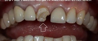
Vertical crack
The vast majority are fixed on the front teeth. But it also happens on molars. It can stretch over the entire surface and, when neglected, affects the root. The integrity of the surface part is damaged, caries appears, quickly turning into pulpitis - such a crack opens the way to infection.
Horizontal crown crack
Statistics show: when the crown of a tooth is cracked like this, the destructive process is slower, and there is no accelerated danger to the pulp. If the crack is already large and there are prerequisites for chipping, then the tooth can be restored only by removing the damaged part and strengthening the tooth with a crown.
Oblique oblique
The pulp is often damaged. Treatment depends on the complexity of the specific situation. In an advanced state, only tooth extraction followed by implantation and prosthetics is possible.
Fracture of the tooth crown into pieces
A common option: separation - the crown of the tooth is cracked into 2 parts. The fragments put pressure on soft tissues and injure them. Food particles end up in the crack, causing pain. Treatment is urgently required; in case of serious pathologies, removal is practiced.
"Web"
Small cracks affect only the enamel of the tooth. At this stage, the treatment prognosis is favorable.
Damage to the masticatory tubercle
The pointed part of the tooth is injured. The result is a small chip. The pulp remains largely intact. It is possible to restore the tooth using modern filling materials or install a crown or veneer.
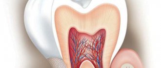
Features of restoring the coronal part of a tooth with a pin
The length and shape of the orthopedic structure (pin) is selected individually, taking into account X-ray data. Before installation, the root canal is processed and expanded, and then a seat is formed in it. After applying cement to its walls, a pin is screwed in, which can be made of ceramic or fiberglass. The first option is very durable and is made individually, the second is elastic, and therefore imitates dentin well, eliminating the risk of the root cracking. This allows you to place it even in the canal of the root that was treated.
| Restoration of the crown part of a tooth using pin structures: | |
| advantages: | flaws: |
|
|
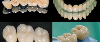
Features of restoring the coronal part of a tooth with a core inlay
The stump tab is able to provide the tightest possible fit to the remaining tissues, which eliminates the risk of developing secondary caries. It is able to withstand intense loads, so the prosthesis on it will serve for a long period of time. Depending on its features, the design can be:
- Solid cast – with a core and one or two pins for restoring premolars with one or two canals;
- Collapsible - having removable pins in the amount of two to three pieces for prosthetic teeth that have several channels.
Due to the fact that the inlay is made according to an individual impression in a dental laboratory, it corresponds to the anatomical structure of the tooth.
| Restoring the coronal part of a tooth with a core inlay: | |
| advantages: | flaws: |
|
|
Treatment of cracks in tooth crowns
If the damage does not cause inconvenience and was discovered by chance, then a visit to the doctor is required. The defect should not be viewed as purely cosmetic.
Treatment depends on the type of chip and its severity. For minor damage, the doctor may suggest restoring the tooth crown with durable materials. In more serious cases, canal cleaning or tooth extraction will be required. In each specific option, the decision is made individually.
In case of micro-manifestations, remineralization is recommended, followed by surface coating with composites and varnish. It is possible to install veneers, and in case of significant damage, crowns.