Reason for breakage
The canals in our teeth have a very complex structure; they are curved, very thin and branched in different directions. To clean the canal to the very end, the doctor uses special devices, which are also very thin and flexible. Unfortunately, it is impossible to completely protect yourself from item damage. Even the most experienced professional cannot be 100% insured against such an incident.
The process of removing a foreign object is quite difficult, so not every master can cope with the task. The final decision on the need to remove a part should be made only after an adequate assessment of all the pros and cons.
There are many nuances, for example, if part of the instrument is lost in a living tooth and at the final stage of treatment, no complications should arise in the future. The patient will not encounter an allergic reaction, inflammatory processes or increased sensitivity to various irritants in the future.
Broken instrument in the root canal. Does it always need to be taken out?
The phrase that, in my personal conviction, in 99% of cases causes shock is “You have a broken instrument inside your tooth.” Immediately, most patients begin to go through all 5 stages of accepting the inevitable. "How? How did he end up there? The scoundrel dentist forgot it there? So what's now? How to live further? Let's go in order.
All root canal treatment is performed using rotating instruments . This is the standard of modern dental treatment. They allow you to remove the “nerve”, expand the root canal, and remove old filling material. The rotation of this tool is ensured by a special machine - an endomotor, in which the speed and rotation power values are set for each specific tool. Each instrument goes through all stages of sterilization: it is “scratched” with brushes, then “fried” in a special sterilizer. Let's not forget that the anatomy of each root canal in the same tooth and the same patient is unique. So this is what I'm getting at: tool failure is often a combination of many factors coming together. From improper load on the tool, metal fatigue to unpredictable anatomy. I very much doubt that a doctor who has broken an instrument in the root canal and realized this (and this is an important point - sometimes millimeters of the tip of the instrument are broken, which is quite problematic to notice without additional devices) feels a sense of joy and pride in his work. You can interview all the doctors involved in root canal treatment, and I am sure that they will answer with one voice - they all broke instruments. The easiest way to avoid breaking instruments is to stop working in root canals.
What to do next?
The most logical solution is to get the fragment out by any means. This is a foreign body. But it's not that simple. Most studies indicate that the instrument fragment itself is not the cause of apical periodontitis (inflammation behind the apex of the tooth), and this is the main goal of treatment. Prevent the occurrence of periodontitis. So, is it always necessary to unconditionally get it out of there?
It all depends on the starting conditions. The instrument can be “left” in any part of the root canal. The most infected part of the tooth is the entry point into the root canals and their initial part. This is due to the fact that carious microorganisms come to the “nerve” from above and gradually move down, simultaneously infecting everything in their path. Our goal is to fight infection. So, if an instrument breaks at the end of treatment before filling, when everything has already been washed and cleaned at the very “bottom” of the root canal, or if an instrument breaks during the initial (!) root canal treatment, you don’t have to remove it. Why? Because the instrument itself is sterile, the root canal at the “lowest” point is also relatively clean, especially after all stages of treatment have been completed. Most often, this will not affect the treatment prognosis in any way. Plus, when trying to remove it, a complication may arise in the form of mechanical damage to the wall of the root canal . And this is much more unpleasant than a sterile instrument “walled up” in the wall of the root canal.
In most cases, an attempt is made to bypass it - this is the least traumatic for the remaining tooth tissue. If the instrument is broken in the middle part, then after such manipulation, it, as a rule, “jumps” out on its own when the root canal is washed. If the attempt to bypass the tool is successful, but it is not possible to “pull it out”, in this case, it also does not need to be removed. Let me remind you that our main goal is to fight infection in the root canal. If the root canal goes the full length, then the “war” with the instrument will not bear any fruit.
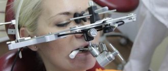
It’s another matter when the instrument “lives” in an infected tooth. What then? Then you need to try to extract it. For this, of course, additional magnification is used, because it is impossible to see a tiny fragment at a depth in the dark. The instrument is freed from tooth tissue using ultrasound and, as a rule, is also washed out of the root canal with solutions.
To summarize, a broken instrument is not a tragedy, not a doctor’s negligence, and, moreover, not a reason for tooth extraction. In most cases, broken fragments can be removed conservatively and root canal treatment can be performed in full.
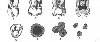
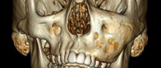
The photographs show a clinical case of treatment of a 1.6 tooth, when the broken fragment of the instrument was circumvented and removed.
And the actual fragments themselves:
Possible complications
It is worth noting that a piece of the instrument itself does not pose any threat to the patient’s health. This is explained by the fact that such items are made from hypoallergenic materials. However, the part may prevent complete cleaning of the dental canal, which will negatively affect the outcome of the entire treatment. As a result, inflammation may develop, severe pain will occur, and in rare cases, tooth extraction may be necessary.
Parts remaining in the canal can negatively affect recovery after treatment. In rare cases, the patient may develop an allergy; sometimes it is accompanied by a strong tooth, swelling and redness of the soft tissues and mucous membrane located next to the diseased tooth.
The final prognosis of endodontic treatment will depend on many factors, for example, on the stage at which the working instrument broke, as well as on the degree of preparation and sufficient treatment of the canal with medications at this stage.
The main ways that fragments of dental instruments get into dental canals
A broken instrument in a tooth canal can appear due to the direct influence of several reasons, and is directly related to the treatment procedure. Most often, this situation occurs when a dead nerve or blood vessels are removed - a fragment of an instrument remains in the tooth, which, at the dentist’s discretion, can be removed or left, depending on the patient’s indications. Also, the reason that an instrument remains in the tooth canal may be:
- When a dentist passes curved and narrow canals, dental instruments may not withstand the stress caused by their bends.
- When using only hand tools, tip fractures occur due to a metallurgical defect or due to metal fatigue.
The process of removing debris from the canal
Even if the fragment does not affect the outcome of treatment in any way, a conscientious doctor will definitely inform his patient about the problem. This is necessary so that if the slightest discomfort is detected, a person immediately goes to the clinic, and does not treat the tooth with folk remedies at home. If the instrument breaks at the initial stage of the intervention before the infection is eliminated, the dentist must try to remove the part from the canal.
This will require a special dental microscope; without it, it will be simply impossible to detect such a small detail in a thin canal. At the first stage of the procedure, the doctor must create direct access to the foreign particle; for this he can use a dental bur.
Next, ultrasonic instruments with different tips come into play; it is with their help that the fragment can be removed from the dentin with minimal consequences. After this, the canal is thoroughly cleaned and sealed again, and medications are placed inside if necessary. A temporary filling is first placed on the tooth to ensure that there is no inflammation. After some time, you can put a permanent filling.
previous post
The dangers of teeth whitening at home
next entry
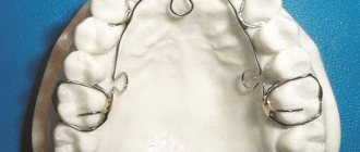
Types of pins and their main features
The pins are either fiberglass or metal. Fiberglass is used when installing tile crowns. Such an instrument does not show through the crown. It is quite easy to remove it if necessary. However, metal knitting needles are stronger, which is why they are used more often. Sometimes they are also called anchor.
Anchor pins are made from materials such as titanium, palladium or stainless steel. There are also pins made of brass and precious metals. They are used in the most difficult cases. Anchor pins are very popular because they make it possible to restore an almost completely destroyed tooth. It is only important that its root is intact.
The pins are fixed in the canal with phosphate cement. The use of ultrasound can help destroy it. This has to be done when repeated caries begins around the pin. Excessive strength of the material often causes displacement of the pin when chewing food, which can lead to damage to the prosthesis. In this case, the entire structure will need replacement or correction. Most often, the violation is detected already at the stage when it is already quite difficult to cure the tooth. In this case, you have to remove the pin along with the tooth. In some cases, the tooth can be saved. In this case, a small part of the dental tissue is removed around the pin using a drill, and then the pin is carefully unscrewed. After removal, re-treatment is carried out, the canals are properly processed and fillings are placed again.
Parapulpal pin wires are non-metallic. They are made from gold or special stainless steel, and then they are necessarily coated with polymer. Such knitting needles are used for retention of fillings and their reinforcement. A special feature of parapulp pins is that they are installed in hard tissue without touching the pulp. The absence of actions inside the tooth cavity ensures the complete elimination of the likelihood of contracting any infection or the development of inflammatory processes in the oral cavity. The disadvantage of such tooth strengthening means is the limited possibilities of use. The fact is that these products are located very close to the surface. They often break teeth and need to be reinstalled.
Dentists divide pin instruments into two groups: standard and special. The first ones are used to eliminate minor damage, and are available in the form of cylinders or cones. Special devices are made in a variety of shapes. Pin devices are also distinguished by fastening methods. Among screw materials, the main drawback is the danger of the tooth part breaking off during screwing.
Types of foreign bodies
Usually, foreign objects get into dental pockets during complex treatment. It can be:
- Broken endodontic instruments.
- Anchor or fiberglass pins.
- Parts of stump inlays.
Foreign bodies are any objects that should not be in the root canal after treatment is completed. If any body remains in the mouth of the tooth, it will interfere with the quality of the filling and will cause the formation of cracks and the development of inflammation. In this case, there is a risk of tooth loss. Therefore, foreign objects must be removed in a timely manner.