Molars are divided into canines, incisors, premolars, and molars. The first three types of teeth erupt in place of similar milk teeth. In turn, molars do not have predecessors and appear behind the temporary ones, therefore their second name is accessory.
Each jaw has 16 teeth - four incisors, two canines, four premolars and six molars. In some cases, third molars, better known as wisdom teeth, do not erupt because they do not have buds in the jaw - then instead of 16, a person has 14 teeth above and below. Experts explain this phenomenon by a reduction, or simplification, of the dental system caused by changes in dietary style.
Anatomical features of the upper teeth
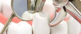
- The central incisor (the largest of all eight) has a chisel-shaped crown with a convex surface and one cone-shaped root. The only root canal in 75% of cases is straight.
- The lateral incisor with the same chisel-shaped crown and the same convex surface has a characteristic depression in the enamel - a “blind fossa”. There is one root canal, but more often it is deviated to the side.
- The canines on the upper dentition are often larger than those on the bottom. They are characterized by a crown pointed on all sides and the longest cone-shaped root. Canines have a single root canal, which can be straight (45%), distally deviated (30%) or vestibular deviated (12%).
- The first premolar with a prism-like crown is characterized by a convex lingual surface. On the chewing side there are tubercles, and between them there is a fissure. In the upper dentition, these teeth are always larger than in the lower one. The root has extended longitudinal grooves, which divide it in 60% of cases into two parts - buccal and palatal. There are often also two root canals.
- The second premolar with the same prism-shaped crown has predominantly one straight cone-shaped root with expanded lateral surfaces. Sometimes the root bifurcates closer to the top. There are often two root canals.
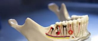
- The first molar is the largest tooth in the dentition. The rectangular crown has a diamond-shaped chewing surface with four cusps and an H-shaped fissure between them. There are usually three root canals, but sometimes there are 4 (25%) or 5 (1%).
- The second molar with a classic cube-shaped crown has 4 cusps and an X-like fissure on the chewing side. The tooth has 3 roots and three (87%) or four (13%) canals.
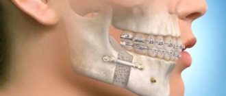
Tactics for impacted supernumerary central incisor in the upper jaw
Supernumerary teeth are teeth that are additional to the normal number of teeth and can be located in any segment of the jaw. They are classified according to morphology, location and number.
The incidence of supernumerary teeth varies from 0.1 to 3.8%. The ratio of men to women is 2:1. The literature indicates that about 80-90% of all supernumerary teeth occur in the upper jaw, with about half of them in the anterior segment. Supernumerary teeth can be found in both the permanent and primary dentition, but are approximately 5 times more common among permanent teeth. When collecting data on 2000 schoolchildren, it was revealed that 0.8% have supernumerary teeth in the temporary dentition, and 2.1% have supernumerary teeth in the permanent dentition.
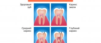
As a rule, supernumerary teeth are asymptomatic and are detected as an incidental finding during an X-ray examination. The tactics of behavior when such an anomaly is detected is removal or preservation, followed by frequent inspections.
Removal of supernumerary teeth is recommended in the following cases:
- There is a delay in the eruption of permanent teeth due to the presence of a supernumerary
- Impaired eruption or displacement of an adjacent tooth
- There is an associated pathology
- There is a need for orthodontic treatment to straighten the teeth
- Their presence prevents the formation of alveolar bone or implant placement.
- There is a violation of function and aesthetics
Description of a clinical case
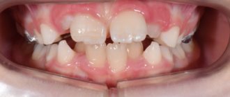
A 9-year-old patient was admitted to the clinic with complaints about the appearance of the anterior segment of teeth. The patient was in the early stage of mixed dentition with an unerupted (missing) upper left central incisor. Oral hygiene is not satisfactory, many temporary teeth have been previously treated.
Visual inspection
The patient has an oval face shape, normal complexion and tone, a slightly convex profile with normal vertical height and lip profile. The lip line corresponds to a slight asymmetry of the dental arch (Photo 2).
Photo 2: Photos before treatment 2a. frontal view 2b. frontal view with a smile 2c. profile view
Intraoral examination
The patient has a U-shaped dentition of medium size. In the oral cavity there is a mixed occlusion with closure of the incisors according to Class 1, delayed eruption of the upper right central incisor, displacement of the right lateral incisor and the left one in place of the right central one. The center line is shifted to the right by 2 mm. With occlusion, a crossbite is formed on the right without displacement of the lower jaw when closing (Photo 3). Palpation of the transitional fold in the area of the right upper incisor determines the bulging of the mucous membrane. Palpation of the mucous membrane from the palate also determines the bulge.
Photo 3: Intraoral view before treatment 3a. front view 3b. buccal occlusion on the right 3s. buccal occlusion on the left
X-ray evaluation
Panoramic, occlusal, and periapical photographs revealed the presence of an impacted upper right central incisor with a normal crown, a deficient root development, which was surrounded by two supernumerary tooth buds delaying eruption (Figure 4). The buccolingual position of unerupted supernumerary teeth can be determined using parallax. And the use of occlusion and panoramic images can replace vertical parallax.
Photo 4: Radiographs before treatment 4a. panoramic tomography 4b. palatal view of central incisor 4c. X-ray of the maxilla in the occlusal plane
Diagnosis
Based on clinical and radiological studies, a diagnosis of supernumerary teeth was established.
1. The tubercular type is characterized by rare natural eruption, often preventing the timely eruption of the affected teeth
2. The conical type has a higher eruption percentage than the cusp type
Treatment plan
The plan was to widen the dental arch to correct the crossbite, create space for the impacted incisor followed by extraction of supernumerary teeth under local anesthesia, and orthodontic treatment to realign the impacted incisor.
The likelihood of ankylosis of the incisor and, in this case, the need for its removal during surgery was discussed with the patient, and voluntary informed consent was obtained from the parents.
Treatment
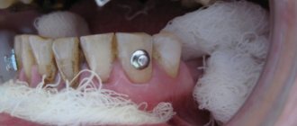
Treatment began with correction of the hygienic condition of the oral cavity, including brushing teeth with fluoride-containing toothpastes, using dental floss, and nutritional advice was also given. After achieving the proper level of hygiene, alginate impressions were made to fabricate the quarthelix appliance in order to eliminate the crossbite and obtain additional space for the right central incisor (photo 5a). At the next visit, quarthelix was activated and edgewise equipment was fixed (APC II 3M 0.018 MBT). The braces are attached to fully erupted teeth: right premolars, right lateral incisor, left central incisor, left lateral incisor, left premolars (Figure 5b). Once sufficient space has been obtained for the right central incisor (Figure 5c), the supernumerary teeth are removed under local anesthesia.
Photo 5: Fixed hardware stage before surgery 5a. Intraoral image of the upper dentition with installed quarthelix 5b. Intraoral photograph of the installed brace system 5c. Intraoral image after 4 months: formation of space for the eruption of the central incisor
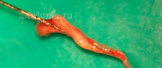
The palatal flap is elevated (6a) and both buds are successfully removed (6b and 6c). The impact central incisor is opened on the vestibular side and secured on a gold chain. The area is treated and sutured. The gold chain from the central incisor is passively attached to the archwire (Figure 6d). The patient was seen every 4 weeks to tighten the gold chain, thereby creating a force to facilitate eruption of the right central incisor (Figures 7a and 7b). After three visits, the right incisor erupted into the oral cavity. This cutter is fixed with 0.014 nickel-titanium wire to a base wire of 0.016×0.022 stainless steel (photo 7c).
Photo 6 (a,b,c,d) 6a. intraoral photograph of the maxilla with a raised mucosal flap 6b. removed cuspal supernumerary tooth 6c. removed sawn conical supernumerary teeth 6d. intraoral photograph after surgery. A gold chain is passively attached to the arch wire
Photo 7: Photographs of the installed equipment after surgery 7a. initial thrust of the central incisor 7b. partially erupted right central incisor 7c. placing a bracket on incisor 7d. fully erupted central incisor
Eight weeks later, the impacted right central incisor was placed in the dental arch (Figure 7d). Over the next few weeks, detailed correction and completion of treatment were carried out (photos 8a and 8b). Retention was achieved using a Howley retainer.
Photo 8: Intraoral photo of treatment results
Treatment results
The patient's treatment was completed after one year. The impacted right central incisor was successfully aligned (Figure 8). All treatment goals were achieved. The patient needs constant monitoring during the growth and development of all teeth; the need for orthodontic therapy cannot be excluded after the eruption of all permanent teeth.
Discussion
The etiology of the occurrence of supernumerary teeth remains unclear. Today, there are several theories depending on the supernumerary teeth themselves. One theory suggests that the supernumerary tooth is the result of a dichotomy of the tooth germ. Another, well supported in the scientific literature, is the hyperactivity theory, which proposes that supernumerary teeth result from local, independent hyperactivity of the dental lamina. Heredity may also play a role in the occurrence of this anomaly, since supernumerary teeth often appear in families rather than spontaneously in the general population. However, Mendelian inheritance is not traceable. Mesiodensity may be suspected if there is asymmetry in the emerging incisors. An early diagnosis allows you to eliminate the problem and avoid possible complications. Extraction of supernumerary teeth while still in a mixed dentition allows spontaneous alignment of the teeth to occur, which will significantly reduce the amount of intervention in the future. This clinical case presents a patient in late mixed dentition, with space loss, midline displacement and delayed eruption of the central incisor, which required surgical intervention and orthodontic correction. Extraction is not always the treatment of choice for supernumerary teeth. In cases where supernumerary teeth are asymptomatic, do not affect other teeth, and are discovered incidentally, they should be preserved and kept under observation.
The fixed technique made it possible to cause eruption of the incisor and correct the appearance of the anterior group of teeth.
In rare cases, when eruption cannot be caused orthodontically (incorrect location, ankylosis), there are two options: surgical reposition or removal followed by placement of an implant. Replacing a tooth with ankylosis with an implant is the best option, as it reduces the risk of root resorption, discoloration and periodontal complications. However, any of the treatment options should be considered strictly individually.
Authors: Dr. Tasneem AL Farhan, Dr. Kholoud Al-Foudari, Dr. Nour AL Hasan
Anatomical features of the lower teeth
- The central incisor is the smallest tooth in the “adult” bite and the smallest among the incisors. The root is quite short, in 65% of cases there is one root canal, less often - two.
- The chisel-shaped lateral incisor is always larger than the central one. One or less often two root canals – both narrow. “Adult” incisors on the lower jaw are less susceptible to damage than others, so dentists rarely turn to them for the treatment of caries in children and adults.
- Fang - similar in structure to the upper one, but is smaller in size. In 96% of cases it has a single root canal of normal structure.
- The first premolar has a rounded crown in cross-section and two characteristic tubercles on the chewing side. One root is slightly flattened.
- The second premolar is very similar in crown shape to the canine, and is always larger than the adjacent premolar. The surfaces of the single root are smooth and slightly shiny. Two roots occur in only 3% of cases or less.
- The first molar with a cubic crown has five cusps on top of the crown, separated by an F-like fissure. In 88% of cases, three root canals are formed.
- The second molar is smaller than the first, but completely replicates its anatomical features. Root canals are curved and have poor patency. In 85% of cases, the tooth has three canals, in 10% - four.
The anatomical characteristics of teeth are similar in all people, but each person may have individual characteristics, for example, the absence of wisdom teeth or an increased number of root canals in a particular tooth.