Home / Articles / Impacted fang
It has long been known that teeth can fall out too early and humanity is trying to combat this problem in every possible way. Recently, another problem of a slightly different nature has appeared - teeth began to erupt ahead of time, and this leads to another problem - an impacted tooth. Most often, this happens in childhood, while adults are faced with impacted teeth at the time of the eruption of the so-called “wisdom teeth” or eights. What is an impacted molar or canine tooth, why does it need close monitoring, and should it be removed?
Causes of impacted teeth
- Due to injury or damage to the jaw;
- If the child’s primary fangs were pulled out prematurely;
- Incorrect location of the tooth germ;
- Dental crowding, which means teeth are too close together;
- Inflammatory diseases of baby teeth and gums;
- The presence of “extra” teeth (supercomplete);
- Poor nutrition;
- Rickets;
- Exhaustion or weakness of the body.
The main prerequisite for the appearance of retention is the peculiarities of the child’s development in the embryonic period (lack of minerals in the body, thickening of the mucous membranes, low fetal growth rate).
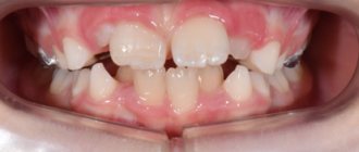
Why is the “vampire smile” formed?
In dentistry, the incorrect position of the eye teeth is called dystopia. It can manifest itself in different ways:
- fangs can protrude forward in relation to the dentition;
- hide behind adjacent teeth;
- be shortened or too long;
- unfold in the jawbone;
- be impacted, that is, not fully erupted.
Such anomalies arise because they erupt at the age of 9–12 years, later than other teeth, and in the jawbone, for various reasons, they may simply not have enough space:
- if the place is occupied by another tooth, the fang begins to grow “in the second row”;
- if the baby inherits large teeth from one of the parents, and a small jaw from the other, the fangs will become curved due to lack of space;
- If a change in bite occurs with a shift in timing, the eye tooth may protrude forward.
Types of location of an impacted tooth
| Fine | The tooth has a vertical position and grows upward very slowly. Baby teeth and canines can grow properly. “Wisdom teeth” are rarely positioned normally. |
| Horizontally | The impacted tooth forms a right angle to the healthy one, located horizontally. This situation is detrimental to a healthy “neighbor”. In such a situation, it is necessary to remove the impacted tooth, which is performed only surgically. |
| Obliquely | An impacted tooth grows at an angle toward the cheek or tongue. If such an anomaly does not cause inconvenience to a person, then removal is not necessary. |
Contraindications to the procedure
The list of contraindications for removing a fang on the upper jaw includes:
- Cardiovascular pathologies – myocardial infarction (suffered earlier six months ago), bacterial endocarditis, hypertension.
- Diseases of the urinary system - pyelonephritis in the acute stage, glomerulonephritis, renal failure.
- Diseases of an infectious and viral nature - hepatitis, acute respiratory infections, influenza and other pathologies transmitted by airborne droplets.
- Exacerbation of psychological problems - manic psychosis, epilepsy.
- Pregnancy at 1, 2 and 9 months.
- Poor clotting .
- Cancer – acute leukemia.
- Malignant tumors located in the maxillotemporal zone.
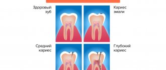
Symptoms of tooth impaction
In most cases, an impacted tooth is detected on an x-ray at the dentist or at an appointment with an ENT doctor. Retention in a child can be determined if one of the elements of the jaw row is absent for a long time or if a baby tooth is not replaced by a permanent “brother” for too long. The eruption of an impacted tooth is usually accompanied by inflammatory processes. The child may be bothered by swelling of the gum tissue and jaw, pain and aches, numbness and tingling in the retention zone.
On palpation of the gums, thickening and hardening are noted, reminiscent in shape and outline of an unerupted tooth.
Features of the procedure
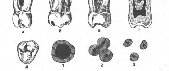
The canines on the upper jaw have one root located laterally. In cross section, part of the element resembles the outline of a triangle. In 30% of cases, the apex of the canine root has a curved structure. The outer part of the root is thicker than the inner one, but both sides of the alveoli are thicker in width than the incisors. Due to the listed features of the structure of the fangs, some difficulties arise when removing them from the socket.
During the operation, the doctor should place the fingers of the left hand in the same way as when removing incisors on the upper jaw. When removing an element from the right side, the patient's head should be slightly turned to the left and vice versa. This position of the patient will be more comfortable at the time of surgery.
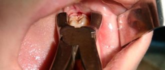
The canines on the upper jaw are removed from the socket using wide forceps. During the operation, the element swings alternately towards the lips and palate with rotation around the longitudinal plane of the fang. The doctor makes the first movement of the instrument towards the outer wall of the alveoli, since it has a thinner structure.
Preparation and stages of tooth extraction
The next dislocation is performed towards the palatine alveolus, and then the specialist performs rotations. The procedure consists of successive rocking and rotation of the element until the nerve fibers and tissues surrounding it are completely ruptured.
Due to the anatomical features of the canines on the upper jaw, when extracting them, a specialist has to make a lot of effort.
Removal of impacted teeth
If there are no complaints at all about the impacted tooth, and it does not cause any inconvenience (cysts do not form near it, the mucous membranes of the mouth are not injured), then removal may not be necessary. But if the impacted tooth is not removed, then it is necessary to undergo an X-ray examination of the mouth 2 times a year.
However, there are a number of cases when it is necessary to remove an impacted tooth:
- Retention causes constant inflammatory processes in the oral cavity;
- The occurrence of various paradental or follicular carpal formations, abscess, osteomyelitis in the gum tissue;
- Injuries and scratches of the oral mucosa that are caused by retention;
- An impacted molar or canine is also dystopic (improperly positioned in the dental arch) towards the cheeks or tongue.
The main factors on which the decision is made to remove impacted teeth are:
- The likelihood of injury during surgery,
- The location of the canine or molar and ease of access to them,
- Possibility of complications.
In most cases, the operation is painless and eliminates the possibility of pain even after the anesthesia wears off.
The removal of an impacted tooth itself takes place in several stages:
- pain relief by local anesthesia,
— an incision in the gum above the impacted canine and removal of a flap of tissue of the mucous membrane and periosteum,
- sawing out the bone to the dental crown using a drill,
- removal of a fang or molar from the bone using forceps (special biomaterials are left in its place).
Removal of premolars located on the upper jaw, canines and molars of the lower jaw has its own characteristics:
- Due to the fact that the roots of the teeth of the upper jaw are sometimes located close to the sinus, their removal must be carried out with the utmost care.
- In the case where the maxillary sinus has nevertheless been opened, medical treatment of the hole is not carried out, and the place where the bone has been broken is immediately sutured.
- Then a biomaterial is placed inside, which will promote rapid and painless overgrowth of bone tissue.
- The previously removed flap of mucous tissue and periosteum is returned to its place and sutures are applied.
- When removing impacted teeth in the lower jaw, the dentist must determine their location as accurately as possible, calculating their proximity to the mandibular canal. The safest way to access such teeth is through the vestibule of the mouth.
Types of tooth extraction
In medical practice, all removal procedures are divided into:
Easy removal
The doctor manages to extract the entire tooth using special forceps; its duration is up to 5 minutes. This is how single-rooted teeth or teeth affected by periodontitis are removed.
Difficult removal
To extract a tooth, the doctor uses auxiliary tools (most often an elevator), and the entire extraction process takes up to 15 minutes. It is most often performed when a part of the root or crown is broken during the removal of multi-rooted teeth.
Atypical removal
It has no clear boundaries in terms of time and extraction method and implies a complex and multi-stage procedure. For this, the doctor can use a bur, an elevator, forceps, a chisel, surgical sutures and needles to close the wound. Often such removal requires from 30 minutes to 2 hours.
This division is reflected both in the cost of the removal itself (due to the various labor costs of the doctor) and in the rehabilitation period:
Simple removal results in minimal damage to surrounding tissue. At the same time, squeezing them with an instrument (and stopping full blood circulation in this area) lasts no more than 5 minutes, and does not in any way affect the subsequent healing of the wound. If you follow the recommendations, the risk of complications is minimal.
Complex removal involves the use of more aggressive instruments and prolonged compression of the walls of the bone alveolus. After it, there may be difficulties with the formation of a blood clot and the development of alveolitis of the “dry socket” type, which is supervised by taking anti-inflammatory drugs and applying turunda with iodoform.
Atypical removal is a surgical intervention in which the doctor violates the integrity of the mucosa, periosteum, or a certain part of the bone. Pressure on tissue can last up to 2 hours, which greatly affects tissue microcirculation. Sometimes a hemostatic sponge is used to stop socket bleeding during such removal. The wound is sutured, which ensures the tightness of the hole and protects it from the development of alveolitis, but it is important to maintain a hygienic regime to avoid infection on the soft tissue.
The doctor’s further tactics, his prescriptions and recommendations for the recovery period depend on how the tooth was removed.
Start of orthodontic treatment
Before installing braces, the patient must undergo professional teeth cleaning by a hygienist. The specialist cleans the teeth, removes all plaque (soft and hard), checks the condition of the gums, polishes the enamel and gives recommendations on how to care for your teeth while wearing braces. Patients who decide to start treatment with braces receive a starter hygiene kit, which includes an orthodontic brush, toothpaste, brushes, dental floss, monobrush, and wax.
Damon metal braces are fixed:
Treatment with braces at this stage is aimed at expanding the dentition in the areas of the 2nd and 4th teeth (special springs are installed on the brace system).
After 5 months, the next stage begins - the return of the fangs to the dentition. The second arch holds the anterior incisors in a stable position.
Stage of wearing orthoelastics (12 months after fixing braces):
Removal of lower incisors
Removing the lower incisors is the easiest procedure. Due to the fact that the incisors have only one flat root. The presence of additional roots in these teeth is extremely rare. Removal occurs under local anesthesia. During this procedure, the patient takes a sitting position. The doctor's hands securely fix the patient's jaw. In such cases, the surgeon uses beak-shaped forceps. First of all, the tooth begins to dislocate in the labial side, and then in the lingual side, a slight rocking is allowed.
Is it possible to put braces on fangs?
Dystopia can and should be corrected for both a five-year-old child and an adult. The question of how this can be done will be decided based on the complexity of the anomaly formed, the degree of its neglect and, of course, the age of the patient.
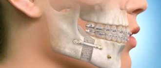
If the reason for the lack of space is an erupted wisdom tooth, you will have to get rid of it to get the necessary free space in the jaw. You can install metal or sapphire braces, which will move the canine to the place of the least valuable, previously removed first premolar. Finally, surgery may be performed to reposition the displaced eye tooth.
Even the best braces in Moscow will not make the correction process quick. The easiest way to return the fangs to the correct position is for 14-15 year olds. How long do adults need to wear braces ? At least 1.5–3 years - it all depends on the type of orthodontic structure, the material of the clasps and the patient’s dental characteristics.
Molar removal
The lower molars have branched roots. One of the roots has a long, wide shape with a curved line. The second one is thin and has a deviation towards the back. There is a possibility that the roots will move closer to each other or move in different directions. Removing such teeth can be a difficult procedure. The surgeon uses forceps equipped with wide cheeks. To ensure a secure grip, the tongs have protrusions or spikes. Cases of tooth crushing and parts being extracted separately cannot be ruled out. It is not advisable to perform rotational movements during this procedure.
From medical practice, it is worth noting that wisdom teeth can sometimes have more than three roots. The roots may diverge to the sides and be twisted. In most cases, wisdom teeth are abnormal, which makes them difficult to remove.