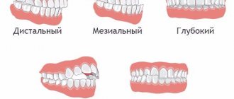
The replacement of baby teeth with permanent ones is a natural physiological process that every child experiences. It usually begins in preschool age and proceeds without difficulty. But parents need to approach it responsibly, since any violations when changing teeth are fraught with unpleasant consequences. This may be the formation of a malocclusion, uneven teeth, or the occurrence of a diastema (interdental gap).
In this article we will consider the correct sequence of tooth replacement, possible violations, and situations when consultation with a pediatric dentist is necessary.
Why and when does baby teeth fall out?
As the child grows, the dentofacial apparatus also develops. At approximately 4–6 years of age, the preparatory stage begins, during which interdental spaces appear. This prepares the place for permanent teeth in the primary dentition.
The process of replacing baby teeth with permanent teeth is a slow process, taking approximately 6 or 7 years (until adolescence). Active growth of the roots of permanent teeth activates the process of bone resorption - this is the destruction (dissolution) of milk roots, which leads to loosening and tooth loss. Standard diagram of the sequence of changing baby teeth in a child.
- Central lower incisors (5–7 years). They fall out one by one with a short time interval.
- Lateral incisors (7–8 years).
- First molars on the upper jaw, second molars on the lower jaw (9–11 years)
- Cone-shaped teeth (canines) at the age of 9–12 years.
- Second molars on the upper jaw and first molars on the lower jaw (10–13 years)
Not everyone's third molars (wisdom teeth) erupt. This is considered the norm. Most often, the optimal age for them is considered to be from 17 to 25 years.
Reference!
The later the first teeth erupted in infancy, the later they will be replaced in preschool age.
By the age of 15–16, a teenager’s permanent bite should number 28 units.
What diseases can be diagnosed by a panoramic dental photograph?
A panoramic photograph of teeth is an effective method for x-ray diagnosis of a number of dental diseases. Among them:
- benign and malignant neoplasms;
- fractures, cracks and dislocations;
- deep caries;
- bite pathologies;
- periodontitis;
- improper teething;
- cancer of the maxillary sinuses, etc.
What deviations from the norm can there be when baby teeth fall out?
Each child develops individually. Sometimes the timing of the replacement of certain teeth may differ from the generally accepted norm. Minor deviations are allowed up to 12 months. But sometimes the change in the primary bite begins too early or, on the contrary, the temporary teeth do not want to fall out.
Causes:
- long-term breastfeeding;
- severe infectious diseases in infancy;
- pathologies of intrauterine development.
Reference!
Dentists believe that a safer deviation is a late change in the primary occlusion than vice versa.
Let us consider in detail the common violations and their causes.
Early tooth loss
We can talk about such a pathology if a child’s baby teeth begin to fall out before the age of 5. Possible reasons:
- advanced multiple caries;
- injury;
- gum disease;
- manual loosening of the tooth.
In all these cases, consultation with a pediatric dentist is required.
Important!
Parents should record the time of tooth loss. If after 4 months the permanent tooth does not begin to emerge, then the help of a doctor is required.
In case of early unnatural tooth loss, it is advisable to conduct an X-ray diagnosis. This will help to identify possible damage to the permanent root rudiment in time and begin treatment. Otherwise, the child will need prosthetics in the future.
Late change from primary to permanent occlusion
The deadline for starting the process of changing baby teeth is 8 years. But this is considered a late shift. The disorder may be caused by:
- heredity;
- metabolic disorders;
- infectious diseases suffered in early childhood;
- mental disorders.
If after 8 years a child has not lost a single baby tooth, this is a reason to consult a doctor.
Preparing for a panoramic dental photograph
The procedure does not require special preparation. You can consume drinks, food and medicine - this will not affect the x-ray in any way. Immediately before the procedure, metal jewelry must be removed from the body. It is not recommended to drink alcohol.
When visiting a dentist, you must take panoramic and x-ray photographs from the last few years, as well as a medical history. With their help, the doctor will be able to track visible changes in the patient’s health status or compare treatment results.
X-ray radiation poses a possible risk to pregnant women. According to some studies, it can negatively affect the intrauterine development of the fetus. Pregnant women should tell their dentist about their pregnancy. In this case, carrying out OPTG depends on the ratio of possible risks and benefits.
Causes of late eruption after loss of baby teeth
Normally, after a baby tooth falls out, it takes 1-2 months for the permanent tooth to erupt. This is the longest period. In most cases, the rudiments of a permanent tooth can already be seen at the site of the lost tooth.
But, if a child’s toothless smile persists for 3 months or more, then this is a cause for concern for parents.
Let's consider why such dental pathology occurs:
- Retention
– a common condition that mainly affects the incisors and canines. They cannot erupt due to dense gums or because they rest against neighboring teeth. There are complete and partial retention. With the full form, a healthy root is visible in the picture, but it is completely under the gum. With partial retention, only part of the crown is visible. In this case, surgical assistance is required.
- Edentia
– a congenital pathology in which there is a lack of rudiments of permanent dental units. Can be complete or partial (sparse teeth). A rare disease. Orthopedic treatment is required as early as possible.
- Impact
– delayed eruption in this pathology is associated with a mechanical obstacle, that is, the child has a supernumerary of dental units. In this case, the permanent root simply does not have room to erupt. Impaction can only be detected using a panoramic x-ray of the jaw.
The sooner the child is examined by a doctor, the higher the chance of having an even and complete dentition.
How is a panoramic dental x-ray performed?
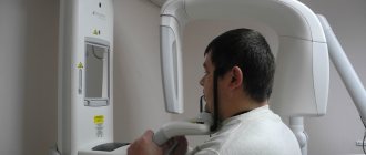
Panoramic photography of teeth is the specialty of a dental radiologist. It is carried out using an orthopantomograph - a modern device that exposes the body to a minimal amount of x-ray radiation.
To take a photo, you need to stand or sit near the device. Next, the patient must wear a protective apron that protects the chest from radiation. When the patient is ready, the doctor starts the orthopantomograph. The entire procedure lasts no more than 15 minutes and is completely painless.
During the process you must remain stationary. After the panoramic photo is taken, you need to wait a few more minutes. During this time, the specialist will prepare the image and provide it to the patient. The entire procedure lasts no more than 15 minutes, including the time required for preparation and waiting.
Possible problems when changing baby teeth
Common dental pathologies when changing a primary dentition to a permanent one include:
- Shark teeth. A phenomenon in which baby and permanent teeth are located parallel to each other, in 2 rows. This arrangement can interfere with the normal development of the dental system. But in most cases, the temporary root becomes loose, and the “extra” tooth falls out on its own. If this does not happen, removal at the doctor's office is recommended.
- Increased pain. Sometimes a change in the milk bite is accompanied by increased body temperature, redness of the gums and severe pain. These symptoms usually accompany early or late change of teeth. Inflammatory diseases of the oral cavity may also be the cause.
- The appearance of a hematoma.
In rare cases, when molars erupt, a hematoma occurs on the gum in the form of a small bubble with an accumulation of blood. This occurs due to severe eruption, which leads to rupture of blood vessels. The gums may be pale in color. Pain and discomfort occurs. If suppuration occurs, medical attention is required.
If pathological phenomena do not go away for a long time, and the child is irritable and complains of pain, be sure to visit the dentist.
Additional specialists and procedures
A patient who has received an appointment for a panoramic dental x-ray may need the help of the following specialists:
- dental surgeon;
- maxillofacial surgeon;
- oncologist;
- otolaryngologist;
- periodontist;
- orthodontist, etc.
OPTG is an important, but not the final stage of diagnosis. To confirm the diagnosis and develop effective therapy, in most cases a number of additional studies are prescribed. If tumors are present, a tissue biopsy is performed. A laboratory examination of pathological discharge is also performed.
If an orthopantomogram indicates problems with the canals or subgingival part of the tooth, it is necessary to open the tooth (filling) and further treatment. In some cases, tooth extraction may be indicated (if it cannot be treated).
What not to do when baby teeth fall out
Incorrect actions of children and parents can lead to the formation of malocclusion, increased pain, or the formation of crooked teeth. To avoid this, it is recommended to adhere to the following rules:
- Don't help baby teeth fall out if they are straight and not loose. Even if, according to the child’s age, it is high time for him to have a toothless smile.
- Don't loosen your teeth with your hands. And do not pull them at home, for example, with a thread.
- Baby teeth are quite fragile in preschool age. Therefore, your child should not indulge in solid foods. The crown may break, but the root will remain in the gum.
- If a tooth falls out, do not allow your child to touch the socket with his tongue or hands. Gentle rinsing with antiseptic solutions or herbal decoctions is recommended.
If you have a fever, cough or lethargy, do not rush to resort to antiviral drugs. Poor health can be a harbinger of the imminent change of baby teeth.
Types of X-rays for examining children
Examinations of the teeth and jaw may require different areas to be examined or results obtained from different types of images. Depending on the indications for x-rays, a certain type of procedure is used.
Types of x-rays of a child's teeth and jaw.
- Sight shot. Obtaining an image that displays a separate area (one or two teeth with closely spaced soft tissues).
- Panoramic shot. An image showing a large area of the dentition along with the jaw bone and the rudiments of molars.
- 3D pictures. A modern method of obtaining three-dimensional images of the upper and lower jaw.
There are also differences in technique. There are internal or external diagnostic methods. If it is necessary to obtain a targeted image, internal x-rays are used; in other cases, the external method is used.
Tips for parents
During the period of teeth change, you should enrich your baby’s food with foods that are rich in calcium. To prevent inflammation in your mouth, make it a habit to rinse your mouth every time you eat or have a snack. As permanent teeth erupt, offer your baby soft or liquid foods. Avoid hot drinks.
If slight bleeding occurs when a baby tooth falls out, do not use alcohol tinctures. Apply a cotton ball to the wound and do not allow food for 2 hours. In order to promptly identify possible irregularities in the eruption of the permanent dentition, it is recommended to record the date of baby tooth loss.
Beautiful, straight and healthy teeth in adults are the result of proper care in childhood. Therefore, parents should teach their child proper oral hygiene from the first tooth.
