The human body is a multi-stage structure, each organ and system of which is closely interconnected with each other and with the environment. And so that this connection is not interrupted even for a split second, a nervous system is provided - a complex network that permeates the entire human body and is responsible for self-regulation and the ability to adequately respond to external and internal stimuli. Thanks to the well-coordinated work of the nervous system, a person can adapt to the factors of the external world: any, even minor, change in the environment causes nerve cells to transmit hundreds of impulses at an incredibly high speed so that the body can instantly adapt to new conditions. Internal self-regulation works in a similar way, in which the activity of cells is coordinated in accordance with current needs.
The functions of the nervous system affect the most important processes of life, without which the normal existence of the body is unthinkable. These include:
- regulation of the work of internal organs in accordance with external and internal impulses;
- coordination of all units of the body, from the smallest cells to organ systems;
- harmonious interaction between humans and the environment;
- the basis of higher psychophysiological processes characteristic of humans.
How does this complex mechanism work? What cells, tissues and organs make up the human nervous system and what is each of its sections responsible for? A brief excursion into the basic anatomy and physiology of the human body will help you find answers to these questions.
Stages of nervous system development
In evolution, the nervous system has undergone several stages of development, which became turning points in the qualitative organization of its activities.
These stages differ in the number and types of neuronal formations, synapses, signs of their functional specialization, and in the formation of groups of neurons interconnected by common functions. There are three main stages of the structural organization of the nervous system: diffuse, nodular, tubular. The diffuse nervous system is the most ancient, found in coelenterates (hydra). Such a nervous system is characterized by a multiplicity of connections between neighboring elements, which allows excitation to freely spread throughout the nervous network in all directions.
This type of nervous system provides wide interchangeability and thereby greater reliability of functioning, but these reactions are imprecise and vague.
The nodal type of nervous system is typical for worms, mollusks, and crustaceans.
It is characterized by the fact that the connections of nerve cells are organized in a certain way, excitation passes along strictly defined paths. This organization of the nervous system turns out to be more vulnerable. Damage to one node causes dysfunction of the entire organism as a whole, but its qualities are faster and more accurate.
The tubular nervous system is characteristic of chordates; it includes features of the diffuse and nodular types. The nervous system of higher animals took all the best: high reliability of the diffuse type, accuracy, locality, speed of organization of nodal type reactions.
The leading role of the nervous system
At the first stage of the development of the world of living beings, interaction between the simplest organisms was carried out through the aquatic environment of the primitive ocean, into which the chemical substances released by them entered. The first oldest form of interaction between the cells of a multicellular organism is chemical interaction through metabolic products entering the body fluids. Such metabolic products, or metabolites, are the breakdown products of proteins, carbon dioxide, etc. This is the humoral transmission of influences, the humoral mechanism of correlation, or connections between organs.
The humoral connection is characterized by the following features:
- lack of an exact address to which a chemical substance entering the blood or other body fluids is sent;
- the chemical spreads slowly;
- the chemical acts in minute quantities and is usually quickly broken down or eliminated from the body.
Humoral connections are common to both the animal and plant worlds. At a certain stage of development of the animal world, in connection with the appearance of the nervous system, a new, nervous form of connections and regulation is formed, which qualitatively distinguishes the animal world from the plant world. The higher the development of an animal’s organism, the greater the role played by the interaction of organs through the nervous system, which is designated as reflex. In higher living organisms, the nervous system regulates humoral connections. Unlike the humoral connection, the nervous connection has a precise direction to a specific organ and even a group of cells; communication is carried out hundreds of times faster than the speed of distribution of chemicals. The transition from a humoral connection to a nervous connection was not accompanied by the destruction of the humoral connection between the cells of the body, but by the subordination of nervous connections and the emergence of neurohumoral connections.
At the next stage of development of living beings, special organs appear - glands, in which hormones are produced, formed from food substances entering the body. The main function of the nervous system is both to regulate the activity of individual organs among themselves, and in the interaction of the body as a whole with its external environment. Any impact of the external environment on the body appears, first of all, on receptors (sensory organs) and is carried out through changes caused by the external environment and the nervous system. As the nervous system develops, its highest department—the cerebral hemispheres—becomes “the manager and distributor of all the activities of the body.”
Treatment
Human nerve endings, the recovery scheme of which corresponds to the treatment of inflammatory processes, respond to all types of therapy: drugs, physiotherapeutic procedures, surgery and some folk remedies:
- Medication – first of all, analgesic agents are prescribed to relieve pain symptoms. For neuritis of infectious origin, topical antibiotics and antiviral drugs are prescribed. A frequent effect is exerted by a glucocorticoid drug, which is capable of universally removing fermentation in the area of inflammation. A sharp attack of radiculitis is temporarily relieved with diclofenac-based medications. Vasodilating and decongestant drug groups are used. Complexes of vitamins B and C and nicotinic acid are often added to the course of treatment. To improve neuromuscular conduction, the appropriate pharmacological group is used: to enhance contractile function - cholinomimetic drugs, to relax muscles - muscle relaxants. The latter have a positive effect in the fight against intercostal neuralgia. For the treatment of diabetic polyneuropathy, the correct external insulin dosing system is calculated. Anticonvulsants are used to restore limb nerves.
- Physiotherapeutic – the entire range of physiotherapeutic treatment procedures is suitable for the treatment of neuralgia. When restoring nerve fibers of the face and head, ultra-high-frequency therapy is used, which has an anti-inflammatory effect on the affected area of the nerve. At the same time, it increases blood circulation and lymph flow, which gradually restores nerve conductivity. Ultraviolet irradiation is used to relieve acute inflammation. Diadynamic currents, otherwise known as “Bernard currents,” are prescribed as impulse therapy for neuralgia of various localizations. Laser therapy, which acts at a depth of up to 3 cm from the skin, has a positive effect in the treatment of nervous disorders. This allows the nerve plexuses located close to the surface of the body to be affected. Laser physiotherapy is actively used in the restoration of the trigeminal and facial nerves, lumbosacral plexus, cervical and brachial nerve branches.
- Exercise therapy and massage. Therapeutic exercise and massage have both a therapeutic and preventive effect in the event of neuritis. The main goal is to strengthen the spinal nerve branches by placing stress on the spine.
Human nerve endings (the PNS circuit allows you to influence nerve endings through muscle tone) respond well to massage. When treating neuralgia of the face and head, special massage schemes with acupressure and vibration techniques are used.
When massaging the occipital region, avoid sharp chopping movements and tapping. A course of facial gymnastics is prescribed. Exercise therapy and massage techniques are the safest for humans due to the natural approach to healing, as opposed to the drug method.
- Surgical treatment – operations on nerve fibers and nodes are approached in two cases: lack of results in other treatment methods and traumatic consequences. The latter predominate in neurological surgery and consist of complex multi-stage operations. Microsurgical techniques are used using high-precision optics, special instruments and ultra-thin suture material. In the case of non-traumatic development of trigeminal neuritis, a neurosurgical operation is used - microvascular decompression. The effect is achieved by installing a Teflon protector between the damaged nerve and the vessel, thereby restoring the connection between the periphery and the central nervous system. To relieve a patient from inflammation of the sciatic nerve, they resort to removing the provoking factor - an intervertebral hernia or part of the affected vertebra.
- Folk remedies - the main goal of folk medicine is pain relief and relaxation of the nerve ending. Topical application involves fresh leaves of geranium, aloe, and horseradish. Leaves are applied to the sore spot and wrapped in several layers of warm cloth. For internal use, decoctions of chamomile, peppermint, sage and willow bark are used. Dried and crushed parts of plants are poured with boiling water and infused under a lid. They relieve pain and also have a general sedative and anticonvulsant effect. To treat disorders of the facial and trigeminal nerves, a hard-boiled egg helps - it is peeled and cut in half before it cools, then applied to the diseased area.
The human body is equipped with a dense system of nerve endings that are responsible for the full functionality of the body.
If the circulatory system plays a transport role, then the peripheral nervous system, which schematically repeats it, is responsible for motor and sensory tasks. Disturbances in the nerves, regardless of their location and scale, lead to various consequences - from mild inconvenience to paralyzing and painful manifestations.
Structure of the nervous system
The nervous system is formed by nervous tissue, which consists of a huge number of neurons - a nerve cell with processes.
The nervous system is conventionally divided into central and peripheral.
The central nervous system includes the brain and spinal cord, and the peripheral nervous system includes the nerves that arise from them.
The brain and spinal cord are a collection of neurons. In a cross section of the brain, white and gray matter are distinguished. Gray matter consists of nerve cells, and white matter consists of nerve fibers, which are processes of nerve cells. In different parts of the central nervous system, the location of white and gray matter is different. In the spinal cord, gray matter is located inside, and white matter is outside, but in the brain (cerebral hemispheres, cerebellum), on the contrary, gray matter is outside, white matter is inside. In various parts of the brain there are separate clusters of nerve cells (gray matter) located inside the white matter - the nuclei. Clusters of nerve cells are also located outside the central nervous system. They are called nodes and belong to the peripheral nervous system.
Nervous tissue
Nervous tissue is the main tissue that forms the nervous system and creates the conditions for the implementation of its many functions. Nervous tissue is of ectodermal origin; it is not customary to divide nervous tissue into any types of tissue. It has two main properties: excitability and conductivity.
Neuron
The structural and functional unit of nervous tissue is a neuron (from ancient Greek νεῦρον - fiber, nerve) - a cell with one long process - axon (Greek axis - axis), and one / several short ones - dendrites (Greek dendros - tree ).
I hasten to inform you that the idea that a short process of a neuron is always a dendrite, and a long process is always an axon, is fundamentally wrong. From a physiological point of view, it is more correct to give the following definitions: dendrite - a process of a neuron along which a nerve impulse moves to the body of a neuron, axon - a process of a neuron along which an impulse moves from the body of a neuron.
Neurons have 4 properties:
- Reception (lat. receptio - acceptance) - capable of perceiving incoming signals (dendrites)
- In response to signals, they are able to switch to a state of excitation or inhibition
- Conduction of excitation (from the dendrite to the neuron body, then to the end of the axon)
- Transmission of a signal to other objects - a neuron or an effector organ
In physiology, an effector organ (from the Latin efferes - efferent) is often called an executive organ or a target organ (muscles, glands). The effector organ carries out certain “orders” of the central nervous system (CNS) or endocrine glands
The processes of neurons conduct nerve impulses and transmit them to other neurons, effectors, due to which muscles contract or relax, and the secretion of glands increases or decreases.
Myelin sheath
Nerve fibers are divided into myelinated and unmyelinated. A nerve fiber is one or more processes of neurons (can be either axons or dendrites) with a surrounding sheath.
Unmyelinated nerve fibers are found predominantly in the autonomic nervous system (conduction speed 1-2 m/s). Myelin - form the white matter of the brain and spinal cord, nerve fibers of the somatic nervous system (5-120 m/s).
In myelinated nerve fibers, the processes of neurons are covered with a myelin sheath (70-75% composed of lipids (fats)), which ensures isolated transmission of the nerve impulse along the nerve. If there were no myelin sheath (imagine!), nerve impulses would propagate chaotically, and when we wanted to move our arm, our leg would move along with our arm.
There is a disease in which one’s own antibodies destroy the myelin sheath of the nerve fibers of the brain and spinal cord (such malfunctions of the body also occur). This disease - multiple sclerosis, as it progresses, leads to the destruction of not only the myelin sheath, but also the nerves - which means muscle atrophy occurs and the person gradually becomes immobilized.
The myelin layer is represented by several layers of glial cell membrane (lemmocyte, Schwann cell), which twist around the axial cylinder (neuron process). This twisting is clearly visible in the picture of a healthy nerve, just above 
The myelin layer of the fiber sheath is regularly interrupted at the junction of neighboring lemmocytes - nodes of Ranvier. The myelin sheath provides isolated and faster conduction of excitation (saltatory type, Latin salto - I gallop, jump).
Neuroglia (Greek νεῦρον - fiber, nerve + γλία - glue)
You have already seen how important neurons are; their high specialization leads to the emergence of a special environment - neuroglia. Neuroglia (glial cells, gliocytes) are an auxiliary part of the nervous system that performs a number of important functions:
- Supporting - supports neurons in a certain position
- Regenerative (Latin regeneratio - rebirth) - in case of damage to nerve structures, neuroglia promotes regeneration
- Trophic (Greek trophe - nutrition) - with the help of neuroglia, neurons are nourished: neurons do not come into direct contact with blood
- Electrical insulating - lemmocytes (Schwann cells) curl around the processes of neurons and form the myelin sheath
- Barrier and protective - isolate neurons from the tissues of the internal environment of the body
- Some gliocytes secrete cerebrospinal fluid - cerebrospinal fluid (from the Latin liquor - liquid)
Neuroglia consists of different cells; there are tens of times more of them than neurons themselves. In the peripheral part of the nervous system, the myelin sheath, which we studied, is formed precisely from neuroglia - Schwann cells (lemmocytes). Between them, the nodes of Ranvier are clearly visible - areas devoid of the myelin sheath between two adjacent Schwann cells.
Classification of neurons
Neurons are functionally divided into sensory, motor and intercalary.
Sensory neurons are also called afferent, centripetal, sensory, perceptive - they perceive irritations, convert them into nerve impulses and transmit them to the central nervous system. A receptor is the terminal ending of sensory nerve fibers that perceive a stimulus.
Interneurons are also called intermediate, associative - they provide communication between sensory and motor neurons, transmit excitation to various parts of the central nervous system, and participate in information processing and the generation of commands.
Motor neurons are also called efferent, centrifugal, or motor neurons - they transmit a nerve impulse (excitation) to an effector (working organ). The simplest example of the interaction of neurons is the knee reflex (however, there is no interneuron in this diagram). We will study reflex arcs and their types in more detail in the section on the nervous system.
Synapse
In the diagram above, you probably noticed a new term - synapse (Greek sýnapsis - connection). A synapse is the point of contact between two neurons or between a neuron and an effector (target organ). At the synapse, the nerve impulse is “transformed” into a chemical one: special substances are released - neurotransmitters (the most famous is acetylcholine) into the synaptic cleft.
Let's look at the structure of a synapse in the diagram. It is made up of the presynaptic membrane of the axon, next to which there are vesicles (Latin vesicula - bubble) with a neurotransmitter inside (acetylcholine). If a nerve impulse reaches the terminal (end) of the axon, then the vesicles begin to merge with the presynaptic membrane: acetylcholine flows out into the synaptic cleft.
Once in the synaptic cleft, acetylcholine binds to receptors on the postsynaptic membrane, thus excitation (nerve impulse) is transmitted to another neuron. This is how the nervous system works: the electrical transmission path is replaced by a chemical one (at the synapse).
Poison curare
It is much more interesting to study any subject with examples, so I will try to please you with them as often as possible. I cannot hide the story about the poison curare, which the Indians have used for hunting since ancient times.
This poison blocks acetylcholine receptors on the postsynaptic membrane, and, as a result, the chemical transfer of excitation from one neuron to another becomes impossible. This leads to the fact that nerve impulses cease to flow to the effectors, including the respiratory muscles (intercostal muscles, diaphragm), as a result of which breathing stops and the death of the animal occurs.
Nerves and ganglia
When the processes of neurons (nerve fibers) come together, they form bundles of nerve fibers. Nerve bundles unite into nerves, which are covered with a connective tissue sheath. If the bodies of neurons are concentrated in one place outside the central nervous system, their clusters are called a nerve ganglion - or ganglion (from the ancient Greek γάγγλιον - node).
In the case of complex connections between nerve fibers, they speak of nerve plexuses. One of the most famous is the brachial plexus.
Nervous system diseases
Neurological diseases can develop anywhere in the nervous system: the clinical picture will depend on this. If the sensitive pathway is damaged, the patient ceases to feel pain, cold, heat and other irritants in the area of innervation of the affected nerve, while movements are fully preserved.
If the motor link is damaged, movement in the affected limb will be impossible: paralysis occurs, but sensitivity may remain.
There is a severe muscle disease - myasthenia gravis (from the ancient Greek μῦς - “muscle” and ἀσθένεια - “powerlessness, weakness”), in which one’s own antibodies destroy motor neurons (motor neurons).
Gradually, any muscle movements become more and more difficult for the patient, it becomes difficult to speak for a long time, and fatigue increases. A characteristic symptom is observed - drooping of the upper eyelid. The disease can lead to weakness of the diaphragm and breathing muscles, making breathing impossible.
© Bellevich Yuri Sergeevich 2018-2021
This article was written by Yuri Sergeevich Bellevich and is his intellectual property. Copying, distribution (including by copying to other sites and resources on the Internet) or any other use of information and objects without the prior consent of the copyright holder is punishable by law. To obtain article materials and permission to use them, please contact Yuri Bellevich
.
Reflex activity of the nervous system
The main form of activity of the nervous system is the reflex. Reflex is the body’s reaction to changes in the internal or external environment, carried out with the participation of the central nervous system in response to irritation of receptors.
With any irritation, excitation from the receptors is transmitted along centripetal nerve fibers to the central nervous system, from where, through the interneuron along centrifugal fibers, it goes to the periphery to one or another organ, the activity of which changes. This entire path through the central nervous system to the working organ, called the reflex arc, is usually formed by three neurons: sensory, intercalary and motor. A reflex is a complex act in which a significantly larger number of neurons take part. Excitation, entering the central nervous system, spreads to many parts of the spinal cord and reaches the brain. As a result of the interaction of many neurons, the body responds to irritation.
Spinal cord
The spinal cord is a cord about 45 cm long, 1 cm in diameter, located in the spinal canal, covered with three meninges: dura, arachnoid and soft (vascular).
The spinal cord is located in the spinal canal and is a cord that at the top passes into the medulla oblongata and at the bottom ends at the level of the second lumbar vertebra. The spinal cord consists of gray matter containing nerve cells and white matter consisting of nerve fibers. Gray matter is located inside the spinal cord and is surrounded on all sides by white matter.
In a cross section, the gray matter resembles the letter H. It distinguishes the anterior and posterior horns, as well as the connecting crossbar, in the center of which there is a narrow canal of the spinal cord containing cerebrospinal fluid. In the thoracic region there are lateral horns. They contain the bodies of neurons that innervate internal organs. The white matter of the spinal cord is formed by nerve processes. Short processes connect sections of the spinal cord, and long ones make up the conductive apparatus of bilateral connections with the brain.
The spinal cord has two thickenings - cervical and lumbar, from which nerves extend to the upper and lower extremities. 31 pairs of spinal nerves arise from the spinal cord. Each nerve begins from the spinal cord with two roots - anterior and posterior. The dorsal roots are sensitive and consist of processes of centripetal neurons. Their bodies are located in the spinal ganglia. The anterior roots - motor - are processes of centrifugal neurons located in the gray matter of the spinal cord. As a result of the fusion of the anterior and posterior roots, a mixed spinal nerve is formed. The spinal cord contains centers that regulate the simplest reflex acts. The main functions of the spinal cord are reflex activity and conduction of excitation.
The human spinal cord contains reflex centers for the muscles of the upper and lower extremities, sweating and urination. The function of excitation is that impulses from the brain to all areas of the body and back pass through the spinal cord. Centrifugal impulses from organs (skin, muscles) are transmitted through ascending pathways to the brain. Along descending pathways, centrifugal impulses are transmitted from the brain to the spinal cord, then to the periphery, to the organs. When the pathways are damaged, there is a loss of sensitivity in various parts of the body, a violation of voluntary muscle contractions and the ability to move.
Diagram of nerve endings on the human body
Each major part of the human body has a special system of nerve construction.
Back
The formation of the sensory roots of the neural tube plays a fundamental role in the formation of peripheral nerves. When they merge in the 6th week of embryonic development, the formation of spinal nerves occurs. They represent 31 pairs of innervating formations connecting the spinal cord with certain areas of the body.
Of these, cervical - 8 pairs, thoracic - 12 pairs, lumbar and sacral - 5 pairs each and a pair of coccygeal. The size of the nerves depends on the metameric region for the innervation of which a particular block of spinal nerves is responsible. Due to the high concentration of receptors and muscle fibers in the limbs, they are connected to the PNS by the lower cervical, lumbar and sacral nerve branches.
When leaving the intervertebral foramen, each nerve of the spinal cord is divided into 4 branches: anterior, posterior, meningeal and communicating branches. The latter connects the nerve process with the sympathetic trunk.
The meningeal, or meningeal, returns to the spinal cord trunk and supplies it with nerve conduction. The remaining branches extend metamerically to the dorsal muscle groups, limbs, and skin. The anterior branch forms the intercostal group of nerves.
Ribs
Each of the 12 pairs of human ribs is surrounded by muscles of the same name. The nerve supply continues from the anterior branch of the spinal nerves. Intercostal nerves penetrate the outer and inner sides of muscle tissue. Moreover, 11 pairs of nerves pass between the ribs, the last pair is called the subcostal. They occupy a concomitant position with the artery and vein.
The upper 6 pairs represent the cutaneous branch and end in the cutaneous layer of the chest wall. Their task is to innervate the intercostal muscles, muscle fibers of the sternum, abdomen, and lower back. The lower 5 pairs reach the level of the oblique and transverse abdominal muscles and end in the anterior wall of the peritoneum. The intercostal muscles are responsible for nerve conduction of the segments of the thoracic region and abdomen.
Leg
The lumbar plexus of spinal nerves passes into the main nerve of the lower limb - the femoral nerve. It innervates the lumbar muscles, as well as the muscles of the thigh and the articular part of the knee. The femoral nerve reaches the foot with a long subcutaneous branch and ends in the skin of the big toe.
Along the way, it branches to the anterior cutaneous part of the thigh, then to both surfaces of the lower leg. Along the way, the peroneal nerve follows, dividing into the common, deep and superficial branches.
The posterior surface of the leg is equipped with a continuation of the sacral roots - the sciatic nerve, which is the largest nerve ending in terms of branching area. It extends to the entire gluteal region, thigh muscles and hip joint.
Hand
The human nerve endings, the diagram of which, according to the anatomical atlas, is concentrated in the hands, originate in the complex brachial plexus. The nerve trunks are responsible for the innervation of the shoulder girdle and the free part of the upper limbs. The brachial nerve continues down to the skin of each finger, passing in the grooves of muscles and tendons.
It is accompanied by branches of the radial nerve, which innervates the triceps and ulnar muscles. The nerve conductor of the flexor muscle group is the median nerve, which arises from the brachial plexus and descends along the artery, simultaneously bending around it.
Human nerve endings
The ulnar nerve is responsible for the coordination of the hand and the precise regulation of impulses when moving the arm. The sensory and motor functions of the thumb involve 3 nerves at once: the radial, ulnar and median. The area in the brain that processes the operation of the thumb is much larger than the areas that control the other fingers.
Head
The peripheral system of nerves of the head is represented by the cervical nerve plexus, which branches into 3 types: muscle, sensory and phrenic nerves. The muscular branch innervates the muscles of the neck and head.
The particular importance of this branch is to accompany the motor function of the cervical muscles. The phrenic nerve does not ascend to the head, but descends to the chest and is involved in the nerve conduction of the diaphragm.
The scalp is supplied with two sensory branches of nerves. The first - the large auricle - innervates the auricle, earlobe and external auditory canal. The second, the lesser occipital nerve, provides conduction to the occipital region and part of the auricle.
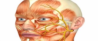
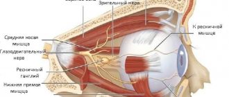
Face
The facial area is connected to the PNS via the trigeminal nerve. The first of its three branches conducts impulses in the frontal zone, the upper eyelid and the nasal mucosa. The second plexus works in the infraorbital region of the face and provides nervous regulation of the lower eyelid, sinuses, upper lip, cheek mucosa, as well as the gums of the upper teeth and the teeth themselves.
The third branch of the trigeminal nerve is the mental nerve. It supplies innervation to the lower corners of the mouth, lower teeth and gums, muscles of mastication, salivary glands, and the anterior portion of the tongue.
The facial nerve supplies the facial muscle tissue with nerve endings. It is divided into 5 main groups of nerves: temporal, maxillary, buccal, mandibular and cervical. The motor innervation of the lower lip is regulated by the mandibular branch.
The muscles that elevate the edges of the sinuses and the upper lip are innervated by the zygomatic branch of the facial nerve. For nerve conduction of the lacrimal gland and taste areas of the tongue, the intermediate nerve is responsible, which adds a sensory character to the motor facial nerve.
Evolution of the vertebrate brain
The formation of the central nervous system in the form of a neural tube first appears in chordates. In lower chordates, the neural tube is preserved throughout life; in higher chordates, vertebrates, a neural plate is formed on the dorsal side during the embryonic stage, which is immersed under the skin and folded into a tube. In the embryonic stage of development, the neural tube forms three swellings in the anterior part - three brain vesicles, from which parts of the brain develop: the anterior vesicle gives rise to the forebrain and diencephalon, the middle vesicle turns into the midbrain, the posterior vesicle forms the cerebellum and medulla oblongata. These five brain regions are characteristic of all vertebrates.
Lower vertebrates - fish and amphibians - are characterized by a predominance of the midbrain over other parts. In amphibians, the forebrain somewhat enlarges and a thin layer of nerve cells is formed in the roof of the hemispheres - the primary medullary vault, the ancient cortex. In reptiles, the forebrain increases significantly due to accumulations of nerve cells. Most of the roof of the hemispheres is occupied by the ancient cortex. For the first time in reptiles, the rudiment of a new cortex appears. The hemispheres of the forebrain creep onto other parts, as a result of which a bend is formed in the region of the diencephalon. Beginning with ancient reptiles, the cerebral hemispheres became the largest part of the brain.
The structure of the brain of birds and reptiles has much in common. On the roof of the brain is the primary cortex, the midbrain is well developed. However, in birds, compared to reptiles, the total brain mass and the relative size of the forebrain increase. The cerebellum is large and has a folded structure. In mammals, the forebrain reaches its greatest size and complexity. Most of the brain matter is made up of the neocortex, which serves as the center of higher nervous activity. The intermediate and middle parts of the brain in mammals are small. The expanding hemispheres of the forebrain cover them and crush them under themselves. Some mammals have a smooth brain without grooves or convolutions, but most mammals have grooves and convolutions in the cerebral cortex. The appearance of grooves and convolutions occurs due to the growth of the brain with limited dimensions of the skull. Further growth of the cortex leads to the appearance of folding in the form of grooves and convolutions.
Symptoms of inflammation of nerve endings
In the neurological clinic, half of the diseases are pathologies of the peripheral nervous system. Moreover, in most cases they do not threaten human life, but become the main prerequisites for loss of ability to work.
This is due to structural changes in the nerve fiber, which disrupt not only motor and autonomic functions, but also affect sensory conduction. The main symptom of such manifestations is severe pain and is commonly called neuritis or neuropathy.
Nerve endings are schematically similar to the branches of the circulatory system, therefore the localization of their pathological changes occurs in any segment of the human body. However, the symptoms are characteristic of the specific location of nerve damage.
Spinal nerve disorders
The most common (especially in old age) are neurological diseases in the back area:
| Name | Brief description of the pathology |
| Inflammation of the sciatic nerve | The disease is characterized by aching, stabbing, sharp or dull pain starting from the lower back. A shooting sensation spreads down the lower limb all the way to the toes. May be accompanied by numbness, swelling, and redness of the skin. At the site of nerve damage, the temperature may increase or decrease relative to the rest of the body. Nerve damage accompanied by a feeling of cooling and “pins and needles” is called sciatica. |
| Radiculitis | Changes in the spinal cord roots cause pain in the lumbosacral region. A lingering burning pain from the buttock to the foot is likely. The peculiarity of radiculitis is that it increases sensations during sudden movements: it is difficult for a person to sit down, stand up, or bend the body. Radiculitis of the cervico-brachial area is identified, which spreads the pain syndrome to the neck, shoulder blade and shoulder. Makes itself felt when coughing and raising hands. In severe cases, it completely immobilizes the neck and head. |
| Radiculopathy | In another way, radicular syndrome, which occurs due to compression of the roots of the spinal nerves, but manifests itself mainly not in the back. Sharp tingling of internal organs, in the arms and legs, neck and head - symptoms of radicular disorders. As a rule, it has an increasing character over time. |
Intercostal neuralgia
Accompanied by girdling pain from the back to the chest. When moving, shooting spasms, both prolonged and point-like, are felt. There is stiffness and stabbing pain when breathing and coughing. Neuralgia of the intercostal nerves is often accompanied by involuntary muscle contractions, sweating and numbness of the skin around the chest.
Nerve lesions in the legs
Serious consequences come from disorders of nervous activity in the lower extremities:
| Diseases | Symptoms |
| Polyneuropathy | Toxic damage to the nerves of the lower extremities is accompanied by impaired sensitivity of the feet. Itchy and tingling sensations and convulsive manifestations occur. The gradual spread of affected areas of nervous tissue leads to muscle atrophy, up to necrosis. Accompanying feelings of hypothermia or sweating, sometimes together. |
| Tibial nerve neuritis | There is a decline in motor processes in the foot, the function of toe flexion decreases and, as a result, the patient loses the ability to stand on his toes. There is atrophy of the posterior surface of the leg and the lower surface of the foot. Painful pulling sensations occur in the popliteal region and heel tendon |
| Peroneal nerve neuropathy | The main symptom is the inability to stand on the heel. The foot hangs down, the function of extending the toes and ankle joint does not work. The predominant sensations are numbness in the muscles of the anterior surface of the leg and the back of the foot. The pain syndrome is observed during walking and intensifies with increasing dynamics. |
Hand nerve lesions
No less serious problems are caused by pathologies in the nervous activity of the upper extremities:
- Neuritis of the median nerve. The lesion leads to the inability to flex the hand and dysfunction of the interphalangeal joints of 1-4 fingers. The sensitivity of the palm decreases with its gradual flattening. The muscles of the forearm, as well as the skin of the lower part of the arm, atrophy. Accompanied by vegetative-vascular disorders in the form of sweating of the palm, bluish skin and destruction of the nail plates.
- Radial nerve neuropathy. It is characterized by deformation of the hand due to depletion of small muscles. Aching pain occurs with decreased sensitivity in the 4th and 5th fingers. The patient does not have the ability to bend the thumb and rest the entire palm on it. There is a functional disorder of the elbow and wrist joints. Radial neuritis is also called drop hand syndrome.
Disturbances in the nerves of the head and face
Damages to the nerves of the head are also quite numerous:
- Trigeminal neuralgia. Distributes throughout the entire head and face area. Accompanied by a burning, sharp pain with attacks from several seconds to a couple of minutes. This is characterized by the removal of the pain signal from the site of nerve damage. When a symptom occurs, psychological stiffness occurs, fear of making any movement, and possible holding of breath until the nerve relaxes. Externally it is expressed in profuse lacrimation and mucous secretion from the nose. Convulsive contractions of the facial muscles are often observed.
- Neuritis of the facial nerve. The most pronounced symptom is facial asymmetry, and the curvature occurs in the healthy direction. Characteristic peripheral paralysis is accompanied by changes in skin folds, the inability to close the eye - the eyeball rolls up. When chewing, food remains behind the cheek, and the motor functions of the tongue are weakened. Accompanied by disturbances of taste and hearing, lacrimation and salivation increase or decrease, and dry mouth often occurs.
- Neuralgia of the occipital nerve. The pain syndrome affects one half of the back of the head in accordance with the affected branch of the nerve. The shootings are localized in the ear, neck, and shoulder blade. They intensify with any dynamic movements, coughing and sneezing. The patient is forced to fix the position of the head and avoid sudden turns. There may be a feeling of numbness in the occipital region. A chronicle of jerking headaches gradually develops.
- Slader's syndrome. Neuritis of the pterygopalatine ganglion causes a sharp spasm in the area of the eye, jaw, and nasal septum. The main distinguishing symptom is pressing pain in the palate and tongue with profuse salivation. Spreads to the ear, neck and half of the head. The development of dizziness with sensations of noise and ringing in the ears is typical. There is a high probability of redness on the corresponding side of the face with an increase in the volume of lacrimation. At least an hour passes before the nerve begins to relax.
This is the most common part of neurological disorders of the peripheral system. The causes can be trauma, infection, intoxication, compression and many other disorders. All prerequisites can be acute or chronic, and pain sensitivity depends on the duration of the disease.
Brain
If the spinal cord in all vertebrates is developed more or less equally, then the brain differs significantly in size and complexity of structure in different animals. The forebrain undergoes particularly dramatic changes during evolution. In lower vertebrates, the forebrain is poorly developed. In fish, it is represented by the olfactory lobes and nuclei of gray matter in the thickness of the brain. The intensive development of the forebrain is associated with the emergence of animals onto land. It differentiates into the diencephalon and two symmetrical hemispheres, which are called the telencephalon. Gray matter on the surface of the forebrain (cortex) first appears in reptiles, developing further in birds and especially in mammals. Truly large forebrain hemispheres become only in birds and mammals. In the latter, they cover almost all other parts of the brain.
The brain is located in the cranial cavity. It includes the brainstem and telencephalon (cerebral cortex).
The brainstem consists of the medulla oblongata, pons, midbrain and diencephalon.
The medulla oblongata is a direct continuation of the spinal cord and, expanding, passes into the hindbrain. It basically retains the shape and structure of the spinal cord. In the thickness of the medulla oblongata there are accumulations of gray matter - the nuclei of the cranial nerves. The posterior pons includes the cerebellum and the pons. The cerebellum is located above the medulla oblongata and has a complex structure. On the surface of the cerebellar hemispheres, gray matter forms the cortex, and inside the cerebellum - its nuclei. Like the spinal medulla oblongata, it performs two functions: reflex and conductive. However, the reflexes of the medulla oblongata are more complex. This is reflected in its importance in the regulation of cardiac activity, the condition of blood vessels, respiration, and sweating. The centers of all these functions are located in the medulla oblongata. Here are the centers for chewing, sucking, swallowing, saliva and gastric juice. Despite its small size (2.5–3 cm), the medulla oblongata is a vital part of the central nervous system. Damage to it can cause death due to cessation of breathing and heart activity. The conductor function of the medulla oblongata and the pons is to transmit impulses from the spinal cord to the brain and back.
In the midbrain there are primary (subcortical) centers of vision and hearing, which carry out reflexive orienting reactions to light and sound stimuli. These reactions are expressed in various movements of the torso, head and eyes towards the stimuli. The midbrain consists of the cerebral peduncles and quadrigeminalis. The midbrain regulates and distributes the tone (tension) of skeletal muscles.
The diencephalon consists of two sections - the thalamus and hypothalamus, each of which consists of a large number of nuclei of the visual thalamus and subthalamic region. Through the visual thalamus, centripetal impulses are transmitted to the cerebral cortex from all receptors of the body. Not a single centripetal impulse, no matter where it comes from, can pass to the cortex, bypassing the visual hillocks. Thus, through the diencephalon, all receptors communicate with the cerebral cortex. In the subtubercular region there are centers that influence metabolism, thermoregulation and endocrine glands.
The cerebellum is located behind the medulla oblongata. It consists of gray and white matter. However, unlike the spinal cord and brainstem, the gray matter - the cortex - is located on the surface of the cerebellum, and the white matter is located inside, under the cortex. The cerebellum coordinates movements, makes them clear and smooth, plays an important role in maintaining the balance of the body in space, and also influences muscle tone. When the cerebellum is damaged, a person experiences a decrease in muscle tone, movement disorders and changes in gait, speech slows down, etc. However, after some time, movement and muscle tone are restored due to the fact that the intact parts of the central nervous system take over the functions of the cerebellum.
The cerebral hemispheres are the largest and most developed part of the brain. In humans, they form the bulk of the brain and are covered with cortex over their entire surface. Gray matter covers the outside of the hemispheres and forms the cerebral cortex. The human cerebral cortex has a thickness of 2 to 4 mm and is composed of 6–8 layers formed by 14–16 billion cells, different in shape, size and functions. Under the cortex is a white substance. It consists of nerve fibers connecting the cortex with the lower parts of the central nervous system and the individual lobes of the hemispheres with each other.
The cerebral cortex has convolutions separated by grooves, which significantly increase its surface. The three deepest grooves divide the hemispheres into lobes. In each hemisphere there are four lobes: frontal, parietal, temporal, occipital. The excitation of different receptors enters the corresponding receptive areas of the cortex, called zones, and from here they are transmitted to a specific organ, prompting it to action. The following zones are distinguished in the cortex. The auditory zone is located in the temporal lobe and receives impulses from auditory receptors.
The visual zone lies in the occipital region. Impulses from the eye receptors arrive here.
The olfactory zone is located on the inner surface of the temporal lobe and is connected to the receptors of the nasal cavity.
The sensory-motor zone is located in the frontal and parietal lobes. This zone contains the main centers of movement of the legs, torso, arms, neck, tongue and lips. This is also where the center of speech lies.
The cerebral hemispheres are the highest division of the central nervous system, controlling the functioning of all organs in mammals. The importance of the cerebral hemispheres in humans also lies in the fact that they represent the material basis of mental activity. I.P. Pavlov showed that mental activity is based on physiological processes occurring in the cerebral cortex. Thinking is associated with the activity of the entire cerebral cortex, and not just with the function of its individual areas.
| Brain department | Functions | |
| Medulla | Conductor | Connection between the spinal and overlying parts of the brain. |
| Reflex | Regulation of the activity of the respiratory, cardiovascular, digestive systems:
| |
| Pons | Conductor | Connects the cerebellar hemispheres to each other and to the cerebral cortex. |
| Cerebellum | Coordination | Coordination of voluntary movements and maintaining body position in space. Regulation of muscle tone and balance |
| Midbrain | Conductor | Approximate reflexes to visual and sound stimuli (turns of the head and torso). |
| Reflex |
| |
| Diencephalon | thalamus
hypothalamus
| |
Structure and types of the nervous system: structural classification
To simplify the structure of the nervous system, in medicine there are several classification options depending on the structure and functions performed. Thus, anatomically, the human nervous system can be divided into 2 broad groups:
- central (CNS), formed by the brain and spinal cord;
- peripheral (PNS), represented by nerve ganglia, endings and nerves themselves.
The basis of this classification is extremely simple: the central nervous system is a kind of connecting link in which the analysis of the incoming impulse and further regulation of the activity of organs and systems is carried out. And the PNS serves to transport the received signal from the receptors to the CNS and the subsequent activator, but from the CNS to the cells and tissues that will perform a specific action.
central nervous system
The central nervous system is a key component of the nervous system, because it is here that the main reflexes are formed. It consists of the spinal cord and brain, each of which is reliably protected from external influences by bone structures. Such thoughtful protection is necessary, since each part of the central nervous system performs vital functions, without which it is impossible to maintain health.
Spinal cord
This structure is contained within the spinal column. It is responsible for the simplest reflexes and involuntary reactions of the body to stimuli.
In addition, spinal cord neurons coordinate the activity of muscle tissue, which regulates defense mechanisms. For example, upon feeling an extremely hot temperature, a person involuntarily withdraws his palm, thereby protecting himself from a thermal burn. This is a typical reaction controlled by the spinal cord.
Brain
The human brain consists of several sections, each of which performs a number of physiological and psychological functions:
- The medulla oblongata is responsible for the vital functions of the body - digestion, respiration, blood movement through the vessels, etc. In addition, the nucleus of the vagus nerve is located here, which regulates the autonomic balance and psycho-emotional reaction. If the nucleus of the vagus nerve sends active impulses, a person’s vitality decreases, he becomes apathetic, melancholic and depressed. If the activity of impulses emanating from the core decreases, the psychological perception of the world changes to a more active and positive one.
- The cerebellum regulates the precision and coordination of movements.
- The midbrain is the main coordinator of muscle reflexes and tone. In addition, neurons regulated by this part of the central nervous system contribute to the adaptation of the sensory organs to external stimuli (for example, pupil accommodation at dusk).
- The diencephalon is formed by the thalamus and hypothalamus. The thalamus is the most important organ that analyzes incoming information. The hypothalamus regulates the emotional background and metabolic processes; there are centers responsible for the sensation of hunger, thirst, fatigue, thermoregulation, and sexual activity. Thanks to this, not only physiological processes are coordinated, but also many human habits, for example, the tendency to overeat, the perception of cold, etc.
- Cerebral cortex. The cerebral cortex is a key link in mental functions, including consciousness, speech, perception of information and its subsequent comprehension. The frontal lobe regulates motor activity, the parietal lobe is responsible for bodily sensations, the temporal lobe controls hearing, speech and other higher functions, and the occipital lobe contains the centers of visual perception.
Peripheral nervous system
The PNS provides interconnection between organs, tissues, cells and the central nervous system. Structurally, it is represented by the following morphofunctional units:
- Nerve fibers, which, depending on the functions performed, are motor, sensory and mixed. Motor nerves transmit information from the central nervous system to muscle fibers, sensitive nerves, on the contrary, help to perceive information received through the senses and transmit it to the central nervous system, and mixed nerves are involved to one degree or another in both processes.
- Nerve endings, which are also motor and sensory. Their function is no different from fiber structures with the only nuance - the nerve endings begin or, conversely, end the chain of impulses from the organs to the central nervous system and back.
- Nerve ganglia, or ganglia, are clusters of neurons outside the central nervous system. The spinal ganglia are responsible for transmitting information received from the external environment, and the autonomic ganglia are responsible for transmitting information about the state and activity of the internal organs and resources of the body.
In addition, all peripheral nerves are classified depending on their anatomical features. Based on this characteristic, there are 12 pairs of cranial nerves that coordinate the activity of the head and neck, and 31 pairs of spinal nerves that are responsible for the torso, upper and lower extremities, as well as internal organs located in the abdominal and thoracic cavities.
Cranial nerves originate from the brain. The basis of their activity is the perception of sensory impulses, as well as partial participation in respiratory, digestive and cardiac activity. The function of each pair of cranial nerves is presented in more detail in the table.
| No. | Name | Function |
| I | Olfactory | Responsible for the perception of various odors, transmitting nerve impulses from the olfactory organ to the corresponding center of the brain. |
| II | Visual | Regulates the perception of visual data by delivering impulses from the retina. |
| III | Oculomotor | Coordinates the movement of the eyeballs. |
| IV | Block | Along with the oculomotor pair of nerves, it takes part in the coordinated movement of the eyes. |
| V | Trigeminal | Responsible for sensory perception of the facial area, and also participates in the act of chewing food in the oral cavity. |
| VI | Abductor | Another nerve that regulates the movements of the eyeballs. |
| VII | Facial | The nerve that coordinates facial contractions of the facial muscles. In addition, this pair is also responsible for taste perception, transmitting signals from the papillae of the tongue to the brain center. |
| VIII | vestibulocochlear | This pair is responsible for the perception of sounds and the ability to maintain balance. |
| IX | Glossopharyngeal | Regulates the normal activity of the pharyngeal muscles and partially transmits taste sensations to the brain center. |
| X | Wandering | One of the most significant cranial nerves, the functionality of which determines the activity of internal organs located in the neck, chest and abdominal wall. These include the pharynx, larynx, lungs, heart muscle and digestive tract organs. |
| XI | Dorsal | Responsible for contractions of muscle fibers of the cervical and shoulder regions. |
| XII | Sublingual | Coordinates the activity of the tongue and partially forms speech skills. |
The activity of the spinal nerves is classified much more simply - each specific pair or complex of pairs is responsible for its assigned area of the body with the same name:
- cervical - 8 pairs,
- infants - 12 pairs,
- lumbar and sacral - 5 pairs respectively,
- coccygeal - 1 pair.
Each representative of this group belongs to mixed nerves, formed by two roots: sensory and motor. That is why the spinal nerves can both perceive an irritating effect, transmitting an impulse along the chain, and intensify activity in response to a message from the central nervous system.
Cerebral cortex
The surface of the human cerebral cortex is about 1500 cm2, which is many times greater than the inner surface of the skull. This large surface of the cortex was formed due to the development of a large number of grooves and convolutions, as a result of which most of the cortex (about 70%) is concentrated in the grooves. The largest grooves of the cerebral hemispheres are the central one, which runs across both hemispheres, and the temporal one, which separates the temporal lobe from the rest. The cerebral cortex, despite its small thickness (1.5–3 mm), has a very complex structure. It has six main layers, which differ in the structure, shape and size of neurons and connections. The cortex contains the centers of all sensory (receptor) systems, representatives of all organs and parts of the body. In this regard, centripetal nerve impulses from all internal organs or parts of the body approach the cortex, and it can control their work. Through the cerebral cortex, conditioned reflexes are closed, through which the body constantly, throughout life, very accurately adapts to the changing conditions of existence, to the environment.