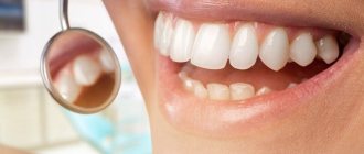
What is curettage? This is the cleaning of periodontal pockets from subgingival plaque. The structure of the gum allows plaque to accumulate between the tooth and its tissue. If oral hygiene is insufficient, plaque will accumulate there, eventually hardening and turning into tartar. And this threatens tooth loss, serious inflammation and other consequences. The need for curettage occurs when the distance between the tooth and the edge of the gum is more than 3 mm. The procedure is carried out open and closed.
What is curettage
Normally, between the gum and the cervical area of each tooth there is a dentogingival junction that protects the tissues from the spread of pathological processes, microorganisms, and food particles. In the absence of adequate treatment for gum inflammation, destructive processes and local disruption of microcirculation lead to the destruction of the dentogingival junction, a periodontal pocket is formed - the space between the tooth root and the connective tissue of the gums, where a favorable environment is created for the active activity of pathogenic bacteria.
Thus, gingivitis, which most often manifests itself as bleeding and swelling of the gums, develops into a more serious dental pathology - periodontitis, the treatment of which is no longer possible without surgical interventions. The causes of the occurrence and spread of the inflammatory process may be poor hygiene, constant trauma to the teeth, and some somatic diseases.
Curettage is a procedure for cleaning the cavity between the gum and tooth root. Only after treating the root part of the tooth with careful removal of all pathological contents (granulations, plaque, stone), it is possible to create conditions for successful healing of the lesion and restore the dentogingival junction. Curettage of the periodontal pocket is the basis of periodontitis therapy, which is confirmed by positive reviews from doctors about the procedure. The method of curettage is selected by a specialist depending on the severity of periodontitis, the main criterion of which is the depth of the periodontal pocket.
Causes of periodontitis and its consequences
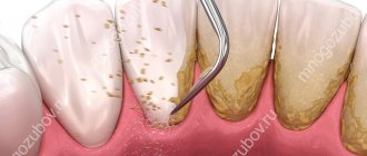
Many people are too negligent about oral hygiene. This leads to the fact that plaque and hard deposits begin to form on the teeth, indicating the appearance of the first signs of periodontitis. These processes are accompanied by bleeding and inflammation of the gums. The primary signs of the development of periodontitis also include tooth mobility and the appearance of purulent discharge from under the gums.
Subsequently, the soft plaque on the teeth mineralizes and becomes tartar, which promotes the release of toxins. These, in turn, cause gum inflammation and other adverse consequences.
Such consequences, first of all, include the gradual resorption of bone tissue around the tooth. In its place, granulation tissue appears, which contains many microbes that absorb the bone. This process contributes to a significant acceleration of bone tissue atrophy.
Another unpleasant consequence is the formation of periodontal pockets. They represent an area with destroyed bone tissue and a cavity that is filled with granular tissue, pus and dental plaque. To detect this pathology, x-rays or probing are required. It is worth adding that the periodontal pocket is often called the dental or gingival pocket.
Identification of a deep periodontal pocket (from 3 mm) means that the patient will no longer be helped by drug therapy. This is due to the fact that the destruction process has already taken an irreversible form.
The ineffectiveness of drug and therapeutic treatments can be explained by several reasons:
- Even the most qualified doctor cannot guarantee the removal of all subgingival deposits by inserting an ultrasonic tip under the gum. The lack of confidence in the 100% result of therapy is due to the fact that no specialist will be able to control what is happening in the periodontal pockets. That is why a large amount of destructive sediment remains there.
- This procedure is considered quite labor-intensive and expensive. However, it does not guarantee complete elimination of the problem.
- If the specialist manages to completely remove all subgingival deposits from the pocket, favorable conditions for the development of periodontitis will still remain in the oral cavity. Therefore, even removing all deposits will not stop the progression of the infection.
In such situations, the only effective treatment option is surgery. Only in this case will the removal of all subgingival deposits, granulation tissue and periodontal pockets be ensured.
Surgical methods for treating periodontitis
There are 3 main methods of surgical treatment of this disease:
- open curettage;
- closed curettage;
- flap surgery.
Closed curettage of periodontal pocket
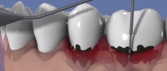
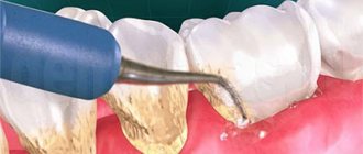
Closed curettage is a treatment technique performed in cases of mild or moderate pathology. Its peculiarity is that pockets are cleaned without incisions or gum detachment. After diagnosis, during which dental X-rays are necessarily studied and the depth of each pathological pocket is measured using periodontal instruments, local anesthesia is performed. The patient rinses the oral cavity with an antiseptic solution, and after complete anesthesia of the gums in the corresponding area, the doctor sequentially cleans each side of the tooth. Closed curettage of periodontal pockets is performed using a set of special hand instruments - scalers, curettes. The specialist inserts the instrument to the depth of the pocket and, using lever-like scraping movements, cleans the surface of the root and gums, as well as the bottom of the formation. Thus, the following are removed from the pathological pocket:
- hard subgingival calculus;
- granulations that have grown in the pocket;
- soft coating.
After basic cleaning with a periodontal bur, the accessible part of the tooth root is polished to prevent the accumulation of plaque on it and improve the conditions for restoring the periodontal attachment. The cleaned area is washed with an antiseptic. In one session, closed curettage can be performed in the area of 5-6 teeth.
When to do gum curettage?
Indications for curettage of periodontal pockets are: 1) the presence of chronic generalized periodontitis, 2) local form of periodontitis. In both cases, periodontal pockets will appear along the surface of the tooth roots, the formation of which is associated with the resulting destruction of the dentogingival attachment (i.e., the attachment of the marginal gum to the neck of the tooth).
Destruction of the periodontal attachment in chronic generalized periodontitis occurs against the background of irregular oral hygiene, which contributes to the deposition around the necks of teeth - soft microbial plaque and hard tartar. Microorganisms in the composition of dental plaque trigger an inflammation process in the gums, which at the first stage is manifested by swelling and redness of the gums, bleeding when brushing teeth. Already during this period, gradual destruction of the periodontal attachment begins (with the formation of periodontal pockets).
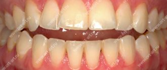
Chronic generalized periodontitis –
1) Formation of periodontal pockets –
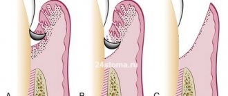
As soon as the periodontal attachment is destroyed, so-called “periodontal pockets” are formed between the gum and tooth. At first they are relatively small. Shallow periodontal pockets up to 4.0 mm deep correspond to mild periodontitis, and closed curettage may be indicated for precisely such patients (to prevent the progression of periodontitis).
Periodontal pockets are localized between the gum and the root of the tooth (Fig. 2). One wall of the pocket is the surface of the tooth root, on the surface of which microbial plaque and subgingival tartar are attached. The other wall is the inner surface of the unattached gum, which is covered with pathological epipelium and granulations. Thus, the typical contents of a periodontal pocket are dental deposits, granulations, and pathological epithelium.
Scheme of the structure of the periodontal pocket -
As we said above, in the absence of treatment, the depth of the pockets gradually increases. This occurs due to the destruction of the bone plate around the tooth, as well as periodontal fibers, due to which the tooth root is firmly attached to the bone. This gradual process inevitably leads to the appearance of tooth mobility, the increase in the degree of which in the future will be directly related, among other things, to the continuing increase in the depth of periodontal pockets.
2) Destruction of bone tissue around the tooth –
As was seen above in Fig. 2, the periodontal pocket “grows” deeper due to the destruction of periodontal fibers between the root surface and the bone plate, which also leads to the destruction of the bone graft itself (alveoli). Moreover, it should be noted that bone tissue does not simply disappear without a trace, but is replaced by the so-called inflammatory granulation tissue, which contains a large number of microbial cells, osteoclasts, etc. On an x-ray, the absence of bone around the teeth looks like this (Fig. 3b).
Clinical situation No. 1 –
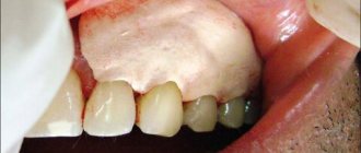
Next, in Figures 4-6, you can see exactly what a deep periodontal pocket looks like under the gum in the interdental space between the canine and premolar in the lower jaw. As we can see from the outside, the gums look quite healthy, and only on an x-ray (Fig. 5) can we see a slight darkening between the roots of the teeth, indicating the presence of destruction of bone tissue. But we have a completely different look after the gums are detached and granulations are cleaned out of the pocket between the teeth (Fig. 6).
In Fig. 6 we see a deep bone defect of a large volume, as well as subgingival calculus on the surface of the tooth root (indicated by an arrow). Such deep periodontal pockets can no longer be sanitized using the closed curettage technique, which is carried out without gum detachment and therefore does not imply visual inspection of bone defects in the depths of the periodontal pocket (i.e., the doctor will simply blindly try to clean out what to clean out from under the gums).
Therefore, for pockets larger than 4.0 mm, only open curettage or flap surgery should be used. For medium and deep periodontal pockets, only these methods allow you to remove all granulations and dental deposits from under the gums, carefully scrape out the pathological epithelium from the inner surface of the gums, and also replant synthetic bone tissue in the area of deep bone defects (with the hope of partially restoring bone levels ).
Why is it impossible to do without curettage when treating periodontitis (in principle) -
With shallow periodontal pockets up to 4.0 mm, provided that you have good equipment (a good scaler, a Vector device) and the painstaking work of a periodontist, it is still possible to stop the progression of chronic periodontitis. The biggest challenge will be removing subgingival deposits from periodontal pockets, which is difficult to do even in shallow pockets, given that the work is carried out almost blindly.
When deep periodontal pockets have formed (more than 4 mm deep) and granulation tissue has formed at their depth, the process essentially becomes irreversible, despite any conservative local and general anti-inflammatory therapy. You can drink antibiotics, rinse your mouth with antiseptics, smear your gums with anti-inflammatory gels, irradiate your gums with a laser, but all this will be ineffective. Why?
- Firstly , it is impossible in principle to completely remove dental plaque from deep periodontal pockets. The fact is that the doctor inserts the ultrasonic attachment under the gum “blindly”, i.e. he makes movements without seeing what exactly is happening in the periodontal pockets. Therefore, some dental plaque necessarily remains in the pockets.
- Secondly , if deep periodontal pockets have formed, then in them, even after removal of dental plaque and anti-inflammatory therapy, conditions are created for the development of infection and further progression of periodontitis. The fact is that the pockets are filled with granulations, without surgical removal of which it is impossible to stop further destruction of the bone tissue around the tooth.
It turns out that you can still try to remove subgingival dental deposits and granulations in shallow pockets using ultrasonic cleaning and closed curettage techniques. It will be possible to do the same thing efficiently in deep pockets only by performing open curettage or patch surgery.
Open curettage
The open curettage technique is more complex and requires making incisions in the gums. Thanks to gum detachment, deeper access is provided, and the doctor performs manipulations in the periodontal pocket with a wide view of the entire pathological focus. This allows the use of the open method in the treatment of moderate periodontitis, when the depth of the pathological pocket can reach 5 mm.
The operation is carried out as follows:
- After rinsing the mouth with an antiseptic and anesthesia, the dentist makes an incision at the tops of the gum papillae and peels away the tissue to expose the tooth roots for the duration of the procedure.
- With periodontal instruments, all surfaces are cleaned of necrotic tissue and plaque, vascular and epithelial formations are scraped out, and the bottom of the pocket is processed.
- Root polishing is carried out.
- The surgical field is washed with antiseptics, the gums are returned to their original position, and sutures are applied. If necessary, hemostatic sponges are used.
- The operation is completed by applying a bandage with therapeutic components to the treated area, designed to protect against oral fluid and injury.
During normal healing, the sutures are removed on average after 10 days. A course of local and general treatment with medications is prescribed. The patient takes a selected antibiotic and anti-inflammatory drugs. At home, baths and rinses are performed, and dental ointments are used. After thorough cleansing of pathological formations, drug therapy helps reduce signs of inflammation, eliminate pathogenic microflora, and promote tissue regeneration.
Is it painful to do curettage?
Patients' concerns about pain during this procedure are completely unfounded. Open and closed curettage is performed under local anesthesia. Anesthetics used in modern dentistry are effective and safe for human health. They are well tolerated by patients (with rare exceptions). Some people are allergic to certain types of pain medications. Therefore, the doctor must find out whether the patient has a history of any allergic reactions before the operation begins.
Periodontist's recommendation: do not delay visiting a doctor if symptoms of periodontal disease appear. The sooner treatment is started, the faster your gums will be healthy. Do not be afraid of pain; during surgical procedures you will be provided with effective local anesthesia. The branches of our clinic use only health-safe anesthetics that have been tested in leading clinics in the world.
The network of dental clinics “Smile” offers gum curettage services. Contacting our branches has a number of significant advantages:
- treatment is carried out by highly qualified doctors;
- all surgical and dental procedures comply with international standards;
- We provide family and cumulative discounts;
- pricing for our services is transparent to patients;
- We work according to a schedule convenient for you: every day until 21:00 (on Sunday until 16:00).
You can make an appointment at any of the branches of our clinic in Moscow, located within walking distance from metro stations:
- Alekseevskaya (VDNKh district, etc. Mira), address: st. 3rd Mytishchiskaya house 3, building 2;
- Shelepikha, address: Shelepikhinskaya embankment, building 34, building 1.
The modern equipment of the clinic and the experience of our doctors allow us to effectively and painlessly perform gum curettage, regardless of the severity of the disease. We guarantee the effectiveness and safety of treatment. We will keep your gums healthy!
Laser curettage
Dental pockets can be cleaned using a laser device. Instead of curettes and scalers, manipulations in the periodontal pocket are carried out using a diode or erbium laser beam. The procedure is prescribed for mild periodontitis. Using a dental laser attachment, the doctor inserts a beam into the space between the gum and the surface of the tooth root. When a laser acts on pathological growths (granulation tissue), they coagulate - destruction under the influence of high temperature and evaporation. An important feature of the technique is the absence of bleeding during curettage of periodontal pockets with a laser, which is due to the instant cauterizing effect of the beam.
To better eliminate pathogenic flora, the laser method is combined with photodynamic therapy. To do this, before the procedure, the gums and pocket area are treated with a preparation based on chlorophyll, a plant pigment. Its molecules accumulate in periodontal cells affected by microorganisms and form photosensitizer compounds. Half an hour after applying the pigment, the doctor acts on the colored tissue with a visible spectrum laser, which activates the resulting photosensitizers. The compounds destroy bacterial cells and elements affected by them, actively releasing oxygen. Since the chlorophyll in the drug has a selective effect, the laser does not affect healthy tissue. According to patient reviews, the procedure is almost painless; a tingling sensation may be felt.
Advantages of the laser method:
- low risk of gum injury;
- fast healing;
- no bleeding.
Laser cleaning is the method of choice for a group of patients who have contraindications to standard curettage methods - patients with diabetes mellitus, serious cardiovascular diseases, and bleeding disorders.
Indications for curettage:
- feeling of tension (pulsation, bloating) in the gums;
- putrid odor from the mouth that does not disappear after brushing your teeth;
- swelling, redness and soreness of the gums;
- bleeding gums under any impact, including while brushing your teeth;
- tooth mobility;
- increase in interdental spaces;
- deposition of tartar under the gum;
- destruction of periodontal tissue and jaw bone tissue (formation of replacement tissue in their place).
Curettage has contraindications. The procedure is not performed if the pocket depth is more than 6 mm, there is an abscess and pus in the periodontal pocket, as well as with thinned gums affected by fibromatosis. Obstacles to curettage include tooth mobility of degrees III–IV, infectious diseases (influenza, tonsillitis, acute respiratory infections) and any diseases in the acute stage.
Deep curettage
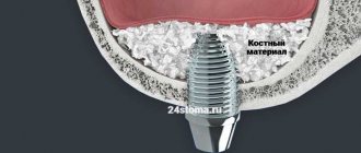
Severe periodontitis is a serious pathology, accompanied by the presence of periodontal pockets deeper than 5 mm, in which bone pockets often appear. With such signs, more extensive interventions are indicated to cleanse subgingival deposits, especially if the lesion has spread to a large number of teeth. The large depth of the gaps between the gums and the tooth root requires visual control to completely remove the pathological contents, so a radical operation is performed with peeling off a flap of tissue in the area of half or the entire jaw to the depth of the lesion.
After exposing the roots, they undergo deep treatment: removal of epithelial growths, any deposits, affected cement, as well as altered bone tissue. The treated area is washed with antiseptics and stitches are applied. In the presence of severe bone loss, osteoplastic materials are used to promote regeneration. Healing of oral tissue continues for a month.
Flap surgery on the gums for periodontitis –
Flap surgery on the gums allows not only to remove all subgingival dental plaque, but also to remove inflammatory granulation tissue from under the gums. It allows, in principle, to eliminate periodontal pockets, and also, in some cases, to stimulate partial restoration of bone volume (in vertical bone defects). The difference between flap surgery and open curettage is that the incision during flap surgery is made 1-1.5 mm from the edge of the gum (24stoma.ru).
Then this 1.5 mm strip of gum, which wraps around the necks of the teeth, is simply removed. Firstly, this allows us to reduce the size of the flaps (gum size), because with severe bone resorption and after removing a large number of granulations, excess gum will be observed. We will not have enough underlying tissue volume to stretch the gum back to the necks of the teeth. That is, as a result, we will receive partial exposure of the roots of the teeth, but at the same time, periodontal pockets between the gums and the roots of the teeth will be completely absent. And this is the most important thing if we want to stop the progression of periodontitis.
The decrease in gum level after surgery can be somewhat reduced if synthetic bone tissue preparations are used for grafting into bone defects. The effect will be even better if special bioresorbable membranes are also used. At one time, the operation is performed on a segment of no more than 6-8 teeth, and takes at least 2.5 hours. This is a complex procedure that requires good surgical skills.
Carrying out flap surgery (animation and video) –
Vacuum curettage
Vacuum curettage is a combined method; its technique is similar to the closed method of curettage of periodontal pockets, but together with standard curettes, hollow nozzles connected to a vacuum apparatus are used. Using a compressor, a vacuum is created in the device’s container, capable of sucking out mucus, deposits, and granulations from the space between the root and the gum. To improve visual control, the vacuum method can be performed in conjunction with gingivotomy (dissection of the gums).
Stages of the vacuum method:
- injection pain relief;
- scraping out accessible deposits from the tooth root with curettes;
- scraping granulations from the gums;
- bottom treatment with a vacuum nozzle;
- washing the cleaned area with antiseptics;
- applying a protective bandage for two days.
The vacuum method can be used in the area of 2-4 teeth in one session. As a result of the procedure, congestion is reduced, lymph and blood circulation is normalized. During the action of vacuum, microhematomas are formed, which help accelerate regeneration processes and, as a result, more rapid restoration of the periodontal junction.
How to do curettage
Curettage is a surgical operation.
Plaque, stone, and pathological tissue are removed from the periodontal pocket. If necessary, osteoplastic surgery is performed. This is necessary if bone resorption has already begun. Curettage must be carried out in the treatment of periodontitis and periodontal disease, as this is one of the most effective methods for eliminating inflammation in the gums. According to the technology of the procedure, there are two types of it:
- Open;
- Closed.
Indications and contraindications for surgery
Curettage is indicated for:
- mild (closed method) and moderate periodontitis (open method);
- some forms of gingivitis, accompanied by abundant subgingival deposits;
- absence of bone pockets.
Since curettage is a type of surgical intervention, it has a number of restrictions on its use in patients with severe systemic diseases, as well as under some unfavorable conditions that prevent operations in the oral cavity.
Contraindications related to the condition of local tissues include:
- discharge of pus from the gums;
- severe gum atrophy, thinning or changes in the walls;
- high tooth mobility;
- infectious lesion of the mucous membrane.
Contraindications related to general condition:
- bleeding disorders, diabetes mellitus (laser technology is recommended instead of standard technology);
- diseases of the cardiovascular system in the stage of decompensation;
- mental illnesses in which the patient is not able to adequately perceive the doctor’s manipulations during the procedure;
- feverish condition.
When visiting dentistry, before the procedure, diagnostic measures are required to select the optimal therapy and exclude contraindications to this treatment. Before carrying out a specific method of curettage of a periodontal pocket, a specialist will explain in detail what kind of cleaning method this is and give professional recommendations for the postoperative period.
Flap surgery and curettage: price 2022
So how much does curettage of periodontal pockets cost in mid-price clinics in Moscow? For closed curettage, the price will start at an average of 700 rubles per tooth. Carrying out closed curettage with a laser will be slightly cheaper and will cost from 500 rubles per tooth.
The cost of open curettage for a segment of 6-7 teeth will be about 14,000 rubles (including the cost of economy-class bone material). The cost for each tooth starts from 1,500 rubles, but this price does not include the use of bone material. As for flap surgery, a segment of 6-7 teeth will cost you about 18,000 - 20,000 rubles, taking into account the cost of economy-class bone material.
Dental care after curettage
The duration of recovery and the nature of recommendations during the recovery period depend on the chosen method of intervention.
The healing process when using the closed curettage technique for periodontal pockets takes about a week. Immediately after the operation, the pockets are filled with a blood clot, the presence of which is very important for normal tissue repair, so rinsing it out is prohibited. Brushing your teeth is not recommended for 2-3 days; a brush with soft bristles is used throughout the entire treatment period. Hot, irritating and solid foods should be avoided. At home, oral baths are made with solutions of antiseptics and anti-inflammatory drugs.
Immediately after open curettage, an ice pack is applied to the treated gum area. Drinking is allowed after 2-3 hours; to maintain the integrity of the protective bandage, it is recommended to use a straw. You will have to wait 6-8 hours to eat. In the first 2-3 weeks, increased sensitivity to temperature stimuli may occur. Brushing your teeth should be done with brushes with super-soft bristles, eating soft foods and taking baths with medications selected by your doctor. If you have a bad habit, smoking is prohibited for 8-9 weeks; during this period, physical activity and visiting a bathhouse or sauna are also not recommended.
Performing any curettage method requires the dentist to have excellent manual skills, attentiveness and careful attitude towards the gum tissue. At the Academy Dent clinic in Moscow, the procedure is performed by a specialist with many years of clinical experience in periodontology. Comprehensive equipment with advanced equipment allows the use of only modern operating techniques, and an individual approach at each stage of therapy and diagnosis makes treatment comfortable and painless for the patient.
How open curettage is done at the Grandmed St. Petersburg clinic
Cleaning periodontal pockets with peeling off part of the mucous membrane is a full-fledged surgical operation that requires thorough preparation. In addition to radiographic diagnostics, blood is given for a general analysis - this will help identify contraindications associated with the presence of chronic diseases and prevent the development of complications in the postoperative period.
Operation stages:
- Preparation . The patient undergoes professional dental cleaning of the oral cavity with ultrasound, after which anti-inflammatory therapy is prescribed, and, if necessary, splinting. Immediately before the procedure, the oral cavity is treated with an antiseptic solution.
- Anesthesia. Open curettage is done under local anesthesia. Pockets up to 6 mm deep can be treated; up to 8 teeth can be treated in one session.
- Operation . The mucous membrane in the area of the necks of the teeth is cut along the upper points of the interdental papillae and, to gain access to the roots, is peeled off to no more than the depth of the pocket. If the edge of the gum is severely deformed, the incision site is slightly shifted, and the resulting strip of mucous membrane is removed at the end of the operation, since due to structural changes it will no longer be able to fit tightly to the neck of the tooth. The surgeon carefully cleans the surface of the roots from stones, and the gums from pathologically altered tissues (granulation tissues and epithelium grown into the pocket). The cleaning is completed by polishing the supra- and subgingival surfaces of the teeth with special attachments. In case of large losses of bone tissue volume, an osteogenic preparation is injected into the pockets to compensate for it. All manipulations are carried out under the visual supervision of the surgeon; hand instruments (curettes and scalers) are used during the procedure.
- Stitching . The wound is sutured in the area of the interdental papillae. If during the operation a flap of deformed gum was cut off, the mucous membrane is pulled up to the necks of the teeth, fixed and a protective bandage is applied with anti-inflammatory drugs (according to indications).
- Removing stitches. This manipulation is usually performed after the gums have completely healed, that is, on the 10th day after surgery. Complete restoration of the gums after open curettage should be expected 2–3 months after surgery.