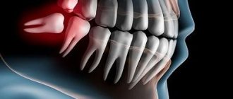
Normally, a person grows 52 teeth over a lifetime: 20 milk teeth and 32 permanent teeth. Supernumerary teeth are “extra” dental units that appear in addition to some incisors, canines, premolars or molars. In various medical sources, the phenomenon is called hyperdontia, supradontia or polyodontia. A superset can include 1–2 teeth or reach several hundred dystopic structures, be true or false.
Supernumerary teeth: causes of anomaly
The main cause of polyodontia is a violation of the formation or development of tooth germs. Doctors are considering several theories for this phenomenon:
- Heredity – hyperdontia develops as a result of genetic abnormalities that can be transmitted from parents to children. In this case, the pathology should manifest itself in the patient’s closest blood relatives.
- Splitting of the root germ of the tooth in the embryonic period of development. Usually observed after the mother becomes ill during pregnancy. Characteristic of single supernumerary teeth (1–2 pcs.).
- Failure during the embryonic period of tooth formation with the formation of multiple dental buds (up to several hundred). It is observed when the mother lives in unfavorable environmental conditions, during treatment with dangerous drugs, taking drugs, or alcohol abuse.
In more rare cases, polyodontia develops as a result of odontoma, a benign tumor in the area of the jaw arches. The disease is typical for children and young people during the period of active growth of tooth germs. Such a tumor structure may contain several tens or even hundreds of highly deformed dentin-enamel formations.
Provoking factors in the prenatal period:
- malfunction of genes - primarily the Msx1 gene (responsible for the formation of tooth germs);
- viral infections;
- taking teratogenic and potentially dangerous drugs;
- smoking, drug use, alcohol use;
- poor environmental conditions;
- exposure to radiation - not only direct exposure, but also increased general background radiation in the place of residence/work.
Reference! Some scientists consider the presence of supernumerary teeth as an atavism. Thus, it has been scientifically proven that various species of the genus Homo could boast a set of teeth of 36–44 pieces. However, this theory does not explain in any way the appearance of multiple rudiments covering all spaces of the sky (a typical example is an Indian boy who had to remove 230 supernumerary units).
Causes of the disease
The causes of polyodontia have not yet been established; there are three hypotheses for the occurrence of the anomaly:
- Atavism. The concept of atavism means the appearance in a person of any signs characteristic of his distant ancestors, but absent in his closest relatives. Proponents of this theory explain supernumerary teeth as an accidental return to the past, when human ancestors had six incisors on each jaw. This version is confirmed by the frequent occurrence of polyodontia in the frontal area of the jaws.
The disadvantage of this assumption is that it explains the appearance of extra teeth near the incisors and canines, but does not explain the reasons for the presence of supernumerary molars and premolars.
- Splitting of the tooth germ. The reason for polyodontia, according to this theory, is disturbances that occur during embryonic development at the stage of formation of tooth buds (the appearance of the buds of permanent teeth occurs in the 10th week of the prenatal period).
The theory explains the occurrence of hyperdontia in various parts of the dentofacial apparatus, but is not able to explain why one person can simultaneously develop congenital hypodontia with an increased number of teeth - their absence associated with underdevelopment of tooth germs.
- Proponents of the third hypothesis combine the previous two, considering supernumerary teeth to be a combination of genetic anomalies and features of embryonic development.
According to statistics, the number of cases of polyodontia is currently increasing; scientists associate this with an increase in the level of ionizing radiation, which leads to mutational changes in the body. Polyodontia of permanent teeth is much more common than supernumerary teeth.
Symptoms of pathology
The main symptom is the presence of teeth beyond the natural set. They can have different positions, shapes and numbers. The process of teething of abnormal teeth is often accompanied by pain, fever, and inflammation of areas of the oral cavity. If measures are not taken in a timely manner, complications may develop:
- redness and swelling at the site of the impacted tooth;
- problems with chewing food and digestion;
- injuries to the mucous tissues of the oral cavity;
- violation of the position of the main teeth (dystopia) and the formation of malocclusion;
- various speech defects - mainly problems with the pronunciation of hissing sounds;
- loosening of adjacent normal teeth;
- deformation of the jaw bones.
In young children and adults, the pathology has its own nuances. They are associated with age-related characteristics of the body and affect the subsequent development of maxillofacial structures. Statistics on the spread of pathology in children and adults:
- 60% of cases are children and adolescents during the period of replacement of milk teeth with permanent ones;
- 35% of cases are adults;
- 5% of cases are small children during the period of growth of baby teeth.
Reference! Supernumerary teeth, as a rule, have a standard structure, sometimes with a smaller crown size. Less common are deformed formations of a teardrop-shaped, lumpy, chisel-shaped form.
Children's dentition and everything about it
All parents carefully search for information about the correct number of teeth in the baby’s mouth in relation to his age. They are often interested in shape and size. It is worth remembering that the information received should not exempt the child from visiting the dentist for preventive purposes. Sometimes there may be congenital manifestations of anomalies in the oral cavity. We are talking about defects in the growth of teeth, as well as their incomplete composition in the upper or lower jaw.
Directly, baby teeth do not remain in the oral cavity throughout life, but only for a limited time, after which they fall out. After them, permanent ones grow up, which stay with the person for life, provided they are treated with care. Primary and permanent teeth have a number of differences, namely:
- The incisors of baby teeth have a more convex shape.
- Permanent teeth have a more developed root system than baby teeth, which fall out without causing discomfort.
- The enamel of primary teeth is thinner, but their pulp capsule is more pronounced. Baby teeth are more susceptible to caries.
The first teeth begin to appear at 4-5 months, but a delay of six months is not a violation. There is a certain sequence in which a baby’s first teeth appear:
- Incisors: central ones appear from 4 to 9 months, and lateral ones are expected from 8 months to 12 months.
- The first molars appear after the child is one year old.
- Clicks are expected from 1 to 2 years.
- By the age of 3, the second molars begin to emerge.
It is worth noting that at the age of three, a baby already has about 20 teeth in his mouth. Then, in their further growth, a pause is expected until the child reaches 5 or 6 years of age.
Features of polyodontia in children
Possible symptoms of hyperdontia in childhood:
- intrauterine formation of teeth (a child is born with several teeth);
- the appearance of teeth in the first months, weeks and even days of life;
- delayed eruption of baby teeth.
This has unpleasant consequences:
- problems with feeding the baby - he does not latch onto the nipple well and sucks weakly;
- swelling of the mucous tissues can spread to the nasal area and cause breathing problems;
- poor closure of the dental arches causes severe salivation;
- a prolonged increase in temperature to 38C is possible.
Hyperdontia is especially harmful during the period of speech formation. Teeth located outside the general row prevent the tongue from taking the correct position when pronouncing most consonant sounds. Without eliminating the pathology, any speech therapist will be powerless.
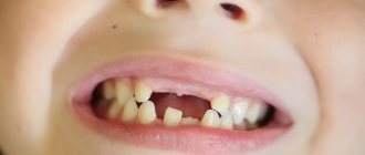
In addition to true hyperdontia, children (less often adults) can develop the so-called false form of the disease. It is characteristic of the period of tooth change and develops during the eruption of molars next to the milk teeth that have not yet fallen out. This is a fairly common phenomenon that usually goes away on its own as the dentition finally forms. In some cases, orthodontic treatment may be required.
Features of polyodontia in adults and adolescents
In adults, after complete replacement of complete teeth, the presence of additional units can lead to additional pathologies:
- chronic rhinitis, sinusitis when the wall of the maxillary sinuses is perforated by the roots of supernumerary impacted structures;
- interdental caries - due to teeth fitting too closely to each other.
Retention and dystopia
Adults are characterized by 2 main types of supernumerary teeth:
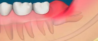
- Dystopic teeth are the name given to teeth with deviations in the direction of growth. The peculiarities of the formation of supernumerary units very often lead to dystopia. This is due to the fact that the space on the dental arch line is limited, and the roots of normal teeth simply push the “intruder” towards the cheek or palate.
- Impacted teeth – impaction occurs when a tooth loses its growth impulse and remains embedded in the jawbone. Impacted teeth can cause the adjacent normal teeth to become loose and cause them to shift and change the bite. Often cause pain.
On a note! According to statistics, hyperdontia accounts for up to 2% of cases of dental problems. Of these, 70% are associated with the appearance of single supernumerary teeth, 25% with a couple of such formations, and only in 5% complex multiple complexes of 3–4 or more teeth can be found.
Types of teeth with hyperdontia
Supernumerary teeth in hyperdontia can be divided into several types according to their location in the mouth:
- Spiny. May form on the upper jaw. Localization - between the central and lateral incisors, within the immediate limits of the main dental arches. They are called spiky because of the presence of very sharp ends.
- Additional premolars. They grow from the cheek side within the normal molar units of the dentition.
- Extra fangs. These grow on the upper jaw, in 99% of cases - on the side of the cheek and lip.
- Additional premolars. The localization area of these additional teeth is the lower jaw, on the cheek side.
Additional and primary teeth may fuse. There are 4 types of such merger:
- Bedding. The enamel of both teeth, twin neighbors, grows in the form of tubercles over the entire tooth surface.
- Root merge. The crown parts are located separately, but the roots are fused together.
- Coronal fusion. The root system of each tooth is separate, but the crown part is fused and has one common enamel “shirt”.
- Complete fusion of twin teeth. The crowns, which have a common enamel coating, and the roots are fused together.
Diagnostic methods
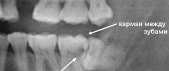
The main indication for diagnosis is the presence of an “extra” tooth in the patient’s oral cavity or obvious signs of its eruption (painful tubercle or swelling in the gum or palate). All of these signs can be identified through a routine visual examination, during which the dentist evaluates the condition of the oral cavity. To confirm the diagnosis and carry out differential diagnosis, an X-ray examination is performed - an orthopantomogram. If it is necessary to examine the dentition in several planes, computed tomography (CT) is additionally prescribed.
On a note! The patient may not be aware of the presence of impacted supernumerary teeth. In this case, hyperdontia is detected only by the results of an X-ray or CT scan of the maxillofacial region.
Removal of impacted teeth
In order for the operation to be successful and polyodontia to be cured without any complications, the doctor must fully examine the patient and plan his further actions.
- To begin with, X-rays and/or computed tomography are performed to determine the exact topography of the anomaly.
- Removal is performed under local anesthesia, but there are cases when general anesthesia can be used on the patient.
- First, the mucous membrane is peeled off, then the bone tissue is opened and the root and crown parts of the tooth are removed.
- If necessary, bone defects are covered with osteoplastic material, and the mucous membrane is sutured.
After tooth extraction, the patient continues treatment at home: takes antibiotics (if prescribed by the attending physician), rinses the oral cavity with antiseptic solutions.
Until the wound heals after surgery, it is not recommended to eat too hot, hard or spicy food. You should also brush your teeth carefully, especially on the operated side.
Do supernumerary teeth need to be removed?
There is no clear answer to the question of what to do with “extra” teeth. Much depends on the shape, position and quantity. The following are subject to mandatory removal:
- baby teeth that interfere with normal growth and formation of permanent teeth;
- strongly dystopic structures - located on the palate or at a large angle to the lateral side of the gums;
- impacted formations that put pressure on the adjacent roots of normal teeth and provoke periodic inflammatory processes of soft tissues.
If the additional tooth does not cause discomfort and does not disrupt the development of the dentition, removal may not be necessary. Moreover, such a superset can play the role of a strategic reserve in case of damage to nearby normal teeth.
How does the removal work?
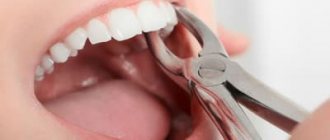
The removal procedure can be simple or complex. In the first case, the doctor is dealing with a fully erupted tooth, the root of which is not intertwined with the surrounding structures. Simple removal steps:
- The oral cavity is prepared - treated with antiseptics, anesthesia is given.
- Grasp the crown with forceps (in some cases, an additional incision of the mucous tissue is required to facilitate access).
- The elevator destroys the retaining ligaments.
- The root is freed and the tooth is removed.
- Clean the hole from tooth fragments and bones.
- Apply stitches (if necessary) and a healing bandage.
Complex extraction is prescribed in the case of impacted, semi-impacted or dystopic teeth with complex root shapes. The procedure is carried out with a deep incision of soft tissues, in especially severe cases - with opening of the jaw bone to gain access to the roots. The latter option is relevant for deep bone occurrence, position in close proximity to the cranial sinuses and orbits, as well as for irregularly shaped roots and a high risk of damage to complete teeth. A complex removal operation is performed by a dental surgeon.
Stages of complex tooth extraction
Complex removal surgery is carried out only after x-ray diagnostics, during which the shape, length and depth of the roots are determined. If the patient has inflammation, he is prescribed treatment with antibacterial drugs.
Technique of the procedure
Wisdom tooth removal is carried out in the following order:
- the gum is separated from the neck of the tooth by making an incision in the soft tissue;
- if necessary, the interroot septum is sawed or sections of bone tissue are cut out at the location of the tooth;
- then, using forceps, the tooth is rocked and pulled out of the socket;
- Sutures are placed on the gum.
Painkillers are not required after the procedure, since the effect of the anesthetics is still present.
Orthodontic treatment
Depending on the situation, it is used after tooth extraction or instead of it. In children, it is used to stimulate the loss of baby teeth and eliminate false hyperdontia, as well as to facilitate and accelerate the eruption of a normal set. In addition, orthodontic treatment with the installation of braces, mouth guards, dental plates or other auxiliary structures can straighten crooked teeth and return them to their proper place.
Before prescribing treatment, the dentist carefully evaluates the condition of the complete and supernumerary structures and, depending on the overall picture and prognosis for the future, can use both formations or leave the strongest and most durable tooth (not necessarily complete).
Types of hyperdontia
There are several of them. And they differ in the degree of inconvenience for their wearer and in the possible consequences. And even according to the subjective feelings of people who have been diagnosed with this disease.
Typical hyperdontia
This pathology is characterized by the presence of extra teeth that began to grow without leaving the dentition in the mouth. Since two normal-sized teeth would be cramped, such teeth, called twins, are reduced in size. This is expressed in the reduction of their coronal part.
The cause of this anomaly is most often heredity.
Atypical or atypical
This pathology with an increase in the number of teeth is characterized by the growth of extra teeth outside the dentition. Usually on the palate, where they can take up almost all the available space. Or the lateral surfaces of the jaw arches, most often from the side of the tongue. It is a very rare form of pathology.
Real (true)
Supernumerary permanent teeth in this type of hyperdontia appear from the formation of excess root buds or when the roots split in the initial stage of their growth. It does not cause significant inconvenience as long as both teeth in a pair of “twins” are healthy. Problems arise if at least one is affected by caries, the treatment of which can be difficult due to the intertwining of the roots of such teeth. For the same reason, there are difficulties with their removal.
False hyperdontia
Probably the most harmless of all, from the “it will go away on its own” series. Although, of course, medical supervision is mandatory in order to avoid developmental complications. It occurs when a child’s bite changes due to the replacement of milk teeth with permanent ones. It occurs when a permanent tooth erupts, when the milk tooth has not yet fallen out. Corrected after the loss or removal of a baby tooth. The decision to remove is made by the doctor.
The long-term coexistence of missing baby teeth and new, permanent ones is not such a rare phenomenon. This happens due to the inattention or even frivolity of parents who missed such an important stage in the development of the child. Especially if the growth of permanent teeth occurs painlessly.