If you ask a dentist what he hates doing most in his job, 8 out of 10 doctors will answer: “Cleaning root canals.” Dislike for the preparatory stage before obturation (filling) is associated with the complex structure of the root canals. At the same time, preliminary X-ray diagnostics do not always show the anatomical features of the root canal of a particular tooth. And dentists are well aware of the sad consequences of poor-quality obturation of root canals, ranging from a small focus of infection to extensive periodontal inflammation and tooth loss.
When working with root canals, dentists use a classification compiled in 1984, which includes eight different root canal configurations. But, despite all the knowledge in this area, each case of endodontic treatment is “an improvisation on a well-learned topic,” where each tooth requires an individual approach to treatment.
In clinical practice, the apical zone is recommended as a physiological level for instrumentation and root canal filling. According to a number of authors, in 75% of cases the apical foramen is deviated from the main axis of the tooth. This means that the apex visible on the X-ray image and the apical constriction are often located at different levels.
Methods for determining the working length of the canal
There are several methods for determining the working length of the root canal:
- Mathematical (tabular) method. Special tables (for example, according to JI Ingle, LK Bakland) provide the lengths of teeth and roots, as well as the ratio of crown and root sizes, the number and frequency of canals in the root, apical openings in the canal, and the direction of bending of the canal. This method is quite popular, but requires taking into account corrections of ± 10-15% for the individual characteristics of the patient.
- The tactile method is based on measuring the length of the instrument inserted until resistance appears in the root canal. The method is subjective and therefore unreliable.
- The red dot (paper point) method involves inserting a paper point into the dried root canal until the tip of the point becomes moist with tissue fluid. The appearance of moisture at the top of the pin indicates that the paper pin has reached the apical foramen, and the length of such a paper pin is taken as the working length of the root canal.
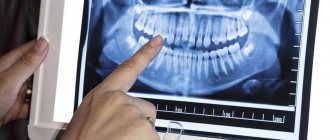
- The X-ray method is the most common and its results are considered the most reliable. However, this method does not provide an idea of the location of the apical constriction and the apical opening of the canal, which often does not coincide with the radiographic root apex and may be at a distance of several millimeters from it.
Endometrium. Practical recommendations for using an apex locator
As an alternative to the X-ray method for determining the length of the root canal, in 1962 dentists were first offered an electronic device - an apex locator. The very idea of electronically measuring the working length of the root canal, based on measuring the coefficient of resistance, was revolutionary.
The first generation devices had limited capabilities: the active electrode could only be inserted into dry channels, and the readings were quite variable.
Technical improvements in apex locators have led to the fact that fourth and fifth generation devices with multi-frequency technology reliably indicate the length of the root canal in 95% of cases.
Accurate determination of the working length of the root canal is one of the most important stages of effective mechanical and chemical treatment of the canal, as well as its obturation. The most widely used method is x-ray.
However, if the projection of the X-ray machine tube or the anatomical features of the canal is incorrect, it is impossible to accurately determine its length only from an x-ray image.
The solution to this problem was the use of apex locators, which, due to the difference in electrical potentials, make it possible to increase the accuracy of determining the working length of the root canal.
The length of the root canal limited by the apical constriction and the canal orifice is considered to be the ideal working length for endodontic treatment.
Scientific work carried out by Ricussi and Langeland indicates that the apical constriction is one of the narrowest areas of the root canal with the smallest cross-section for the neurovascular bundle.
Mechanical and medicinal treatment of the root canal before this narrowing ensures the formation of a minimal wound field and the creation of the best conditions for regeneration. This anatomical feature is described in the literature as the small apical foramen, or Foramen physiologica.
It is this anatomical narrowing that limits the root dentin from cellular cementum. The dentin-cement boundary is the ideal end point for mechanical treatment of the canal and its obturation in the future. The transition of pulpal tissue to periapical tissue is limited by the large apical foramen, or Foramen apicale (Figure 4).
Kuttler was the first to describe the histological and anatomical differences between the small and large apical foramina. Using a histological method, he studied more than 400 tooth root tips and concluded that the distance between the two holes in the age group up to 25 years was 0.52 mm, and in the group up to 55 years - 0.66 mm.
Kuttler's discovery formed the basis of a new philosophy of root canal treatment. Previously, it was customary to develop and fill the root canal up to the radiographic apex. Unfortunately, some doctors still practice this method, arguing that it is better to overfill than underfill.
Determining the working length to the large apical foramen leads to apical perforation, overfilling of the root canal, postoperative pain, and can affect the restoration processes in the periodontium, and the release of dentin filings and putrid decay with pathogenic microorganisms leads to periapical inflammation and, as a consequence, further complications (abscess , periostitis). Conversely, determining the working length before the apical narrowing can lead to incomplete cleaning of the canal from infected dentin, and the remaining pulp tissue may lead to prolonged pain in the postoperative period.
When the root canal is not filled, periodontal fluid penetrates into the root canal system, causing depressurization of the system and activation of the inflammatory process.
The radiographic apex is the most distant point from the cutting or chewing surface of the tooth and does not coincide with the anatomical apex of the tooth, therefore it cannot be considered the end point for measuring the working length of the root canal.
Microscopic and histological studies show that the distance between the small apical foramen and the radiographic apex is 1-2 mm.
Accurate assessment of root canal length is one of the most important steps in endodontic treatment and determines its ultimate success or failure.
The use of an electric apex locator in combination with an x-ray method can improve the accuracy of diagnosis.
History of endometrial development
In 1962, Sunadu created the first apex locator, based on two constant parameters - the electrical resistance of periodontal tissue, which is 6 kOhm, regardless of the anatomical features of the tooth, shape and age, and the fact that periodontal fibers are fixed to Foramen apica-1e .
The first device generated electrical waves of the same frequency and could record tissue resistance. The passive electrode was placed on the patient's lip, and the active electrode was placed on the endodontic instrument.
When the endodontic instrument came into contact with the periodontium, the electrical circuit was closed and the device showed the length of the root canal. However, the use of electric waves of the same frequency presupposed the use of certain preparations for root canal irrigation, otherwise the device gave a large measurement error. The endodontic instrument served as the cathode or anode.
In the moist environment of the root canal, where it is impossible to achieve absolute dryness, there are both positively charged particles, cations, and negatively charged anions.
From the intraroot fluid surrounding the electrode, cations move to the cathode, and anions to the anode. This leads, firstly, to polarization of the electrode, and secondly, to an unstable magnetic flux and inaccurate readings.
The electric field is generated even before the electrode reaches the periodontium, by transmitting a magnetic field through the liquid. The device shows the top of the tooth, although the instrument has not actually reached the top yet.
In cases where periodontal fibers are destroyed due to an inflammatory process, for example in apical periodontitis, it was also not possible to accurately measure the electrical potential.
In 1994, Kobayashi and Suda proposed the Ruth ZeX apex locator (J.
Morita), in which the so-called “ratio method” was used to determine the length of the root canal,2 which made it possible to simultaneously measure the resistance to current of two frequencies (8 kHz and 0.4 kHz) and find the overall resistance coefficient reflecting the position of the file in the canal.
If the file reached the minor apical foramen, the drag coefficient reached 0.67. This measurement is stable and indicates the presence of electrolytes in the tissue.
Apex locators of the fourth and fifth generations (for example, Endo Analyzer Model 8005, Analyst Sibron Dental) are capable of calculating the channel resistance to current of five frequencies (0.5, 1, 2, 4 and 5 kHz).
The principle of interpreting measurements of the electrical resistance value into length readings (from the tip of the instrument in the canal at the level of the small apical foramen) is carried out by recalculation according to the formula embedded in the device program using the comparative method, which gives more accurate and faster readings.
These measurements allow localization of the small apical foramen. Since the periodontium no longer serves as a determining constant for such devices, pathological changes in the periapical tissues or damage to the ligamentous apparatus of the tooth cannot affect the measurement results.
Further studies of similar devices indicate that the accuracy of determining the working length of the root canal with their help is 95% or higher.
Electronic measurement method or How an apex locator works
The electronic method of measuring the working length of the canal using an apex locator does not have the disadvantages of the above methods. Apex location is based on measuring the electrical resistance between the hard tissues of the tooth and the periodontal tissues and oral mucosa. During the procedure, a mouthpiece (passive electrode) is attached to the patient’s lip on the side of the diseased tooth and an endodontic file (active electrode) is inserted into the canal. When the file reaches the periodontal tissue, the electrical circuit is closed and the device beeps.
If you have an apex locator, you can do anything.
More recently, when treating teeth, endodontists were guided during their manipulations using the “red dot method” or the patient’s reaction (when he felt a slight prick and notified the doctor about it with an expressive wince or a short moo).
Today, an apex locator tells (and shows!) everything that happens in the manipulation zone. The patient's discomfort is completely eliminated, and the doctor's confidence in his every action is absolute.
This means that when using an apex locator, you work quickly, accurately, efficiently and comfortably.
What does the apex locator show?
Modern third-generation apex locators work equally accurately both in a dry canal and in a wet one containing blood and other physiological fluids. By default, the apex locator detects three zones:
- Apical constriction zone
- Large apical foramen
- File exit zone beyond apex
The doctor also has the opportunity to set the desired depth of penetration of the file, upon reaching which an audible alarm will sound. Using an apex locator allows the doctor to clean and disinfect the canal to its full depth and carry out precise filling, guaranteeing the absence of unpleasant consequences for the patient in the form of inflammatory processes and subsequent re-treatment of the tooth.
Price range
There are many manufacturers of this equipment on the market, so apex locators have a wide price range, starting from 8,000 rubles. Based on price, four categories can be distinguished:
- Economy class 8,000 - 20,000 rub.
- Middle class 20,00 - 50,000 rub.
- Business class 50,000 - 70,000 rub.
- Premium class 70,000 rubles and above
Regardless of the price, the direct function of any apex locator is the same, so the main differences in different price categories are:
- Display. Color or black and white, graphic or digital type of information.
- Additional functions. “Virtual apex”, channel humidity, demo mode and others.
- Multi-frequency technology. To more accurately determine the apex.
When you can't trust an apex locator
Despite the wide technical capabilities of modern apex locators, in clinical practice dentists are faced with situations where the data from the electronic method of measuring canal depth are inaccurate.
- Root canal with large apical narrowing. It must be taken into account that the result will be less than the actual length of the channel.
- Root canal with increased moisture due to bleeding. For accurate measurements, it is recommended to wait until the bleeding stops and dry the canal.
- Broken crown. The contact of the file and the gums leads to a change in the level of resistance. A tooth structure or gum isolation with a rubber dam is required.
- Cracked tooth. It also leads to current leakage and measurement inaccuracy.
- Working with a canal filled with gutta-percha. Complete removal of gutta-percha residues is required before starting measurements.
- Metal structures in contact with the gum. It is necessary to widen the access at the top of the crown so that the file does not touch metal parts.
- Tooth fragments or remains of pulp tissue. Complete removal of tooth fragments and pulp remains is required.
- Caries of the cervical region. Leads to current leakage through damaged tissue and measurement inaccuracy.
- Canal obstruction. The canal must be completely opened to the apical constriction.
- Root canal with increased dryness. Requires hydration with electrolyte.
Principles of therapeutic dental treatment at the Yauza Clinical Hospital
- Safety and painlessness. Our dental therapists carefully interview and examine the patient, take into account his medical history, allergies, concomitant diseases or body conditions, and individually select all materials, instruments, antiseptics and anesthetics.
- Minimally invasive. Doctors try to preserve as much viable tissue as possible and make intervention minimal, thereby significantly increasing the service life of teeth.
- Atraumatic. All precautions are taken for the safety of the patient (safety glasses, rubber dam, microscope, exclusively sterile instruments and consumables are used). All manipulations are carried out under the control of X-rays and an apex locator, which allows root canals to be treated without complications.
- Aesthetics. Almost all diseases treated by a dental therapist result in the application of a filling. Regardless of its location, it will definitely be made as similar as possible to healthy tooth tissue.
How to choose an apex locator
How to choose an apex locator? Which manufacturer is the most reliable? These questions are often found on dental forums, where doctors share their experiences and impressions of using this or that professional equipment. To ensure that the choice of an apex locator does not drag on endlessly, you need to decide on several important points:
- In what environment (dry, wet, or dry/wet) should the device operate?
- What is the budget for the purchase of the device?
- What additional functions are needed (“virtual apex”, channel humidity level, demo mode, battery indication, sound signal mute).
- What should be the indication of measurement results (numerical or graphic).
Multifunctional dental device EndoEst-3D / EndoEst-3D
EndoEst-3D is a multifunctional diagnostic device.
Functions of the device EndoEst-3D / EndoEst-3D:
- Apex location – measurement of the working length of the tooth canal;
- Electroodontodiagnostics – determination of the clinical condition of the pulp;
- Dentinometry - determination of the thickness of suprapulpal dentin of vital teeth.
Features and Benefits:
- Combining 3 functions in one device – saving financial investments and optimizing the work space;
- Large display, graphic, light and sound indication;
- Adjustable sound settings;
- Low battery indication;
- Energy saving mode. Auto shutdown;
- Demo mode and self-diagnosis of performance.
The electrical and operational characteristics of the device meet the requirements of Russian standards GOST R50444, GOST R50267.0., GOST R 50267.0.2, technical specifications TU 9452-005-56755207-2002, as well as European standards EN61326, EN60601-1-2.
Specifications:
| Supply voltage, V | 6 (4 x 1.5) |
| Electrical protection | type B |
| Measuring range in “Apex locator” and “Dentometer” modes, mm | from 3.0 to 0 |
| Measurement accuracy in the “Apex Locator” mode with a probability of 87%, mm in the range: | (3 - 2) mm±0.5 (2 - 1) mm±0.3 (1 - 0) mm±0.1 |
| Range of adjustment of the apical stop value (relative to the apical narrowing of the canal), mm | from 0.1 to 0.5 |
| Measurement accuracy in the “Dentometer” mode with a probability of 87%, mm in the range: | (3 - 2) mm±0.5 (2 - 1) mm±0.3 (1 - 0) mm±0.2 |
| Range of “diagnostic” currents in the “EDI” mode, µA | from 0 to 199 |
| Continuous operation time when using new batteries, not less | 170 h |
| Switching to power saving mode: | 1st stage, after 15 min. Stage 2, after 240 min. |
| Overall dimensions, mm | 140x100x76 |
| Weight (with batteries), no more than g | 365 |
| Degree of protection against penetration of dust and moisture (IP code) | 51 |
| Environmental parameters during product operation: temperature, °C from +10 to +35 air humidity, no more than 80% atmospheric pressure 101± 3, kPa | |
| Product service life | 5 years |
Composition of the EndoEst-3D kit: “EndoEst - 3D” device – 1 pc. “Signal Line” cable – 1 pc. Oral Hook mouthpiece - 1 pc. Probe clamp (“Probe Princh”) - 1 pc. EDI probe - 1 pc. Power battery 1.5 V (AA) (included in the device) - 4 pcs. Instruction manual - 1 pc.
Multifunctional dental device EndoEst-3D / EndoEst-3D price: RUB 23,500.00
Our recommendations
It is worth noting that 100% accuracy of results can only be achieved in laboratory conditions, so even modern fifth-generation devices show an accuracy of up to 99.1% when working in any environment. And the quality of the results is constant for all manufacturers (European and Chinese). Therefore, when choosing an apex locator, we recommend focusing on ease of use and the mid-price category, in which most manufacturers represented on the Russian dental equipment market operate.
Our online store presents apex locators from Chinese manufacturers in the mid-price range, starting from a model for 23,000 rubles. and ending with the model for 48,700 rubles. with a color display, endomotor, parameter memory function and the ability to connect to a PC.
