According to antiplagiat.ru, the uniqueness of the text as of October 16, 2018 is 99.9%.
Key words, tags: X-rays, tooth extraction, orthopantomogram, periodontitis
A dental cyst belongs to the group of jaw cysts. Jaw cyst is a general concept that unites many pathologies. The formation of a cystic cavity in the jaw can be caused by an inflammatory process, or in rare cases, by a growing tumor. It can be in a “dormant” state and not manifest itself in any way, or, on the contrary, be in an active, acute phase, get sick, affect neighboring healthy tissues, and cause various diseases. In any case, the presence of cysts in the jaw and, in particular, the presence of a dental cyst, requires attention and serious consideration.
Tooth cyst: historical background
The word “cyst” is of Greek origin (“kystis”) and literally means “bubble.” A dental cyst is a cavity-like tumor-like formation. Previously, when X-rays were not taken, it was extremely difficult to see such formations and, as a rule, dentists encountered them during the extraction of teeth from patients. Thanks to the advent of X-ray diagnostics at the end of the 19th century (1895), it became possible to plan the treatment of dental cysts. And today, if a patient regularly visits the dentist’s office, we can successfully diagnose and eliminate dental cysts at those stages when they do not pose a danger.
Contraindications
Cystectomy is contraindicated in cases of intolerance to anesthesia and severe concomitant diseases of the heart, liver, kidneys, lungs, blood, diabetes mellitus, exhaustion, and systemic infections. In addition, partial cystectomy is not performed if a reduction in the working volume of the bladder is unacceptable for stage 0 cancer, as well as in cases where the tumor is located near the junction of the ureters and urethra with the bladder (there is no technical ability to ensure urinary function).
Tooth cyst: anatomy
A dental cyst is a benign cavity tumor-like formation that has a membrane and an internal epithelial lining, the cells of which produce fluid. Modern medicine still cannot accurately answer the question of how and where an incipient cyst takes epithelial tissue from. However, most scientists are inclined to believe that it comes from the remnants of the dental epithelium, the so-called islands of Malasse-Astakhov. And it develops as a result of chronic odontogenic inflammation, when microbes enter the jaw bone through the tooth canal. Further development of the process can go in two directions, the result of which are pathologies: an extensive destructive process that does not have a shell - a non-shell formation (such a process is not a cyst), or a dental cyst itself - a shell-like formation.
Introduction
Open radical cystectomy (RC) has long remained the mainstay of treatment for patients with muscle-invasive bladder cancer (BCC) 1 - 3. It is also indicated for recurrent superficial tumors with a high risk of progression, BCG-resistant carcinoma in situ, T1G3 1. The active development of medical technologies has led to the evolution
of minimally invasive operations in the treatment of bladder cancer. Over the past decades, laparoscopic and robot-assisted RC have been actively introduced. There are publications in the literature about their effectiveness and relative safety with superior results in terms of blood loss, early recovery of intestinal function, hospitalization and rehabilitation time 4 - 9.
Robot-assisted surgery has gained widespread development in urology, mainly due to the frequently performed radical prostatectomies. Over the past decade, robot-assisted access to RC has begun to be used. Until 2010, the number of centers performing this operation was limited. In recent years, the number of publications has increased, including large series, reflecting the growing acceptance of this approach for RC 7 - 8, 10 - 11. However, RC with urinary diversion remains one of the challenging operations in urology. It is associated with a high complication rate even in the hands of an experienced surgical team. As shown by some authors, their number decreases with experience, although they remain high even in specialized urological centers 5, 10 - 12. The operating time for robot-assisted RC, compared with open access, remains quite long. With the accumulation of experience, it is possible to reduce the duration of the intervention by optimizing the methodology for performing individual stages of RC, among which are cystprostatectomy with pelvic lymph node dissection (stage I) and intestinal diversion of urine (stage II). In this article, we present the stages of robot-assisted RC, additionally describing the technique of nerve-sparing RC in men and organ-sparing surgery in women.
Types of dental cysts
It is customary to distinguish between 2 types of dental cysts - follicular and radicular. Follicular cysts are less common than radicular cysts. In the presence of certain factors, it grows from the dental follicle - the membrane around the growing tooth. When it is large in size and involves adjacent teeth, it is called a follicular dentigerous cyst.
Radicular (odontogenic inflammatory) cysts form on the roots of teeth as a result of pathological processes spreading beyond the root apex. By location, radicular cysts can be apical (root), lateral (lateral), apicolateral and interradicular.
Discussion
Robot-assisted RC is gradually taking its place in the surgical treatment of patients with muscle-invasive bladder cancer. Robot-assisted access makes it possible to reduce the number of intra- and postoperative complications, primarily to reduce the amount of blood loss, shorten the length of hospitalization and rehabilitation of patients 5, 8, 10, 11. The complexity of RC is aggravated by the comorbidity of patients, who are often elderly. RC is a long-term intervention that includes three defining stages: bladder removal, pelvic lymph node dissection and urine diversion 4, 6, 8. At the beginning of mastering the robot-assisted RC technique, we sought to comparatively simplify the operation, breaking each stage into small steps. When performing them, we tried to follow a certain order of execution, that is, we moved on to each subsequent stage after completing the previous one. There are several key benefits to this structured approach. Firstly, the RC technique is simplified and its results are improved. By working in one specific area, the surgeon's attention was focused on a single task at that time before moving on to the next step. It is important to achieve adequate hemostasis at all stages of RC, which provides good visualization and reduces the amount of blood loss. Even from our initial experience, we were convinced that a systematic approach to performing RC allowed us to reduce the operation time. Secondly, in our opinion, with a detailed description of the individual steps of the operation it is easy to teach young specialists and conduct adequate monitoring of training.
Open RC with extended lymph node dissection remains the gold standard for the treatment of bladder cancer, providing excellent local tumor control with a 50–70% five-year cancer-specific survival rate 13–16. The role of robotic surgery in the treatment of cancer continues to evolve with increasing popularity and is supported by good short- to mid-term results 10, 12, 17. RC is primarily performed for cancer, and therefore the effectiveness of any approach will be judged on the basis of long-term survival outcomes. Robot-assisted RC has been performed in some clinics for more than 15 years; data from robot-assisted RC with Kaplan-Meier analysis were published for 36 and 60 months, comparable to series after open RC 5, 8, 11. Thus, according to Collins JW et al. 8 The five-year cancer-specific survival rate was 67%.
RC through various approaches is associated with a certain number of perioperative complications and severe morbidity 13-16. According to various authors, with open RC their number ranges from 49% to 64% 6, 10, 13-17. Clavien-Dindo high-grade complications range from 13% to 40%, and 90-day mortality range from 0% to 4.5% 13, 16. Robot-assisted RC also has a high complication rate, but relatively fewer, according to compared with open RC 12, 18-20. Collins JW et al. 8 early complications (0-
30 days) was observed in 54 (47.8 %), grade 3 according to Clavien-Dindo - in 37 (32.7 %) patients. Late complications (more than 30 days) occurred in 30 (26.5%), grade 3
according to Clavien-Dindo - in 20 (17.7%), 1 (0.9%) patient died from pulmonary artery embolization. A recent meta-analysis showed that robot-assisted RC compared with open RC is associated with fewer perioperative complications, a higher number of lymph nodes removed, longer operative time, reduced blood loss and fewer blood transfusions, and a short hospital stay. 12 Further prospective studies of robotic outcome -assisted RC with a long period of observation will allow us to confirm the data of this analysis.
The mechanism of formation and growth of a dental cyst
The cystic cavity, located in the jaw, has external resistance from the surrounding jaw bone. But the epithelial cells of the lining begin to produce fluid, which gradually fills the cavity, creating excess pressure.
This pressure acts on the surrounding bone tissue, causing its gradual peripheral resorption, giving the cyst the opportunity to grow even larger, increase fluid secretion, and therefore put even more pressure on the walls. This is why cysts can grow to very large sizes, sometimes asymptomatically, if not accompanied by periodic inflammation. This may be due to the patient’s good immune status, low pathogenicity of the microflora in the lesion and other factors.
Diversion of urine: incontinence or retention.
It is important to understand the two-step approach to bladder removal. First, the bladder and lymph nodes are removed. Then the urine needs to be drained. This can be achieved in several ways. In general, we distinguish between the options that are incontinence (a continuous stream of urine immediately leaving the body) and retention (urine stored in the body and released when necessary). Biological age, renal function and other diseases, and the patient’s quality of life are decisive when choosing surgical tactics. To determine which option is best for your specific situation, you must know and understand each type of surgery's limitations and side effects.
In addition to your personal preference, the ability to physically and mentally become accustomed to, and be able to handle, such a urinary diversion is important.
Movement of the ureters
Removal of the ureters through the skin (ureterocutaneostomy). By moving the ureters either together or separately through the skin on the side of the abdomen to drain urine away from the kidneys (stoma) (Figure 1), urine can simply flow through the stoma into a pouch. This urine diversion is the simplest. Although it is rarely used. This method is safe and accessible to patients with a complex medical history (previous surgeries, multiple health problems, palliative care) or patients who are unable to care for themselves after surgery. Major complications are rare, but recurrent infections and tightening of the opening (stenosis) are common and may require treatment. Patients often require ureteral stenting, which must be replaced regularly.
Rice. 1 ureterocutaneostomy
Place a portion of the small intestine between the ureters and the skin (ileal conduit). An ileal conduit can be created by placing the small bowel between the ureters and the skin (Figure 2). This "intestinal stoma" creates more distance between the kidneys and the skin and reduces the risk of infection. Another benefit for patients is that this stoma is easier to handle and has fewer complications such as narrowing of the outlet (stenosis). This operation is technically relatively simple and reliable and is therefore the most commonly used.
Rice. 2 Ileal canal
When choosing this type of urinary diversion, you should know that getting used to life with an ostomy requires a lot of time and effort.
Newly formed bladder. Creating a reservoir inside the body
Using the small intestine or colon, or sometimes the appendix, a reservoir is created in the abdomen and then connected to the skin using a valve mechanism (Figure 3). With such a low pressure reservoir, urine can be stored in the body. The purpose of this procedure is to control bowel movements or the backflow of urine into the kidneys (reflux). The reservoir is emptied by intermittent catheterization with a small plastic catheter every 2 to 6 hours. The hole can be anywhere on the lower abdomen or in the navel. If this type of surgery is chosen, you will need to catheterize and empty this reservoir regularly. Liver and kidney function must be adequate due to the reabsorption of urinary components (salt, uric acid, water) in the intestinal reservoir lining, which places additional stress on these organs.
Rice. 3 Reservoir from the intestine
Complications include infection, urinary incontinence, hernia, reflux, tightening of the opening (stenosis), as well as short bowel syndrome, metabolic and electrolyte imbalances.
Since this operation has technical difficulties, especially when creating a valve mechanism, which is not always successful, this method is rarely used.
Implantation of ureters into the rectum (ureterorectoneostomy)
When ureters are implanted into the rectum, urine is stored in the rectal ampulla (Figure 4). The anus and pelvic floor become the urine retention organ and must function properly. This type of diversion causes feces to mix with urine, so bowel movements and bowel movements occur simultaneously. This method has a high infection rate and is therefore rarely used and only under certain circumstances. Short-term complications include recurring infections (including inflammation of the abdominal wall and kidneys), and tightening of the opening in the rectum (stenosis). Long-term complications include urinary incontinence, bowel irritation, and associated colon cancer.
Rice. 4 Ureterorectoneostomy
Possible complications of a dental cyst
As the cyst grows, it can “push aside” nearby anatomical formations, such as the canal of the inferior alveolar nerve, and can disrupt the external contours of the jaw bones (plastic toy syndrome), thereby changing the contours of the face, that is, causing facial asymmetry. It may also involve other neighboring tissues in the pathological process. So, often, retrograde periodontitis develops in the teeth located next to the cyst. And if the cyst grows into the maxillary sinuses, chronic odontogenic sinusitis is formed, which can be asymptomatic for a long time, but, nevertheless, have a detrimental effect on health.
results
Robot-assisted RC with intestinal urine diversion was successfully performed in 10 patients. In this work, we considered only the technique of the RC itself. There was no conversion to open intervention. RC time (from installation of trocars to removal of MP) - from 100 to 240 minutes, average time - 120 minutes. The volume of blood loss ranged from 250 to 800 ml (average 370 ml), and, mainly, blood loss was noted at the stage of mobilization of the prostate and dorsal venous complex, its suturing and intersection. Blood transfusion was performed in 3 patients. Postoperative complications were associated with urinary diversion, so we did not consider them in this work. Pathomorphological examination revealed stage pT2 in 6 patients, stage pT3 in 4 patients. Prostate adenocarcinoma was also detected in 3 patients. The removed lymph nodes were negative in all patients.
Diagnosis of a dental cyst
The choice of treatment method for a dental cyst is based on a multifaceted diagnosis, which may include:
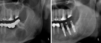
— examination of the patient; - “targeted” x-ray of the tooth, which the patient indicates as the causative tooth; - orthopantomogram (panoramic image of the jaw bones), which allows you to see pathologies and the most likely places of their development in the upper and lower jaws, in the maxillary sinuses, in the canals of the nerve trunks; - in some cases - a computed tomogram, which allows you to see the facial skeleton and skull bones, joints, maxillary sinuses in three projections (frontal, sagittal and horizontal) and in a three-dimensional volumetric image.
Cystotomy and cystectomy in dentistry - what is it and what is the difference
Both operations are used for surgical treatment of cysts. The differences lie in the method of surgery and the final result of the procedure.
Features of the methods:
- During cystectomy, the tumor is radically removed, that is, completely removed. At the last stage, the wound is sutured.
- Cystotomy involves partial removal of the cystic wall and bone. The remaining capsule communicates with the oral cavity. After the procedure, the pressure inside the formation decreases, it flattens, and then gradually resolves.
The choice of method depends on the clinical case. A preliminary X-ray or computed tomography is performed to rule out contraindications.
Advantages and disadvantages of cystotomy:
- advantages - the procedure is easy to perform, low-traumatic, there is no risk of damage to nearby tissues and nerves;
- disadvantages - the duration of postoperative care, as additional cavities are formed, deformations, and jaw defects impair the cleansing of the mouth from microorganisms.
Advantages and disadvantages of cystectomy:
- advantages - complete removal of the capsule, suturing the wound, reparative bone regeneration;
- disadvantages - high trauma, risk of damage to adjacent teeth, nerves, blood vessels, maxillary sinus, accidental removal of a malignant tumor, autolysis of a blood clot in bone tissue cannot be ruled out.
Removal of a dental cyst. Historically significant and modern methods of surgery for removing dental cysts
Initially, the most common method of treating cysts was their removal. For this purpose, 2 fundamentally different methods were used: cystotomy and cystectomy.
Cystotomy (or the PARCH-I method) has historical significance in the development and establishment of medicine. Today this method is practically not used, but previously it was indispensable for removing large cysts. To avoid complications and its severe consequences, a wide channel was created between the cyst cavity and the vestibule of the oral cavity by suturing the edges of the cyst shell with the oral mucosa.
Cystectomy (or PARCH-II method) involves complete excision/removal of the cyst. This surgical intervention is often simultaneously accompanied by the removal of the root apex, the source of the formation of the cyst containing infected apical deltas. Today this method is the most popular.
The main goal of cystectomy is complete sanitation of the cyst cavity, which is not possible without resection of the root apex. The fact is that the root pulp in its apical third has an apical delta with an unclear structure and very fine branching of the canals, even in single-rooted teeth. If such canals can be cleaned during tooth treatment, then during retreatment this is almost impossible, especially considering the narrowness of the lateral deltas. Removing the root apex, in addition, allows you to most effectively clean the cyst cavity behind the root. This is especially true for teeth 12 and 22, which have a more curved root system. The operation is carried out as follows: using a small incision on the gum, the surgeon very carefully reaches the surface of the bone located above the cystic cavity, removes the wall with a trephine or bur, making sure that the neighboring teeth are not damaged, and then removes the cyst shell and performs a resection of the root apex . Next, sanitation of the cystic cavity is carried out and its subsequent revision for residual cyst particles. After this, in some cases, retrograde filling is performed. The final step in the operation to remove cystic formations is filling the resulting cavity with osteoplastic material and suturing the wound.
In the case of the formation of a large cyst, amputation of the apex of the tooth root can be performed, or half of the tooth and damaged root can be removed (hemessection), and in the case of an interradicular cyst, this can be done by corono-radicular separation.
How is the cystectomy procedure performed?
- Examination by a doctor of the damaged area, x-ray diagnostics.
- Conducting anesthesia. The operation is performed under conduction and infiltration anesthesia.
- Carrying out the operation. As a rule, with this intervention the tooth is completely removed.
- The dentist removes the cyst through the resulting hole.
- After curettage, the cyst needs to be filled. A special bone-forming drug is poured into the resulting cavity, aimed at accelerating the healing process. Also suitable for successful healing are xenografts or materials made from bone chips and the patient’s own blood.
- Applying a suture to the wound
- At the end of the cystotomy procedure, complex anti-inflammatory and antibacterial therapy is prescribed, as well as follow-up examinations (images are taken once a month to track the dynamics of bone restoration in the area of the removed tissue).
The cystectomy procedure has no age restrictions. However, each clinical case requires an individual approach and treatment. Your attending physician will tell you in more detail about how the operation is performed, what treatment is needed during the rehabilitation period and other nuances during your consultation.
On a note. For several days after the cystectomy operation, pain (from nagging to moderately acute) and slight swelling of the tissue may be observed in the intervention area. Don't be afraid of this. If swelling and pain are accompanied by an increase in body temperature for 3 or more days, you should consult a doctor.
Quality criteria for dental cyst removal
Despite the fact that cystectomy is a surgical intervention, the main indicator of the quality of treatment will be the sanitation of the root canals, including retrograde filling. Currently, in addition to eliminating the cyst shell itself, the unspoken rule has become the filling of its cavity with one or another osteoplastic materials, because this completely eliminates the possibility of blood clot suppuration, promotes rapid healing of the bone defect and improves the mechanical stability of the tooth.
More about the operation
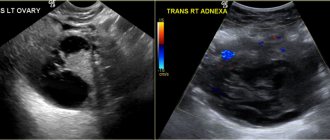
Cystectomy is an organ-saving operation that involves complete excision of an ovarian cyst without damaging healthy tissue.
Despite its benign nature, even a small cystic formation can lead to the development of serious complications - dysfunction of the ovaries and menstrual cycle, adhesive disease, infertility. The intervention can be performed as planned or emergency. The second option is used in the development of emergency conditions - rupture of the cyst, torsion of its stem and some other indications. Cystectomy of the cyst is performed laparoscopically, through several micropunctures in the abdominal cavity. The main advantage of this approach is minimal blood loss, low risk of complications and rapid recovery after surgery.
Indications for tooth extraction with a cyst
As a rule, conservative treatment carried out on time or surgery to remove the cyst allows you to save the tooth. But this is not always possible. A tooth must be removed in the following cases:
— the presence of severe pain symptoms when drug treatment is ineffective; - purulent inflammatory process when it is impossible to drain it; — fracture of the coronal part of the tooth, without the possibility of restoration with anchor pins and/or stump inlays; — obstruction of root canals; — the presence of multiple damage to the roots of the tooth or large damage; the tooth is almost completely destroyed and orthopedic restoration is impossible; - no need for dental treatment due to the presence of a prosthetic plan agreed upon between the doctor and the patient.
Visit to the dentist without pain
Forget the stress of a botched anesthesia, FDC uses painless pain management techniques.
QuickSleeper is an electronic system that provides simple, fast and comfortable pain relief.
VibraJect is a special attachment. It is an effective way to reduce pain and anxiety during local anesthesia. All this helps reduce patient stress from painful injections and unpleasant visits.
Read more about painless anesthesia with QuickSleeper
Price
The cost of treatment or removal of a dental cyst consists, like many other surgical interventions, of a number of factors. The complex of diagnostic procedures performed, types and methods of treatment, conservative preparation of canals, preoperative preparation, the operation itself, the osteoplastic materials used, postoperative observation - all this affects the final cost of treatment in these clinical situations.
A dental cyst is a serious pathology. In all cases, even in cases of complete absence of any symptoms, it poses a health hazard. With regular visits to the dentist, you can be sure: a dental cyst, if there is one, will be detected in time, and subsequent treatment will allow it to be eliminated without leaving a trace, preserving the health of the tooth.
According to antiplagiat.ru, the uniqueness of the text as of October 16, 2018 is 99.9%.
Key words, tags: X-rays, tooth extraction, orthopantomogram, periodontitis
Cystectomy technique
Cystectomy is performed using open (laparotomy) and endoscopic (laparoscopic) techniques. In a classic laparotomy, a layer-by-layer incision is made in the skin of the abdomen to access the surgical field.
Afterwards, tumor-affected bladder and pelvic lymph nodes, sections of the ureters, seminal vesicles and prostate are removed in men, ovaries, fallopian tubes, uterus and vagina in women. If necessary, a new path for urine drainage is formed - for this, the free ends of the ureters are connected or a neocystis and urethrostomy are formed. During the application of postoperative sutures, temporary drains are removed from the abdominal cavity.
If there are no clinical restrictions or contraindications, preference is given to the laparoscopic technique for performing cystectomy. It is carried out through several small incisions in the soft tissue of the abdomen, through which an endoscope - a miniature video camera with a light source - and microsurgical instruments are inserted. The resulting image is displayed under magnification on a monitor screen in the operating room. Due to this, all the manipulations described above are performed with the highest possible surgical precision and atraumaticity.