An inlay is a microprosthesis that fills imperfections in the crown of a tooth, which allows you to restore its shape and return the functions assigned to it. In fact, an inlay is the same filling, but performed by a technician in a laboratory.
The difference from traditional therapeutic treatment is that the material is introduced into the dental cavity not in plastic, but in solid form, which means that the inlay has visible advantages, the main of which are:
- the strength of the connection, which occurs due to the complete contact of the surfaces;
- the likelihood of restoring angles, cusps and contact points depending on age, as well as existing individual qualities;
- prevention of carious recurrence due to the constant volume of the inlay, as well as due to the precise fit;
- durability and wear resistance, which are determined by the high degree of strength of the material used;
- Due to the density of the structure, which was compiled in the laboratory, the insert does not change its color.
It is for these reasons that replacing teeth defects with inlays is a more preferable option than the usual filling.
Classification of tabs.
Tabs are classified depending on the method of expressing chewing pressure and are divided into:
- restorative. These microprostheses restore the chewing pressure that occurs on the periodontal tissues.
- loading. These structures are used for the rehabilitation of the dentition, and they are used as a support for a bridge-class denture.
- distributing. Such inserts redirect chewing pressure during teeth splinting.
Tabs are classified according to the following criteria:
Grouping of tooth cavities under inlays (according to the location of the defect).
Most often, a dental defect in the coronal lobe occurs due to caries. It is for this reason that the division of this disease according to topographic properties is of great importance.
The division was developed in 1891 by G. Black and involves dividing carious cavities into six classes depending on location. Speaking about the main advantage of this work, it should be noted the simplicity with which a doctor can use the classification.
To create the necessary conditions for fixing the tab, the doctor needs to determine which class the cavity belongs to, thereby preventing the secondary appearance of caries.
The only change that occurred with the classification can be considered the change of classes to letter definitions proposed by Boyanov in 1960. In fact, Boyanov proposed designating surfaces that have cavities.
Today the classification is as follows:
- O are cavities on the chewing planes (occlusal surfaces);
- M are cavities located on the medial plane;
- D - this is on the distal surface;
- MO are cavities that cover both the medial and occlusal surfaces;
- MOD are cavities that cover all three surfaces (occlusal, medial, and distal).
Separation by design features.
Depending on the severity of the defect in the coronal part of the tooth and how the doctor plans to place the microprosthesis, inlays replace missing tissue to varying degrees, and therefore, it is customary to distinguish four (main) types of inlay structures.
- inlay - in this design, the microprosthesis is located centrally, but at the same time does not affect the tubercles of the tooth;
- overlay - in this design, the microprosthesis covers up to three tubercles. It is worth noting that such a design, affecting four tubercles, can already refer to three-quarter crowns.
- onley - the tab touches the internal slopes, creating the effect of an overlay;
- pinley - in a similar design, a microprosthesis is strengthened in the tooth using so-called pins, that is, pins that are located in hard tissues. It is worth noting that the manufacture of such systems for chewing teeth involves covering all the tubercles. As for the front teeth, it is possible to install a pinley with full protection of the vestibular surface, as well as the incisal edge. Simply put, pinlay inlays on canines and incisors look like half-crowns with a pin.
Division depending on the material from which the inlays are made.
Depending on the material used, the tabs are divided into:
- metal models made of titanium, since this material takes root almost 100%;
- plastic inserts (nylon, acrylic, polyurethane);
- ceramic, made from materials such as zirconium, porcelain or titanium oxide;
- composite, they are also called ceramic;
- combined (metal-composite or metal-ceramic).
Depending on the chosen material, the doctor prepares the cavity for the tab and determines its features, but at the first stage, the dentist can prepare the cavity for the tab, based on the diagnosis and the drawn up treatment plan. But such an approach occurs only after a complete clinical examination.
What is IROPZ?
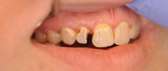
The root cause of damage to the integrity of the tooth remains caries.
During the pathological process, the hard tissues of the teeth are damaged, their shape is deformed, causing disruption of speech and chewing functions, and the appearance of external defects. The effectiveness of actions restoring a damaged tooth is determined by a number of factors:
- the degree of damage to the coronal area, which is under the influence of the carious process;
- timely contact with a dentist;
- correct diagnosis, professionalism and experience of specialists, as well as characteristics of the components and technological methods involved.
Before recreating the anatomical form and function of dental tissue, individual preparation is required for each individual case.
For example, the surgical effect on the hard tissues of the tooth according to Black consists of removing the enamel left without dentin and shortening the cusp damaged by more than 2/3 of the interval from the median fissure to its apex. Such an action will delay a possible fracture of the tooth crown to a later period.
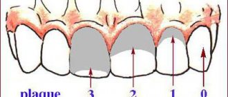
Classification of carious cavities according to Black
When crossing the contact surface with the chewing surface, it is important to try to preserve the area of Black resistance, which provides some stability to the teeth upon completion of the restoration and prevents the filling from chipping due to the load during chewing.
When calculating the coefficient of damage to the occlusion zone of chewing teeth with defects of groups 1-2 and selecting a prosthetic model, it is recommended to use the index of destruction of the occlusal surface of teeth (IROPD), developed in 1984 by the scientist Milikevich V. Yu.
Its use guarantees relatively high accuracy of experiments. Nevertheless, the method is very painstaking; its organization takes a lot of time due to the large number of its stages (taking an impression, selecting and creating a model, calculating the area and calculating the index). Recently, computer technologies have been actively used to calculate the index.
Direct methods for establishing the IROPZ index are mainly reflected in practical healthcare. These include: the visual value of IROPZ and its calculation based on anatomical indications of the occlusion surface. They are often used by specialists in the selection of options and methods of reconstruction.
Computer assessment of IRPH
IROPD in dentistry is defined as the ratio of the “cavity-filling” area to the chewing dental surface. It can be calculated by taking the total area of tooth occlusion as a unit.
With the introduction of the latest techniques and materials, the chances of recreating the coronal zone of a tooth in orthopedic dentistry have greatly expanded, and its functional significance has increased.
The selection of a tooth restoration method is influenced by:
- the existence and position of antagonists;
- degree of bite;
- oral hygiene.
Teeth are restored either by filling or prosthetics (through the use of an inlay, an artificial crown, structures with a pin), using the IROPZ index.
Based on the classification of IROPD, we can consider the main stages of damage to the hard dental membranes:
- 1st degree - from 0.2 to 0.4 - filling is used;
- 2nd degree - IROPD ranges from 0.5 to 0.6 (defect of the occlusion area is more than 50%), to prevent subsequent destruction, the inlay method should be used;
- 3 degree - index in the range of 0.6 - 0.8 (damage exceeds 60%) - filling with the involvement of artificial crowns is recommended;
- 4th degree – index value above 0.8 (destruction of the occlusion zone over 80%) – pin models and crowns are made.
In all these cases, as well as in order to avoid curvature of the dental system (with adjacent teeth with fillings recreating more than half of the chewing zone), prosthetics are recommended.
How are tabs used?
- in the form of an independent design that allows you to restore the shape, function and aesthetics of previously unsuitable crowns with IROPS indicators of 0.3–0.6, that is, in the presence of: caries, if filling turns out to be ineffective or cannot be performed;
- tissue damage of a non-carious nature, for example, with wedge-shaped defects or with increased abrasion.
Methodology for clinical assessment of restoration work
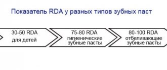
The assessment was carried out according to the USPHS system, which includes anatomical shape, marginal adaptation, surface roughness, marginal staining, color matching, secondary caries, and the presence of sensitivity.
Characteristics of the groups, study performance, number of restorations evaluated, and number of observations are presented according to the reviewed American Dental Association (ADA) Materials Council Requirements Protocol for Enamel/Dentin Adhesive Materials (Chicago, 1994) [10]
Research results and discussions
After 24 months, 68 restorations were re-evaluated in patients in group I (61.8% compared to the first visit).
69.1±5.6% of restorations did not lose their anatomical shape. Minor chips of the surface of the restoration material accounted for 30.9±5.6% of cases (indicator “A”). There is no need to alter the restorations.
According to the criterion of regional adaptation “MA”, a high percentage of high-quality restoration was noted - 85.3±4.3%, corresponding to indicator “A”. Moreover, only in 14.7±4.3% of cases between the restoration and the tooth tissues, the probe can detect the presence of a gap “B”. There were no cases of poor-quality restoration according to this criterion.
The smooth surface of the restoration of group I was preserved in 79.4±4.9% of cases (indicator “A”), in 20.6±4.9% roughness was detected (“B”). There were no cases of poor-quality restoration according to this criterion.
None of the patients in group I during this period of the study had any complaints of increased sensitivity to the effects of thermal irritants, edge staining, color matching or the presence of secondary caries.
Marginal coloration according to indicator “A” was 85.3±4.3%, and for the remaining 14.7±4.3% indicator “B” was determined.
In this group, 100% of the restorations examined retained the absence of secondary caries and color matching (“A”).
In patients of group II, 71 restorations were re-evaluated (76.4±5.0% of the first visit).
The anatomical shape was preserved in 60.6±5.8% of restorations (“A”). In 29.6±5.4% the restorations were classified as “B”, and in 9.8±3.5% the restorations should be redone due to a significant change in the anatomical shape – “C”.
“Excellent” (“A”) marginal adaptation was preserved in 70.4 ± 5.4% of restorations, and in the remaining 18.3 ± 4.6%, the probe revealed the presence of a gap (“B”) between the restoration and the tooth tissues. It is necessary to redo 11.3±3.8%, which corresponded to the “C” indicator.
Discoloration along the margin between the restoration and the surface of the dental structure was recorded in 15.5 ± 4.3% (“B”), and in 14.1 ± 4.1% (“C”), requiring their alteration.
The smooth surface of the restoration material was detected in 69.0±5.4% of cases (“A”), 25.4±5.2% of cases “B”, and 4.2±2.4% of cases “C”.
Patients did not make any complaints about increased sensitivity of teeth due to the action of thermal irritants - 100% (indicator “A”).
Secondary caries was detected in 12.7±4.0% of cases (“B”), which required their reworking in this case.
In terms of color matching, group II – 88.7±3.8% had color matching, and 11.3±3.8% had “B” indicator.
Thus, after 24 months of operation of restorations in teeth with a defect in the IROPD index from 0.4 to 0.7, further deterioration was revealed according to the evaluation criteria AF (anatomical shape), MA (marginal adaptation), MD (marginal coloring), CC (color matching ), SR (surface roughness), RC (secondary caries). Moreover, it was not noted earlier. In this case, the restorations required rework due to significant changes in the anatomical shape, marginal adaptation, marginal staining, roughness and the appearance of secondary caries.
After 24 months, control examination of group III of 62 restorations (60.2% compared to the first visit).
The anatomical shape was preserved in 50.0±6.4% of restorations. Due to minor chips of the restoration material, in 29.0±5.8% of cases the indicator “B” was determined, and in 20.9±5.2% there was a violation of the anatomical shape until dentin was exposed (“C”).
According to the “marginal adaptation” criterion, in 59.7±6.2% of cases the indicator is “A”, in 21.0±5.2% a gap (“B”) is determined between the restoration and the walls of the tooth, and in 14.5± In 4.5% of cases, a deep crack exposed dentin (“C”), and in 4.8±2.7% of cases “D” was detected. The absence of marginal coloring “A” was detected in 62.9±6.1% of cases, and a change in color between the restoration and the surface of the dental structure “B” – in 21.0±5.2%. Indicator “C” was noted in 16.1±4.7%.
In this III group, after 24 months of clinical use, in 71.0±5.8% of cases, the restorations corresponded in color and transparency of the dental structures - CC (color match) (indicator “A”), 24.2±5.4% - “ B", and in 4.8±2.7% they required alteration of the restoration.
Smooth surface according to indicator “A” was in 59.7±6.2% of restorations, in 22.6±5.3% roughness was (“B”), and in 17.7±4.9% restorations had unevenness , a lumpy surface, which did not depend on the anatomical shape (“C”) and required alteration of the restoration.
In 100% of cases, patients did not complain of sensitivity when exposed to a thermal stimulus (“B”).
Secondary caries was diagnosed in 30.6±5.9% of cases.
In group III, after 18 months, further deterioration of restorations was noted in terms of AF (anatomical shape), MA (marginal adaptation), MD (marginal coloring), SR (surface roughness), CC (color matching) and RC (secondary caries). Moreover, CC has not been noted before. At the same time, according to the criteria SR (surface roughness), AF (anatomical shape), MA (marginal adaptation), RC (secondary caries), CC (color matching), poor-quality restorations requiring rework were noted.
Clinical studies of the quality of restorations after 24 months showed a deterioration in their condition from group 1 to group 3 according to the following criteria:
· regional adaptation (p=0.16, differences are not statistically significant), group 1-3 (p=0.007), group 2-3 (p=0.54, differences are not statistically significant);
· marginal staining (p=0.16, differences are not statistically significant), group 1-3 (p=0.02), group 2-3 (p=0.76, differences are not statistically significant);
· color matching (p=0.02), group 1-3 (p˂0.001), group 2-3 (p=0.07, differences are not statistically significant).
Thus, the most effective and more reliable in this case are restorations with a lower IROPV index. That is group I.
Restoration technique
First, the teeth that had undergone restoration were hygienically treated. The color was determined using a color scale.
The carious cavity was prepared with diamond burs using a water-cooled turbine tip. The tooth was isolated. If necessary, a retraction thread, wedges, and matrices were used. The prepared cavity was washed with a 0.05% chlorhexidine solution and dried with a weak stream of air. For acute deep caries, a therapeutic pad based on calcium hydroxide (Dycal, “Dentsply” or Life “Kerr”) was applied to the area of projection of the pulp horns. A gasket is applied. Then the tooth tissue is treated with 35% phosphoric acid gel, washed with a stream of water and dried. The ART Bond adhesive system was applied to the prepared enamel and dentin. Filling material was introduced. After restoration and contouring of the tooth is completed, grinding and polishing is carried out using a diamond head, polishers, finishers and an Enchence polishing set (“Dentsply”).
Clinical assessment was carried out immediately after recovery and after 24 months using a visual-instrumental method according to clinical criteria.
The data obtained during the experiment were statistically processed using the STATISTICA 6.0 software product. To identify the pairwise correlation between the average values of the samples, a correlation analysis was carried out (Spearman's rank correlation index - R was calculated).