The dentofacial system is an important component of the human masticatory apparatus. It meets morphological and functional characteristics, such as number, size, shape, color, structure of dental units. There are a number of genetically determined or external factors that lead to disruption of these characteristics. Dental anomalies can have various consequences: some lead to periodontal pathologies, various forms of caries, others seriously limit chewing function, cause injury to the mucous membrane, and others cause aesthetic problems.
Improper growth of teeth results in speech and bite defects and breathing problems. As a result, the patient’s general health seriously suffers, and psychological problems arise.
Reasons causing anomalies
Anomalies of the dental system are very diverse and include both anomalies of dental units and incorrect formation of the dentition, as well as abnormalities in the development of the jaws. The most frequently diagnosed abnormalities of dental units. They can be caused by a wide variety of reasons, including both exogenous (caused by external factors) and endogenous (formed as a result of the physiological state of the patient).
Endogenous causes
Endogenous anomalies are formed as a result of genetic and endocrine disorders.
- Many structural features of the dental system, leading to improper development of teeth, are genetically determined. This can occur through direct inheritance (abnormal number and shape of teeth, edentia, diastema), through inheritance of a mismatch in the size of the jaw bones, through inheritance of a mismatch in the size of the jaw and teeth (crowding of teeth or sparse arrangement of teeth). Dental anomalies of this type include disorders associated with hereditary and congenital pathologies (clefts of the soft and hard palate, cleft lip, Down syndrome, Vaanderburg syndrome, Seckel syndrome, Shershevsky-Turner syndrome).
- Another part of the endogenous causes of dental anomalies are problems of the endocrine system, such as hypothyroidism (in later stages), hyperparathyroidism, hypocortisolism. Their frequent symptoms may be delays in the eruption and replacement of teeth, and disturbances in the formation of the enamel layer.
Exogenous causes
Exogenous (external) causes of dental anomalies are, for the most part, external unfavorable factors that affect the baby’s body during the formation of tooth germs. There are prenatal (prenatal), postnatal (postpartum) and intranatal (associated with childbirth) periods.
- An example of a prenatal factor is a pregnant woman living in poor environmental conditions. Various developmental disorders of the fetus can also occur as a result of the mother’s poor lifestyle.
- Factors in the intranatal period may include complicated labor, birth injuries, consequences of asphyxia and oligohydramnios.
- In the postnatal period, dental anomalies develop as a complication of pathologies in early childhood. These include childhood infections, rickets, hypovitaminosis, and micronutrient deficiency.
All these reasons are of a common nature. Local factors that affect dental development include everything related to improper oral care, feeding errors or bad habits. Among them are feeding too soft food, prolonged sucking of a pacifier or finger in early childhood.
Childhood injuries and caries with complications lead to dental anomalies.
The death of tooth germs or the development of supernumerary teeth can occur due to osteomyelitis.
Anomalies in the number of teeth
All anomalies in the number of teeth are divided into three types:
- hyperodontia (excess of dental units above normal);
- edentia (complete absence of teeth);
- hypodontia (insufficient number of teeth).
Hyperodontia
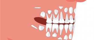
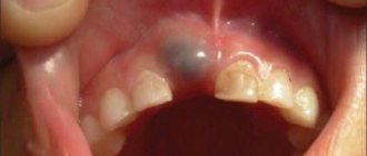
Hyperodontia is a disorder of the formation of the dentition, which is expressed in the presence of supernumerary teeth (over 32 dental units in an adult). The cause of the disease may be a violation of the mechanism of formation of tooth germs in childhood or infancy. Hyperodontia mainly affects the incisors of the upper jaw. Other teeth are involved in the pathological process less frequently. Extra dental units grow small and have a cone shape.
The disease can be true or false.
- With true hyperodontia, the formation of extra dental units occurs in the human jaw.
- False hyperodontia occurs when the loss of primary teeth is delayed, which interferes with the normal growth of permanent teeth.
With polyodontia, teeth may erupt away from the dentition or next to a correctly positioned dental unit, which leads to its gradual displacement. The dentition can be seriously deformed, which leads to serious diction problems.
If supernumerary teeth interfere with the development of normal teeth, they must be removed. Broken teeth are corrected by installing braces.
In those rare cases when a row violation does not occur, the supernumerary tooth is preserved. Prosthetics are used to correct the shape.
Hypodentia
Hypodentia is a deficiency of dental units. This disorder occurs most often due to the death of tooth germs in the fetus. A small number of teeth causes a shift in the midline, the formation of wide gaps between teeth (diastema and three), and shortening of the dentition. All this adversely affects the bite. Possible ways of correction are prosthetics.
Edentia
Adentia is a complete or partial absence of teeth. With edentia, the continuity of the dentition is disrupted, which leads to difficulties in chewing and speaking. The reason is a violation of the development or death of tooth germs under the influence of genetic factors or under the influence of unfavorable external factors during the formation of the dental plate in the fetus (the formation of the germs of permanent teeth in the fetus occurs after the 17th week of intrauterine development). The disorder is corrected using prosthetics or dental implantation.
What can cause caries
There are several suggestions, for example, that tooth decay is transmitted genetically or that lack of hygiene contributes to the development of the disease. All this is true, but does not give the complete picture.
Where a person lives can also influence the occurrence of caries, namely the quality of water in that area.
Nutrition plays an important role in the prevention of caries. There are scientific studies confirming that eating fresh vegetables, greens and foods rich in proteins helps prevent tooth decay. That is, those people who like to chew raw carrots are less susceptible to dental diseases. That's why you should eat meat and a lot of raw vegetables - this is not only smart advice from nutritionists, but sound prevention in the fight against the development of dental caries.
Anomalies of magnitude
There are two forms of this type of deviation, including macrodentia (increase in the size of dental units) and microdentia (small teeth).
Macrodentia
With macrodentia, the size of dental crowns is significantly increased. The cause of the disorder is, in most cases, dysfunction of the endocrine system, which is characterized by the fusion of several rudiments together. The disease is localized mainly in the area of the upper incisors. Large teeth interfere with the process of eruption and growth of neighboring dental units, which leads to their abnormal arrangement and crowding. Giant teeth are found on or outside the dentition line. This is a serious cosmetic defect that causes serious functional impairment and psychological disorders. Pathologically enlarged teeth are removed, and the growth of adjacent teeth is corrected using prosthetics or braces.
Microdentia
Microdentia – teeth that are too small. The disorder mainly affects the upper lateral incisors, but may affect the incisors of the lower or both jaws. The cause of the anomaly has not been studied, but it is reliably known that it develops in genetically predisposed patients. With the problem of small teeth, patients develop too large interdental spaces, which significantly disrupts the aesthetics of the dentition. To correct the pathology, incorrectly growing teeth are removed or covered with crowns.
Optimal parameters
When talking about teeth of reduced size, we should mean their clinical crown, the part that protrudes above the gum.
The difficulty in classifying a particular tooth as “short” (or “small-sized”, so to speak) is that there is no value for the length or width of the crown that can be considered absolutely normal. Everything is relative.
The size of the clinical crown depends on the anatomical features of the jaws of a particular person, his age, face type and even, as it turned out, nationality. Therefore, a crown of a certain size may be “short” for one person, but normal for another.
We'll talk more about all this below. Now let’s talk about what visual signs a person can use to make a conclusion whether he has “short” crowns or not.
One of the characteristic manifestations of “short” teeth is the exposure of the gums by more than 2 mm when smiling. However, this can happen not only due to “short” crowns, but also for other reasons.
By the way, a smile with more than 3 mm of gum exposed is called “gummy”. This is considered a pathology that it is desirable to get rid of.
The patient can also determine the presence of “short” teeth by the following signs:
- Clinical crowns visually appear shorter than they should be and protrude above the gum by a small amount.
- The face looks asymmetrical. Most often this is expressed in a reduced lower third of the face.
- Wrinkles form around the mouth.
- The upper lip looks thinner when smiling compared to the lower lip.
Often reduced sizes are accompanied by the presence of trema and diastemas. Which is quite understandable, in normal-sized dental arches there is an excess of space for teeth of small width.
What is macrodentia and why is the anomaly dangerous?
Come here to learn more about microdentia in primary teeth.
At this address https://orto-info.ru/zubocheliustnye-anomalii/zubov/strukturyi-tverdyih-tkaney/displazija-emali.html all the most important things about enamel dysplasia.
Shape anomalies
There are pathologies in the development of dental units, due to which they acquire an unnatural shape. These disorders are named after the scientists who first described them. The following types of dental shape anomalies are found: spiny teeth, Pflueger teeth, Fournier teeth, Hutchinson teeth.
- Spiked or awl-shaped teeth. With this pathology, the teeth acquire a cone-shaped shape. Wide at the base, they gradually narrow and, towards the cutting edge, become sharpened into a spike shape. This problem is combined with microdentia. There are irregularities and stains on the surface of the teeth. The disease affects the front and lateral incisors. The disease occurs in childhood and is caused by genetic factors in combination with external factors. Among them are vitamin D deficiency and endocrine system problems.
- Hutchinson's teeth are characterized by a modified crown shape of the incisors. Externally, they have the shape of a barrel, since the neck is significantly thickened. The cutting edge of the teeth acquires an arched notch. The enamel layer also suffers, which is present only on the sides and absent in the center.
- Fournier's teeth are a form of systemic hypoplasia of dental units associated with metabolic disorders at the stage of intrauterine development of the fetus. The barrel-shaped shape of the teeth is preserved, but the notch of the incisal edge is absent. The color of the enamel is not disturbed, but the enamel layer is underdeveloped, which can be seen during microscopic examination.
- Pflueger's teeth are a disorder that affects permanent dental units. Their crowns take on a conical shape. Thickening develops in the cervical region, and the chewing surface is significantly underdeveloped. The chewing function of the teeth is completely preserved.
- Turner anomaly. With this pathology, there is no enamel on the teeth. They have an abnormally lumpy surface, and replacement tissue, dentin, forms in the exposed areas.
Systemic hypoplasia with a violation of the shape of the teeth has three degrees of development. The third degree is the most dangerous, in which the crown is severely deformed and the enamel layer decreases. In this case, the defective teeth are removed and replaced with dentures. It is also possible to restore affected teeth using reflective components.
Anomalies of hard tissue structure
An altered shape, abnormal size or color of enamel is formed against the background of an abnormality in the structure of the hard tissues of the dental unit. Among such anomalies are:
- Hypoplasia (underdevelopment of tissue). The initial degree of the disorder is manifested by the presence of chalky spots on the enamel and areas where tissue deficiency is observed. Subsequently, all kinds of pits, grooves, and recesses appear on the surface of the enamel. The defect affects all teeth in the dentition.
- Hyperplasia (excessive tissue formation). Pathology also affects all teeth at the same time. It is characterized by areas where tissue growth is observed - tubercles, sagging enamel. The consequence of the pathology is a change in the shape and size of the teeth, a violation of the occlusion (the line of contact of the upper and lower jaws).
- Anomalies of amelogenesis (enamel formation) are expressed in the presence of brown or yellow spots on the surface of the teeth. Areas where the natural composition of the enamel is disrupted become especially sensitive. Microdentia develops against the background of pathology. The cause of the development of pathology is a deficiency of microelements that take part in the formation of dental tissue. Treatment consists of replenishing this deficiency. Additionally, local remineralizing therapy and physiotherapy are performed.
- Disturbance of dentinogenesis consists of dysfunction of the mechanism of dentin formation. The main symptoms of the disease are as follows: teeth become yellow-brown or grayish in color. The fragile dentin in these areas quickly wears off, causing tooth decay and then tooth loss. The disease occurs in genetically predisposed patients. The problem can only be solved by replacing the units destroyed by the disease with prosthetics.
Sealing
Currently, it is one of the most affordable ways to treat rare teeth. Its wide distribution is due to its low cost and ease of implementation.
The essence of the method is as follows: fillings of the same color as the natural chewing units are applied to the teeth. The procedure is considered a jewelry procedure, since the doctor must literally mold new bone structures. As a result, the teeth become wider and the gaps between them disappear. Only a specialist can visually distinguish fillings from natural dental units.
During the procedure, orthodontists use only high-quality materials. These are ceramics and porcelain.
After filling rare teeth, the functioning of the gastrointestinal tract is normalized and a correct bite begins to form.
Color anomalies
Healthy, young teeth look snow-white due to a thick dentin layer and high-quality enamel, which has sufficient characteristics of whiteness, shine and transparency. Lifestyle, age, genetic factors, and ecology can slightly change the color of teeth from bluish to yellow. Small deviations are not considered pathology, although they indicate a change in the quality of the enamel.
Various pathological processes occurring in the body have a more significant impact on the condition of the enamel. The cause may be carious lesions, the use of medications, or a lack of certain microelements in the body. Teeth can take on a gray, pinkish, brown, purple and even black tint.
When starting to treat a patient, the dentist excludes the development of chronic pathologies. After a course of therapy and stable remission is achieved, other causes of discoloration of the enamel are eliminated. The final stage of treatment is a course of teeth whitening.
Specialists at the Consilium Dent choose the enamel lightening technique together with the patient. The selection criteria are age, condition of the enamel, and the wishes of the patient.
Prices for treatment of caries of anterior teeth in Moscow
The price for treating caries of anterior teeth largely depends on the technique used. Standard therapy with the installation of a light filling will cost from 5,800 rubles. But prices for restoration start at 12,500 rubles. Treatment costs approximately the same without an Icon drill. Treatment of pulpitis on the front teeth involves cleaning the canals, which will add several thousand more to the total amount. If the tooth is seriously damaged and installing a filling is impossible, get ready for serious expenses. The average cost of a ceramic crown is from 25,000–30,000 rubles, and turnkey implantation will cost at least 55,000–100,000 rubles for the installation of one implant.
Position anomalies
Incorrect position of teeth always develops as a consequence of another pathology and forms an incorrect bite. May affect one or both jaws. Incorrect position of teeth makes chewing food and oral hygiene difficult. This can lead to digestive problems and the development of cavities. There are several options for the pathological position of teeth within or outside the dentition:
- External or vestibular position means that the tooth is located outside the dentition, closer to the vestibule of the mouth. This position is typical for canines and incisors and develops as a serious cosmetic defect.
- Oral or internal position. The teeth are deviated inward closer to the tongue. The disorder is typical for canines, incisors and premolars. May cause tongue injuries and dysfunction of the temporomandibular joint.
- Mesial and distal position. Dental units are displaced forward or backward along the dentition, which leads to its shortening.
- Supra and infraocclusion are high or low positions of teeth relative to the occlusal curve. The cause of the disorder is underdevelopment of the alveolar process. It appears due to the presence of some obstacle that interferes with the normal formation of the tooth germ. Effective therapy is surgery.
- Tortoanomaly. The tooth is rotated around a vertical axis. The disorder is typical for incisors, canines, and premolars. The anomaly can cause injury to adjacent teeth. Dental units are rotated into the correct position using orthodontic structures.
- Transposition. The teeth change their location with each other. The disease affects canines that exchange places with premolars or lateral incisors. The disorder develops at the stage of tooth germ formation. The therapy is as follows: problematic teeth are removed followed by prosthetics.
- Crowding of teeth occurs due to lack of space when tooth germs are located too closely. Crowded teeth are closely adjacent to each other and rotate around their axis. Occurs in combination with macrodentia or with an undeveloped basal part. To eliminate the violation, some dental units are removed.
- Diastemas and tremas are wide spaces between teeth. The disorder develops as a consequence of an abnormal shape and size. The problem is treated with orthodontic methods or by installing veneers, which helps restore the aesthetics of the smile.
Misaligned teeth cannot always be cured using braces. Severe pathology, which is characterized by a skeletal disorder of the maxillofacial region, is treated with surgical reconstruction. The pathological fragment is transferred to the desired position and then secured. A fragment containing part of the dentition or the entire dentition can be used.
Installation of crowns
These are non-removable structures that can be used to eliminate dental defects. The installation of crowns is also indicated in case of severe destruction of dental units. The essence of the method is to insert an artificial tooth into the gum using a pin made of metal.
Installing crowns requires some preparation. The patient's natural teeth are prepared and, if necessary, pulp removal is performed. Crowns take several days to make. During this period, the patient is provided with temporary structures. The finished crowns are completely identical to the patient’s teeth. They differ only in width, which makes it possible to hide large gaps between chewing units.
The following conditions are contraindications: loose teeth, allergic reaction to the components used, periodontal pathologies. In addition, crowns are not placed on anyone under 16 years of age.
Diagnostics
An experienced dentist diagnoses dental abnormalities after an external examination and questioning of the patient. To clarify the diagnosis and identify the causes of the disease, a more detailed examination of the dental system is carried out:
- The construction of diagnostic plaster models and odontometric measurements make it possible to accurately identify changes in the size of dental units and their characteristics, which indicate the presence of pathologies.
- The color and transparency of the enamel are determined using a photographic method.
- Computed tomography or teleradiography makes it possible to obtain information about the condition of abnormal dental units in order to determine further treatment tactics.
- Panoramic radiography of the jaws is an important diagnostic method
- Electromyography of the jaws helps to assess the functional state of the facial muscular system.
To carry out differential diagnosis, the patient is referred for consultation to specialized specialists: endocrinologist, otolaryngologist, geneticist. Based on the diagnostic results, the attending physician determines treatment tactics. Depending on the type of disorder and the complexity of the disease, the patient may need the help of a dentist, surgeon, orthopedist, or implantologist.
Prognosis and prevention
Consilium Dent clinic uses advanced methods of therapeutic, surgical, and orthopedic treatment of dental anomalies in patients. By skillfully combining them, experienced specialists are able to restore the aesthetics of a smile and ensure the normal functioning of the dental system, even if the degree of deviation is significant.
Prevention of anomalies in the formation and development of dental units begins during the period of intrauterine development of the fetus and is carried out throughout a person’s life. The main stages are as follows: monitoring the successful course of the prenatal period, caring for harmonious postnatal development, organizing a correct lifestyle while the baby is growing up, caring for dental health and general health throughout life. By conducting an annual preventive examination, the dentist identifies dental anomalies at an early stage, when the pathological process is still reversible.
Expert of the article you are reading: