If you find a slight swelling or edema on a child’s body, you can think about the following conditions: inflammation, injury, cyst. When inflamed, the swollen area is pink or red, feels warmer to the touch than the surrounding tissue, and is painful when touched. Swelling after an injury may be a normal color warmer or cooler than the surrounding tissue, and a bruise may appear after some time. Sometimes, with increased sensitivity to any type of food, cold or other factors, edematous areas with clear boundaries quickly develop on the skin - such edema is called angioedema. They are often accompanied by hives (see Allergic reactions), which is another indication of the allergic nature of the condition. Swelling of the skin may be associated with enlarged lymph nodes (see Enlarged Lymph Nodes). General swelling of a limb or torso may indicate an allergic reaction or kidney disease.
THE DOCTOR'S CONSULTATION
Contact your doctor if the swelling:
- does not go away, grows or arose for no apparent reason
- hot to the touch, painful
ATTENTION!
If swelling occurs for no apparent reason, consult a doctor. In any case, it's better to be safe than sorry.
| ASK YOURSELF A QUESTION | POSSIBLE REASON | WHAT TO DO |
| Do you have one or more painful, raised lesions on your skin with a white center? | Boils (inflammation of hair follicles) | Do not try to open a boil by squeezing or piercing it, as this may spread the infection. Wash the area around the boil frequently with soap and water (not the boil itself!). When the boil opens, apply a bactericidal patch. If there is no improvement or new boils form, contact your pediatrician (or dermatologist ): you may need local treatment and antibiotics. |
| Does your child have several painless raised lesions with a rough surface on their skin? | Warts | Warts are caused by viruses and often go away without treatment. Warts that are a cosmetic defect, as well as those that are often injured, can be removed. Talk to your child's doctor (or dermatologist ) |
| Does your child have a soft, painless lump in the groin? Does it disappear with light pressure? | Inguinal hernia | Contact your pediatrician. If necessary, he will refer you to a pediatric surgeon |
| Does the boy have a soft, painless swelling of the scrotum on one side? Does one testicle look significantly larger than the other? Does the swelling decrease when the child lies down? | Hernia; hydrocele | Make an appointment with your pediatrician . He will determine whether there is dropsy, a congenital condition in which fluid accumulates in the scrotum. If your child has a hernia, your pediatrician will refer you to a pediatric surgeon if necessary. |
| Does your child have a protruding formation on the skin of the sole? Is it mildly painful when walking? | Plantar wart | Seek advice from your pediatrician . If necessary, he will recommend treatment |
| Does your child have many red swellings in the shin area? Is he denying the injury? Has he recently had an infection or is he taking medication? | Erythema nodosum | Go see your pediatrician . If necessary, he will prescribe examination and treatment |
| Does your child complain of pain and swelling in the bone area? | Infection; another disease requiring diagnosis and treatment | Contact your pediatrician immediately . Rest the affected limb. |
FOR INFORMATION
Swelling in the wrist area
Sometimes in children, cysts containing fluid appear under the skin of the wrist on top. This happens like this: synovial fluid - the “lubricant” found inside the joint - leaks through the damaged joint capsule and accumulates in the tendon sheath. Most often this happens in the area of the wrist joint. They occur when there is increased load on the joint, for example, when a child holds a pen incorrectly or writes a lot. As a rule, such cysts are painless and not dangerous. They gradually disappear without treatment. However, if your child's cyst is large, painful, or interferes with normal hand function, treatment may be required. In the latter case, a small operation is performed - the cyst is removed and the “leak” is closed. Do not self-medicate.
A hard, smooth, raised lump, usually about the size of a peanut, just under the skin. Most often it forms on the dorsum of the wrist joint, but can also occur on the ankle or finger.
Types of fibroids
There are two types of tumors: soft and hard.
Soft fibromas in a child are characterized by very slow growth, rather flabby consistency, mobility and clear localization. Such growths occur mainly in the genital area and anus, sometimes forming multiple clusters. Solid neoplasms are smooth or slightly rough pink and flesh-colored nodules with dense contents. Such fibromas are located on a wide base and, as a rule, do not exceed 1 cm in size.
Symptoms
In most cases, the onset of pathology is asymptomatic. Fibroids that form in the dermal layers do not hurt; there is no burning or other negative sensations at first. The presence of the disease can be suspected only in the later stages, when the formed nodules are noticeably visible on the skin, and the child begins to be bothered by severe itching and sensitivity to touch. In some cases, children complain of discomfort in a certain area if the formation comes into contact with clothing and interferes with movement.
Fibroids that affect bone structures can manifest over time in the form of swelling and pain when pressure is applied to the damaged area. Neoplasms on the mucous membranes cause swelling and impaired blood circulation, which leads to pain. If fibroma has formed in the paranasal sinuses, breathing problems, dizziness, and headaches may occur. Plantar elements cause discomfort and pain when walking, and changes in gait.
Obvious symptoms are accompanied by infection of neoplasms, which can be caused by mechanical damage to fibroids. In such cases, the following appears:
- intense pain and redness in the damaged area;
- noticeable swelling of tissues;
- temperature increase;
- discharge of pus and serous fluid from the fibrous cyst.
Possible complications may include various damage to internal organs, bacterial infections, deformations of the bone structure, fractures, and cracks.
Skin syndrome in rheumatic diseases in children
Rheumatic diseases (RD) occupy one of the prominent places in the structure of childhood morbidity. According to consolidated reporting data from the Ministry of Health of the Russian Federation, the prevalence of rheumatic diseases is 5.7 per 100,000 children.
Rheumatic diseases of childhood are presented in the domestic working classification of the Republic of Belarus (1988-1998). These are rheumatic fever (rheumatism), diffuse connective tissue diseases, juvenile arthritis, systemic vasculitis, and other diseases of the joints, bones and soft tissues.
Among the clinical manifestations of a number of RBs, skin changes occupy an important place, the correct assessment and interpretation of which plays an important role in the differential diagnostic search and contributes to reliable and timely recognition of the disease.
This article highlights the features of skin syndrome in some rheumatic diseases of childhood observed by the authors.
Rheumatic fever
Rheumatic fever (rheumatism) is considered as a systemic disease of connective tissue with a predominant localization of the process in the cardiovascular system, developing in connection with acute infection with group A β-hemolytic streptococcus in predisposed individuals, mainly in children and adolescents 7-15 years old.
Diagnostic criteria include polyarthritis, cardiac involvement, chorea, erythema annulare, and rheumatic nodules.
Among the skin manifestations, ring-shaped erythema (annular rash) is observed in 7-10% of children, is usually not accompanied by subjective complaints, does not rise above the skin level, disappears with pressure, is predominantly localized on the skin of the torso, less often on the arms and legs. Ring-shaped erythema, as a rule, is noted at the onset of the disease; when activity subsides, it disappears, but it can also persist for a number of months.
Rheumatic nodules have been very rare in recent years, mainly in children with recurrent rheumatism. These are round, dense, painless formations varying in size from a few millimeters to 1–2 cm. The predominant localization is at the sites of tendon attachment, over bone surfaces and protrusions, in the area of the knee, elbow, metacarpophalangeal joints, in the occipital region and the area of the Achilles tendons. The number of nodules varies from one to several. They persist from 7-10 days to two weeks and less often - up to one month.
Neither erythema annulare nor rheumatoid nodules require special local treatment.
Juvenile rheumatoid arthritis (JRA)
From a modern point of view, JRA is considered as an independent nosological form of unknown etiology, characterized by complex autoimmune processes of pathogenesis, the presence of a large cascade of inflammatory and immunological processes developing on a genetically altered background and accompanied by systemic disorganization of connective tissue with the predominant involvement of joints in the pathological process and the progressive course of the disease.
The most serious variant in terms of the nature of clinical manifestations, course and prognosis is the systemic variant of JRA, which is divided into two forms: Still's disease and Wissler-Fanconi subsepsis.
Still's disease
It occurs, as a rule, in young children, usually begins acutely, is accompanied by hectic fever, a pronounced reaction from the reticuloendothelial system (lymphadenopathy, hepatolienal syndrome), involvement of internal organs in the pathological process (polyserositis), joint damage, and significant changes in laboratory parameters.
A classic sign of the systemic variant is the presence of skin changes in the form of a non-fixed erythematous rash, which is pink, maculopapular, less often pinpoint skin elements located on the chest, abdomen, arms, legs. They disappear with light pressure, are not accompanied by subjective manifestations and significantly intensify with fever and at the height of the activity of the pathological process. The rash often persists for a long time and may appear during exacerbation of the disease.
In 5-10% of children with the polyarticular form of JRA, the appearance of rheumatoid nodules is noted. Nodules of a dense-elastic consistency are usually mobile, not fused with the underlying tissues, the skin over them is sometimes erythematous. The nodules may disappear spontaneously or persist for a long time. Their appearance is considered an unfavorable prognostic sign.
Wissler-Fanconi subsepsis
It is an acute inflammatory condition, the characteristic features of which should be considered hectic fever, persistent rashes, arthralgia or unstable arthritis, hepato- and/or splenomegaly, lymphadenopathy, as well as high leukocytosis with a shift to the left, increased ESR and anemia in the blood. The disease picture resembles sepsis and Still's disease.
A characteristic manifestation of the disease is the presence of a rash, which is characterized by great diversity and polymorphism, often located on the torso and limbs. A typical sign of a rash is the Koebner phenomenon, i.e. the appearance or intensification of a rash in response to mechanical irritation of the skin. The rash is usually not accompanied by subjective complaints. A special variant of skin changes is characterized by a “linear nature”, and more often the stripes are located obliquely or horizontally, localized mainly on the torso and thighs, resembling stretch marks. A hemorrhagic rash is extremely rare.
A distinctive feature of the rash should be considered its stability. The rash worsens significantly as the temperature rises.
Systemic lupus erythematosus (SLE)
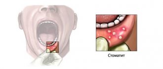
A multisystem autoimmune disease that develops on the basis of a genetically determined imperfection of immunoregulatory processes, which results in damage to many organs and systems. The main signs of SLE are skin changes, nonerosive arthritis, nephritis, encephalopathy, pleurisy or pericarditis, cytopenia (leukopenia and thrombocytopenia), and a positive test for ANA in the blood. There are more than 20 types of skin manifestations in SLE: from erythematous to severe bullous rashes. Most often (in 1/3-1/2 children), especially at the onset of the disease, erythema occurs above the zygomatic protrusions and in the area of the bridge of the nose, resembling a “butterfly”. It is characterized by symmetry and is usually not accompanied by any subjective sensations; worsens with excitement and solar insolation. Erythematous rashes are often located on open parts of the body in places not protected from the sun (face, upper chest), less often over large joints, mainly elbows and knees. Sometimes discoid lesions appear, leaving behind cicatricial atrophy. In the acute period, lupus cheilitis occurs (damage to the red border of the lips). Various vascular changes may also be observed: telangiectasia, palmar, plantar, digital capillaritis, hemorrhagic rashes, as manifestations of vasculitis (petechiae, purpura); livedo reticularis on the skin of the lower, less often the upper limbs and torso. Lupus enanthema or aphthous stomatitis occurs in 1/3 of children.
Scleroderma
In general, the disease is distinguished by clinical polymorphism. The most characteristic is the formation of scleroderma lesions of plaque or stripe shape, localized in various areas, most often on the limbs and torso. The lesions may be hypo/hyperpigmented, with a yellowish or reddish tint. Many children have a fairly pronounced thickening of the skin, which in some of them is combined with thickening of the subcutaneous soft tissues. Most patients have no subjective sensations, but itching and pain may occur. Over time, the lesions undergo a distinct transformation with the formation of residual changes in the form of atrophy and/or skin dyschromia.
In addition to plaque or strip-like skin lesions, rarer varieties are identified: “saber strike” type, Pasini-Pierini atrophoderma, white spot disease, bullous hemorrhagic, etc.
Children with systemic scleroderma have a more pronounced range of signs from the skin and subcutaneous soft tissues, and their relatively greater nonspecificity.
Dermatomyositis. Polymyositis
This is a severe progressive systemic disease of the muscles, skin and microvasculature with less distinct damage to internal organs, often complicated by calcification and purulent infection. The clinical picture includes fever, progressive muscle weakness, up to complete immobility, intense muscle pain, increasing muscular and general dystrophy.
Dermatitis is a pathognomonic sign for the disease, occurs in 3/4 of children with juvenile dermatomyositis, and most often appears within a few weeks of the onset of muscle symptoms. The most typical early skin changes can be considered erythematous spots with a violet tint, primarily in the periorbital (symptom of “glasses”) and perinasal areas, on the eyelids and ears, rashes in the area of the extensor surfaces of the small joints of the hands (Gottron’s papules), equivalents of which are also noted over other joints. Skin symptoms also include erythema with swelling of the periungual ridges, vasculitic spots, livedo reticularis, and ulcerative-necrotic changes. Alopecia and dystrophic changes in the nails may develop. The vascular component is represented by palmar capillaritis, livedo reticularis, and telangiectasia.
Systemic vasculitis
Systemic vasculitis (SV) is a group of diseases characterized by primary destructive and proliferative damage to the walls of blood vessels of various sizes, leading to secondary changes in organs and tissues. Existing classifications of SV take into account the caliber of affected vessels, the nature of inflammation, histological data, and immunological features of the process. Skin phenomena are included in various activity assessment schemes. These include: petechiae/ecchymoses, livedo reticularis, skin necrosis, ulcers, erythema nodosum, nodules along the vessels.
The most significant diseases from the group of systemic vasculitis in terms of the frequency of skin involvement are hemorrhagic vasculitis and polyarteritis nodosa.
Hemorrhagic vasculitis
The most common systemic vasculitis in children, characterized by damage to small vessels with changes primarily in the skin, intestines, and kidneys. Main manifestations: non-thrombocytopenic purpura, arthritis and arthralgia, abdominal pain, gastrointestinal bleeding and glomerulonephritis. In almost half of the cases, a recurrent nature of the process is noted. The prognosis is often favorable.
Almost all patients experience skin changes. Characterized by a symmetrical petechial rash and/or palpable purpura, localized mainly distally on the lower extremities, worsening after the child is in an upright position and observed for several days. These changes are so characteristic that they occupy one of the leading positions among other diagnostic criteria. Along with this, there are erythematous, papular and vesicular elements.
Polyarteritis nodosa
It is a necrotizing vasculitis of peripheral and central arteries of medium and small caliber. Juvenile polyarteritis nodosa is characterized by necrotization of the skin with underlying soft tissues and mucous membranes with the development of polymorphic clinical symptoms. Changes can affect any organ and develop against the background of fairly pronounced general symptoms of the disease (fever and cachexia). In addition, weight loss, skin changes, abdominal and musculoskeletal pain, and involvement of the central nervous system are noted.
Skin changes are observed in more than half of patients and are often one of the first symptoms of the disease. The most typical are painful subcutaneous nodules along the vessels, livedo (reticular or tree-like). Livedo, which occurs in most patients, can persist during periods of remission, becoming more pronounced during exacerbation. Every third patient develops thromboangiitis syndrome with the rapid formation of necrosis of the skin and mucous membranes, gangrene of the distal extremities. Papulo-petechial rashes, vesicles, and bullae may also be observed. The significance of skin changes in the diagnosis of the condition is emphasized by the presence of skin symptoms in the diagnostic criteria of the disease.
Lyme disease
It is a multisystem disease caused by the spirochete Borrelia burgdorferi, which is transmitted by ixodid ticks. Skin changes are represented primarily by tick-borne erythema migrans at the site of the tick bite. Erythema is a red spot growing along the periphery with a diameter of at least 5 cm, homogeneously colored or ring-shaped with clearing in the center. In a third of cases, erythema may be accompanied by itching and pain. Erythema may disappear spontaneously, but progression and chronicity of the process and the appearance of secondary skin erythema are possible. Quite rarely, the appearance of urticarial elements, erythema nodosum, a single benign lymphocytoma of the skin and, in adults, chronic atrophic acrodermatitis are also observed. Other clinical signs include influenza-like syndrome, enlarged regional lymph nodes, manifestations of the nervous system (damage to the facial and other cranial nerves and radiculopathy), heart (atrioventricular block), arthralgia, arthritis, and sometimes eye damage.
Skin changes occur in Kawasaki disease, mixed connective tissue disease, periodic disease and other rare diseases falling under the rheumatological rubric.
Treatment
Treatment of skin changes in most rheumatic diseases comes down to relieving the main symptoms of the disease. One of the leading places in the spectrum of antirheumatic drugs is occupied by glucocorticosteroids. The most intensive method of administering steroids is pulse therapy, in which the dose of the administered drug reaches 20–30 mg/kg per day. The course is three days, repeated administration is indicated if necessary. Doses prescribed orally vary depending on the nosological form and the degree of activity of the process from 1-2 mg/kg (calculated for prednisolone) to 0.5–0.7 mg/kg per day. A dose of 0.1–0.3 mg/kg per day can be considered maintenance. GCS are often prescribed in combination with NSAIDs in standard doses. Among the basic drugs that complement GCS, the drug of choice today (especially for JRA) is methotrexate (calculated dose 10 mg/m2 per week). Azathioprine (1.5 mg/kg per day), cyclosporine A (sandimmune) (3.5-5 mg/kg per day), cyclophosphamide, sulfasalazine, gold preparations, quinoline drugs, and their combinations are also used. A certain role in the treatment of rheumatic diseases is given to immunoglobulin therapy, which is carried out in courses of several cycles of intravenous administration at the rate of 0.4-0.5 g/kg per administration. Recently, there has been a search for new effective and safe drugs, such as necrosis factor inhibitors (Remicade, etanercept, etc.), mycophenolate mofetil, leflunomide, etc.
Local therapy is also used. Applications of DMSO, solcoseryl, indovazin, dollit cream, madecassol, heparin and other local remedies are prescribed. Physiotherapeutic procedures, including spa therapy, also play an important role. In many cases, the treatment complex includes antifibrotic drugs (penicillamine, as well as madecassol, colchicine, enzymes, etc.), glucocorticosteroids, and agents affecting the microcirculation system (pentoxifylline, nicoshpan, etc.). The need to use immunosuppressants (methotrexate, azathioprine, etc.), quinoline derivatives (plaquenil), and systemic enzyme therapy is discussed.
In dermatomyositis, sunscreens, anti-inflammatory and antipruritic drugs, and steroid ointments play a role in the treatment of skin changes. Quinoline derivatives are effective against dermatological manifestations of the disease. If skin changes are resistant, along with the use of GCS and plaquenil, dapsone, mycophenolate mofetil (allows you to gradually reduce the dose of GCS), tacrolimus (topical use for erythematous, edematous changes) can be added to the treatment complex.
Thus, skin changes, often an integral part of a complex picture of disorders throughout the body, occupy a significant place in the practice of pediatricians and pediatric rheumatologists. It is the skin syndrome, first detected at an appointment with a dermatologist or first contact doctor, that should alert you to the possible development of one of the rheumatological diseases.
D. L. Alekseev, Candidate of Medical Sciences N. N. Kuzmina, Doctor of Medical Sciences, Professor S. O. Salugina, Candidate of Medical Sciences Institute of Rheumatology RAMS, Moscow
Causes
The exact culprit behind the development of fibroids in children has not yet been determined. Experts have identified a number of factors contributing to the appearance of tumors. These include:
- unfavorable environmental living conditions (proximity to chemical and cement plants, landfills, contaminated areas);
- frequent injuries, bruises and falls;
- various infectious diseases of acute and chronic form;
- irrational use of potent drugs;
- endocrine pathologies;
- prolonged exposure to direct sunlight.
Allergy to sun or cold
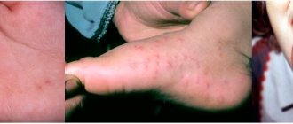
Natural factors can also cause the development of allergic reactions. Photoallergy or sun allergy most often occurs in the warm season - from late spring to early autumn. Sometimes an allergic rash is confused with sunburn, but the allergic nature of the disease is characterized by severe itching, the appearance of small, white pustules, and spotty red rashes even after a short stay in the open sun. In such cases, you need to apply sunscreen, protect exposed skin with clothing made from natural fabrics, and wear a hat3.
Allergy to cold or cold urticaria also occurs. During the cold season, red itchy rashes, peeling, and a feeling of tightness appear on the skin of the face and hands. Symptoms may persist for several hours or several days after exposure to the cold. Typically, allergies develop at temperatures from 10 degrees below zero, but it also happens at temperatures close to zero.
Diagnostics
The examination begins with a visual examination: the doctor listens to the complaints of children and parents, carefully palpates the formation, and collects a detailed medical history. To determine the nature of the lesion, a number of instrumental studies are carried out:
- ultrasound examination of soft tissues;
- radiography;
- computed tomography or magnetic resonance imaging.
A biopsy and cytological examination of biological material help to identify the nature of the pathology and clarify the diagnosis.
Treatment
There are several treatment options, including non-surgical techniques and surgical removal.
The doctor selects the best option based on the nature and location of the fibroid, possible complications, and the wishes of the patient or his parents. Not in all cases it is necessary to monitor the growth of fibroma: if it has a small diameter, does not interfere, does not pose a danger and does not cause cosmetic defects, it is left.
Direct indications of the need to remove tumors are:
- constant injury and irritation of fibroids;
- location on the face, neck, and other areas where the formation causes aesthetic discomfort;
- infection of fibroids and surrounding tissues;
- circulatory disturbance in soft tissues caused by twisting of the formation leg.
Non-surgical treatments include:
- injections of steroids, which leads to resorption of the tumor and subsequent tissue scarring;
- cryodestruction or exposure to cold, which helps to significantly reduce the size of large elements.
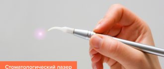
If conservative treatment is impossible or such tactics have not brought results, surgical intervention is indicated. In modern practice, fibroids are excised using a classic, laser or electric scalpel. The procedure takes no more than 60 minutes, is performed under general or local anesthesia and requires minimal preparation. In this case, hospitalization is not carried out; after removal of the tumor, the child is observed in the hospital for several hours and sent home. The rehabilitation period lasts up to 2 months.
Prevention
There is no specific prevention that can eliminate the likelihood of fibroids. You can reduce the chance of neoplasms forming by carefully monitoring the health of your children.
- It is necessary to bring children regularly for preventive examinations.
- It is advisable to avoid prolonged exposure of children to direct sunlight, especially during the period of greatest sun activity.
- It is necessary to promptly and correctly treat infectious diseases, injuries, burns and other damage to the skin or internal organs.
You should not give children medications without the instructions of doctors, or try traditional and non-traditional treatment methods. It is also important to ensure that babies eat properly and get enough physical activity on a daily basis.
Fibroids do not always pose a danger to the body, however, they must be kept under control. Doctors at the SM-Doctor clinic will carefully examine the child and select the optimal method of treating the disease.
Diet
Diathesis in children responds well to treatment if the recommended diet is followed.
During breastfeeding, the baby's mother should adhere to it. Allergenic foods (sweets, citrus fruits, eggs, honey, etc.) are excluded. During the introduction of complementary foods, in order to avoid the development of diathesis, the baby’s skin reactions to the new product should be monitored, and if signs of illness appear, exclude it for three to five months, until the next attempt. After the introduction of complementary foods, the basis of the menu should be cereals, lean meat and fish, vegetables and fruits. The consumption of sweets and foods high in food additives should be limited as much as possible. Contact us!
Specialists at the PsorMak Skin Diseases Center have been successfully treating diathesis in children for many years. Contact us for an initial consultation, our doctors will help you develop a complete understanding of how to properly treat the disease in the acute stage (with skin rashes). In addition, we will help you create a dietary diet and suggest what other measures to correct the child’s lifestyle should be taken to improve his health and effectively combat the disease. Make an appointment by calling +7 (495) 150-15-14,