54077
Crooked, unhealthy teeth can ruin the appearance of an otherwise attractive person. Oral health is important not only for our attractiveness, because the main function of teeth is to grind food. The condition of the stomach and intestines depends on this, which directly affects health and life expectancy .
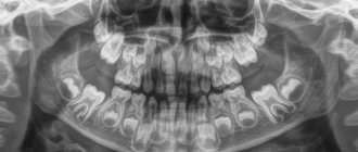
Teeth of ancient people
The dentofacial apparatus of prehistoric and modern humans differs significantly. Ancient people had more than 36 teeth, protruding fangs and a massive jaw. This was explained by the need to chew rough food and raw meat. With the addition of thermally processed foods to the diet, the dentition began to change. The canines were the first to transform, becoming aligned with the bite line. Then the jaw arch narrowed, the interdental spaces disappeared, and the teeth themselves decreased in size. Currently, 32 teeth in humans are the norm, but third molars are considered to be an atavism.
Interesting fact!
The teeth of ancient man cannot be called aesthetic, but they were healthy. According to scientists, cavemen never suffered from caries and other oral diseases.
Diagnostics
The main goal of any diagnosis is to identify the cause of the problem. And make a diagnosis. Likewise, the diagnosis of distal occlusion is designed to identify the cause of closure in the second class. In order to separate the flies from the cutlets during treatment, so to speak. That is, dental causes of closure are in the second class from non-dental (maxillary, skeletal).
The main diagnostic methods are:
- Analysis of TRG (teleradiogram) of the skull in a lateral projection. It allows you to clearly determine all the skeletal (jaw) nuances of the problem. Both with the position of the jaws and with their sizes.
- Analysis of plaster models of jaws (for this, casts are taken). It allows you to clarify the “dental” nuances: the size of the teeth, the length and width of the dentition, the features of the relationship (including closure) of the upper and lower teeth
Analysis of TRG (teleradiogram) of the skull in a lateral projection.
Analysis of plaster models of jaws.
It is not recommended to fall below this diagnostic “bar”. Below - just by eye. But this, as we understand, is not the best option.
Diagnostics is the first and key step in the treatment of distal occlusion. Do you want to know why it’s impossible to do without diagnostics?
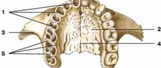
Name of human teeth
Depending on the location and structure, dental units have their own functional characteristics and are called differently.
- Incisors.
On both jaws there are four front teeth in humans - medial and lateral incisors, which are used for biting food. - Fangs.
Sharp teeth designed for chewing hard foods. - Premolars.
"Fours" and "fives" on the left and right sides of each jaw arch grind soft or small pieces of food. - Molars.
Three large outer teeth in each row are aimed at grinding coarse substances. - The canines
and incisors are part of the anterior group, or the “smile zone,” and the human molars are part of the chewing segment.
In addition, teeth are divided into temporary and permanent. In the first case, we are talking about dairy products that appear in children from the fifth month of life to three years. The second refers to the final bite, which is formed between six and thirteen years of age. Milk teeth differ from permanent teeth only in size, but in structure they are identical.
Anatomy of the human jaw
The human maxillofacial region has the following anatomical structure:
- mouth slit,
- vestibule of the oral cavity,
- cheeks,
- lips,
- solid sky,
- soft sky,
- language,
- gums,
- teeth,
- upper and lower jaw.
The oral fissure is limited by the lips, which represent the orbicularis oris muscle and subcutaneous fat.
The cheeks are formed by adipose tissue (Bishat's lump) and bundles of buccal muscle. In the projection of the crown of the upper second molar on the inside of the cheeks there is a papillary elevation of the mucous membrane.
The excretory duct of the parotid salivary gland opens from the papillary eminence.
The oral cavity, cheeks, upper and lower gums and teeth form the vestibule of the oral cavity.
Gums are alveolar processes of the upper and lower jaws, covered with mucous membrane, which cover the teeth in the cervical area.
The mucous membrane of the mouth and the enamel of the teeth are constantly moistened with saliva, which is secreted by paired parotid, sublingual and submandibular glands, as well as many small glands, in a volume of up to 1.5 liters per day. Saliva contains organic and inorganic substances, it contains about 18 amino acids, 50 enzymes, mucin, substances with antibacterial activity (leukins, opsonins, lysozyme).
Saliva promotes the maturation of enamel, remineralization, has a cleansing effect, antibacterial activity and at the same time favors the formation of plaque and tartar.
The hard palate is formed by the palatine processes of the upper jaws and the perpendicular processes of the palatine bones.
The soft palate is formed by muscle fibers covered with a mucous membrane with a large number of mucous glands. On the sides of it there are arches - the palatine lingual and the velopharyngeal, between which there are accumulations of lymphoid tissue, the so-called palatine tonsil.
The tongue is a muscular organ covered with a mucous membrane. Its structure is divided into a wide rear part - root, body, middle part and apex. On the upper mucous membrane of the tongue, there are four types of papillae containing taste buds: filiform, leaf-shaped, mushroom-shaped, and rough.
Upper jaw
The upper jaw is a paired fixed bone. Its structure includes the body, palatine process, frontal process, zygomatic process, and alveolar process.
The palatine process takes part in the formation of the hard palate, the frontal process participates in the formation of the orbit, the alveolar process carries the sockets of the teeth - alveoli, and the zygomatic process attaches the zygomatic bone.
In the body of the upper jaw there is a cavity - the maxillary sinus, containing air and lined from the inside with mucous membrane. In close proximity to it are the apexes of the roots of the large molars (especially the sixth). Therefore, there is a high probability that the inflammatory process that has arisen in the tooth and periodontal tissues can easily spread to the sinus - sinusitis will develop.
Lower jaw
The lower jaw is an unpaired movable bone, shaped like a horseshoe. Its structure includes: a body with dental alveoli; two branches ending in condylar and coronoid processes; the condylar process, which connects with the articular fossa of the temporal bone, participates in the formation of the temporomandibular joint, due to which movement in the lower jaw is carried out.
According to data from dental reference books
A full range of dental services in Istra for adults and children: from consultation to complex operations within one clinic “Doctor Nebolit”
Consultation and appointments daily from 9:00 to 19:00
- +7 (49831) 4-42-12
- Contacts
How many teeth does a person have?
The number of teeth a person has depends on age and anatomical features. The child has a set of 20 primary teeth, which are replaced by a permanent bite of 28 teeth. Third molars erupt, as a rule, after twenty years or do not grow at all, which is not a pathology.
In dentistry, a single numbering of human teeth is adopted. Doctors classify teeth as lower and upper and distinguish the right and left segments of the jaws. Each of them includes two incisors, a canine, two premolars and three molars. The countdown starts from the first front tooth and ends, accordingly, with a figure eight. Sometimes a number is added to the serial number indicating the location zone. For example, the right canine of the top row is numbered 13. This order in the schematic representation is called the formula of human teeth.
Polyodontia
In rare cases, an anomaly such as polyodontia is observed - supernumerary, or extra teeth in a person. Dental units can appear in the primary and permanent dentition anywhere in the jaw, separate from or fused with the main teeth. The defect affects not only the aesthetics of the smile, but also leads to the formation of incorrect occlusion, impairs the quality of chewing food and diction. Most often, supernumerary teeth are removed in childhood or built into the dentition.
Edentia
There is also a deviation of the opposite meaning called edentia - congenital or acquired absence of dental units. The causes of the phenomenon include heredity or improper development of the embryo in the womb. People without teeth cannot fully eat and speak, have a deformed facial contour and weakened immunity.
Causes of anomalies
Let's look at the main reasons why defects occur:
- Genetic factor. Mesial and distal occlusion are most often inherited.
For parents, knowing about the high risk of such a defect occurring in their child, it is easier to control treatment in childhood, during the formation of the maxillofacial system. - Developmental anomalies in the prenatal period. Various pregnancy pathologies can often affect the formation of the fetal dental system.
- Birth injury. Mesial occlusion is caused by displacement or dislocation of the baby's lower jaw during difficult childbirth.
- “Wrong” habits in childhood. These include constant pacifier or finger sucking, improper nipple latching, and improper sucking during bottle feeding. If the hole in the nipple is too large, the child’s lower jaw practically does not work when sucking and remains undeveloped.
- Frequent sinusitis and rhinitis, due to which the child constantly breathes through his mouth. With such breathing, the development of facial bones is disrupted.
- Violation of tooth change. Early removal of baby teeth often causes abnormal maxillofacial development.
- Incorrect prosthetics, lack of prosthetics.
- Hypertonicity of the masticatory muscles due to stress provokes abrasion of the incisors and displacement of the jaws.
- Various injuries of the maxillofacial area.
Dimensions of human teeth
The upper central incisors are twice as wide as their antagonists. The remaining dental units of the same name have approximately equal parameters. The size is determined using special tables with the optimal size and permissible deviations. Experienced doctors calculate proportions by dividing the length of a person’s teeth by the width. A result of about 0.75 millimeters is considered close to ideal. For more detailed diagnostics, other professional formulas and techniques are used.
Size deviations from the norm occur due to improper formation of the jaw, fusion of tooth buds, or genetic predisposition. Teeth that are too large are called macrodentia, and abnormally small teeth are called microdentia. Pathologies are accompanied by problems with bite and chewing functions, but can be successfully corrected by a dentist.
Interesting fact!
The longest tooth in the world belongs to an Indian teenager. The size of its crown is almost four centimeters. About a year ago, the tooth was removed, and the young man was included in the Guinness Book of Records.
The structure of the human tooth
Anatomy
From an anatomical point of view, a human tooth consists of three parts.
- Crown.
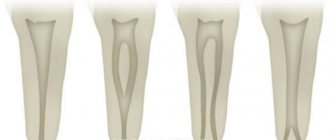
The visible part protruding above the gum. It has four sides: the occlusal, or cutting edge, in contact with the antagonist teeth; contact wall adjacent to adjacent dental units; vestibular and lingual surfaces facing the lips and tongue, respectively. - Root.
Fixed in the socket by connective tissue, located in the recess of the jaw. As a rule, premolars have two roots, and molars have three, four or even five. The remaining dental units have one root canal. - Neck.
It is located between the coronal part and the root of a human tooth, surrounded by periodontium.
Histology
What are human teeth made of? Let's look at the cross-section of the structure of a human tooth.
- Enamel.
A transparent protective coating of the crown, almost entirely consisting of inorganic microelements. - Dentine.
The hard base of the tooth, containing 80% mineral components and 20% organic substances. The shade of dentin is responsible for the color of dental units, as it shines through the enamel. - Cement.
The bone tissue covering the tooth root. Plays the role of a fastening element connecting the tooth to the alveolus. - Pulp.
Soft tissue filled with bundles of nerves and capillaries. Painful sensations during caries are explained precisely by the presence of nerve endings.
Baby teeth
Their formation in a child begins to occur in the womb at the twelfth week . As a rule, the first to appear in a child are the incisors and canines, and only at the very end the molars.
The timing of this process is purely individual and can vary, but in most cases, the formation of the primary bite begins to occur at the age of seven months and ends at three to four years. By this time, the child should have twenty baby teeth.
Compared to permanent teeth, milk teeth have their own characteristics:
- smaller sizes;
- fewer chewing tubercles;
- the roots spread out to the sides.
Despite this, primary and permanent teeth have the same number of roots.
The deciduous row in the jaw consists of ten teeth: four molars, four incisors and two canines. At the age of six or seven years, baby teeth begin to fall out and are replaced by permanent ones.
First of all, the large molar is replaced, and the final formation of the row ends by the age of twelve to fourteen, with the exception of the third molar.
If you find an error, please select a piece of text and press Ctrl+Enter.
Tags: tooth structure
Did you like the article? stay tuned
Previous article
Photos of the smiles of stars and ordinary patients BEFORE and AFTER installation of veneers
Next article
What to do if a wisdom tooth grows into the cheek?
Human wisdom teeth
A “wisdom tooth” is the third outer molar with three to five roots. In structure it is no different from its “neighbors”. To the question “How many wisdom teeth does a person have?” cannot be answered unambiguously. They erupt around the age of twenty, one on each side of both jaws. However, there are people without wisdom teeth. This is a variant of the norm, since in the process of human evolution the need for the “eight” disappeared, and the structure of the jaws underwent corresponding changes. Today, third molars are considered a vestigial organ.