Summary
Reconstruction and rehabilitation of patients with defects of the upper jaw are the most difficult in reconstructive maxillofacial surgery, occupying the minds of oncological surgeons working in this area, microsurgeons, plastic surgeons and orthopedic dentists involved in maxillofacial and somatotropic prosthetics.
The fundamental difference between patients who have undergone anatomical reconstruction of the alveolar process of the upper jaw, as well as nasal support with microsurgical grafts and patients who have undergone maxillofacial prosthetics with obturation structures is an increase in the volume of the respiratory space, improved speech, the absence of atresia, frequent acute respiratory viral infections and, fundamentally, in the absence of regular relocation of definitive orthopedic work [1]. An important difference is also the lack of mobility of dentures on dental implants, which avoids chronic trauma to surrounding tissues.
This article describes the experience of rehabilitation of patients with defects of the upper jaw using fibular and Chinese skin-bone flaps with further plastic surgery of bone and soft tissues in order to create conditions for dental implantation, orthopedic conditions and features of fixed prosthetics for this group of patients.
The developed protocols for the use of a particular graft on a vascular pedicle are described in terms of the type of defect and further restoration of bone joints, as well as measures for the formation of anatomically close to normal conditions for implantation and prosthetic dentistry in patients with a reconstructed upper jaw.
The nuances of implantation in the upper jaw
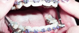
The requirements for the front teeth located in the smile zone are quite high. New teeth should look natural and beautiful, not stand out from others. When planning an operation, the dentist pays attention to the following parameters: 1. Gingival contour. Patients turn to the dentist much faster if one of the front teeth has been lost, since the resulting gap not only causes discomfort, but also disturbs the appearance. Therefore, the condition of the gums allows you to create a beautiful and smooth gum contour without additional procedures. When implanting upper front teeth, dentists often resort to one-stage implantation with simultaneous installation of a crown. A temporary crown allows you to correctly form the gingival contour and prepare the gums for the installation of a permanent prosthesis. If additional procedures are required for implantation, and the operation itself takes place in two stages, then patients are recommended to make an individual abutment and gum former for the front teeth. The gum former is installed in the upper part of the implant for 2 weeks, after which it is replaced with an abutment with a crown. If you use standard components, a gap may form between the gum and the crown. Food particles will accumulate in it, causing an unpleasant odor and inflammation. The gums on the front teeth may sag, exposing the head of the implant. Any visual defects reveal an artificial tooth; the result does not satisfy the patient, and over time can lead to plaque formation and inflammation. Therefore, the issue of aesthetics is also additional protection of the tooth from peri-implantitis.
2. Type of crown. When drawing up a treatment plan, dentists recommend using zirconium or all-ceramic crowns in the area of the front teeth. These materials are highly durable and at the same time, they visually replicate the transparency of enamel and natural shine. Only high-quality materials can convey the transparency of a tooth, so the orthopedic part of implantation of the front teeth will cost slightly more than on the lateral and chewing teeth. To make a ceramic or zirconium crown look natural, it is placed on a ceramic or zirconium abutment. The metal abutment gives a dark gray tint to the tooth and looks unaesthetic.
3. Implant installation. The alveolar ridge must have a certain thickness in order for a sufficiently strong support to surround the implant. Implantation must be as precise as possible. Whenever the implant deviates from the intended trajectory, the bone tissue wall loses strength. If there is excessive load, an incorrectly installed implant can injure surrounding tissues, become loose or fall out. To avoid inaccuracy and medical error, a surgical template is used, which maintains the desired position and prevents deviation into the area of softer tissues. Computed tomography helps to study the bone structure in detail and guide the implant in the right direction, bypassing the anatomical structures of the jaw.
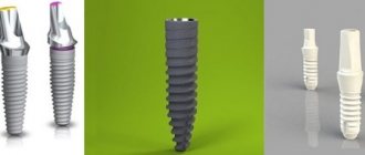
4. Type of implant. For implantation into the bone tissue of the upper jaw in the frontal region, implant models with a special thread design are used. A minimally invasive effect on surrounding tissue preserves the biomaterial. Implants are smaller in length and diameter, which allows you to accurately replace your own tooth root with an artificial one.
Introduction
The upper jaw is the most complex area in reconstructive maxillofacial surgery, being more responsible for appearance than others, and due to the presence of such organs and anatomical formations as the eyes, zygomatico-orbital complex, and teeth.
The upper jaw is a support for the lower jaw from the point of view of the stationary container of antagonists, also a zone of concentration of air cavities and a complex of buttresses. Defects and deformations in this area lead to disfigurement and limit or make impossible vital body functions such as chewing, swallowing, breathing, and vision. Restoration of buttresses is necessary from the point of view of optimizing the transfer of stress of the masticatory load, as well as to strengthen the reconstructed alveolar process of the upper jaw. [3]
Despite the positive results of tissue autotransplantation using microsurgical techniques, creating conditions for fixed and conditionally removable dental prosthetics remains a serious problem. The presence of a skin area with an array of subcutaneous fat makes the creation of an orthopedic bed and the attachment of soft tissues around the completed orthopedic structures almost impossible and reduces the functional significance of the work performed aimed at the rehabilitation of the chewing function. [2]
The article is devoted to the synergistic efforts of a maxillofacial surgeon and an orthopedic dentist to create functionally significant conditions and long-term results in patients with reconstructed upper jaws using skin-bone autografts on a vascular pedicle.
In the world literature available to us on the rehabilitation of chewing function in patients with jaw defects, we did not find any descriptions of the formation of a prosthetic bed, features of the choice of orthopedic structures, or long-term results after dental prosthetics.
Key words: jaw defect, lack of occlusion, obstructive prostheses, dental implantation, fixed prosthetics, integrated implants, micro-surgical autotransplantation, bone grafts, fibula, parietal bone, three-dimensional bone augmentation.
Material and methods
From 2008 to 2014 in the reconstruction of the upper jaw, we used a fibular autograft for the reconstruction of total defects (5 cases) and a radial autograft for the reconstruction of subtotal defects (10 cases). When using a radial skin-bone autograft in the subsequent placement of dental implants, there is a need to recreate the second cortical-spongy layer of the alveolar process on the lingual side, which can be achieved using parietal or mandibular free autoblocks.
With VRGN, relatively small defects of the alveolar process of the frontal part of the upper jaw are noted; more often the problem is the elimination of the defect of the palatal plate. Bone reconstruction requires the alveolar process, represented by genetically modified bone tissue in the projection of the bone cleft. A palatal bone defect does not require repair, since functional loss does not occur in the absence of reconstruction of the bone component. It should be noted that with complete clefts the main problem is communication with the nasal cavity. To eliminate defects of the upper jaw, we have developed a technique for using a free split chin graft and transplanting a fasciocutaneous graft [Application: 2010118686/14, 05/12/2010. Patent No. 2435537 ](4 cases).
In cases where the patient's health condition did not allow microsurgical transplantation, we installed Zygomatic system implants and fixed prosthetics (6 cases).
After a clinical assessment, instrumental diagnostic methods were used to study the pathology of the integrity of the jaw/s: OPTG, TRG, CT (with angiocontrast and soft modes).
If a tumor was present, the extent of resection was assessed and the optimal graft was selected according to the developed algorithm. If the X-ray picture of the tumor was detected, a biopsy was performed and, when the diagnosis was verified, resection of part or all of the jaw was performed. For jaw defects, after clinical and instrumental analyses, treatment planning began.
Preoperative planning was carried out using 3D visualization programs that made it possible to simulate the sizes, shapes, and position of the revascularized autografts relative to the bone structures, taking into account the positioning of the condylar processes of the mandible in the temporal fossae (anterior-superior position in the articular cavities) according to CT studies and the formation of anatomical buttresses for the upper jaw. A special feature of the programs is the absence of distortions in the individual dimensions of the patient’s skull.
We used fibular and radial autografts, since only they allow us to perform 3D modeling of the bone component congruent with the defect in the maxillary defect. Subsequently, when using a radial graft, the technique of transplanting free cortical-cancellous grafts was used to thicken the alveolar process for further dental implantation. Thus, a comprehensive and step-by-step reconstruction of the jaw defect was carried out to create conditions for restoring chewing function with fixed and conditionally removable orthopedic structures, which is important for any jaw defects.
After engraftment and activation of the dental implants, the subcutaneous fat pad of the fibular and radial flaps and the removal of excess skin pad were removed.
Orthopedic rehabilitation consisted of choosing the design of the prosthesis and precise adherence to the accuracy of taking impressions to transfer the complex relief of soft tissues and the position of the implants onto precise plaster models. The usual occlusion and central relationship of the jaws were recorded for the subsequent selection of prosthetic tactics for patients with reconstructed upper jaws. Plaster models were installed into the articulator using a facebow. The existing occlusion and centric relation were assessed. Depending on the type of defect, we selected the desired position of the lower jaw to create optimal chewing function and aesthetic results.
Particular attention was paid to the creation of a prosthetic bed for a future orthopedic design supported by dental implants. In these clinical situations, there is no attached keratinized mucous membrane around the implants in the oral cavity, which makes it difficult to accurately transfer the relief of the soft tissues of the prosthetic bed to create adapted non-removable orthopedic structures.
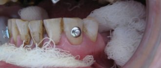
After the manufacture and fixation of temporary crowns on the implants, the presence of tumor-like growths of the mucous membrane around the installed orthopedic superstructures, which were histologically described as polyps, was noted. Patients complained of bleeding and discomfort around the installed crowns. Attempts to change the eruption profile of structures from implant shafts did not produce a positive result.
We used a method of simultaneous surgical correction of the subcutaneous fat area of autografts with the installation of provisional structures supported by dental implants. In a number of cases, we used a removable compression acrylic plate on the gum formers, which held the surgically created profile until permanent prosthetics or installation of provisional fixed and conditionally removable structures.
After 3 months, we made permanent orthopedic fixed or conditionally removable structures.
For the manufacture of orthopedic structures, we used cobalt-chrome alloy, titanium and zirconium dioxide.
As a rule, after the integration of implants and placement of gum formers at the prosthetic stage, we were faced with the need to modernize the prosthetic elements of the implantation system for specific clinical cases.
The gum formers were needed to be longer and predominantly conical in shape. The cylindrical shape of the gum former created parallel walls of the crater of the mucous membrane, which caused rapid collapse of the walls during the placement of the impression coping and caused severe discomfort to patients during manipulations. We attribute this to the presence of excess connective tissue layer, which is located on the autograft bone.
It is important to note that immediately after corrective operations on the skin-fat part of the flaps, orthopedic compression plates were fixed to prevent further growth of the soft tissue component.
Patients were monitored every seven days and appropriate compression plate adjustments were made.
Depending on the chosen permanent structure, a set of further orthopedic manipulations was carried out to make prostheses.
The results were recorded using a Canon D 60 camera, 100 mm lens, and MR-100 ring flash.
Description of clinical observations
Patient U. , 18 years old, was admitted to the clinic on April 1, 2008 with a diagnosis of osteoblastoma of the upper jaw, a condition after subtotal resection of the upper jaw on the left. From the anamnesis: a neoplasm was discovered and removed in early childhood at the Federal State Institution Central Research Institute of Infectious Diseases and Maxillofacial Surgery. The patient was sent to the clinic of the Russian Scientific Center for Surgery named after. acad. B.V. Petrovsky.
Status localis: the configuration of the face in front and profile is changed, the upper lip is retracted on the left. On palpation, a defect in the alveolar process of the upper jaw on the left is noted.
Mouth opening is not limited, there is a through defect in the alveolar process of the upper jaw with teeth 21-28, the patient wears an obturating denture, the denture teeth are in the bite.
Bite according to the second class according to Angyu.
On OPTG and CT: there is a defect of the zygomatic tubercle, alveolar process on the left, absence of the spine of the upper jaw, the base of the pyriform foramen on the left, defect of the tubercle of the upper jaw on the left. An impacted 28th tooth is noted in the remains of the pterygomaxillary articulation.
Treatment tactics: since the defect of the upper jaw was subtotal and through, a radial skin-bone graft on a vascular pedicle was used. When transplanting a radial cortico-periosteal-skin graft, a zygomatic-maxillary buttress was formed using a free split graft from the branch of the mandible on the right. The second stage involved the formation of the alveolar process using parietal grafts.
Rice. 1. Appearance of the patient, existing defect of the upper jaw, bite, obturating prosthesis, CT scanThe patient was noted to have no nasal lining, and to anatomically restore aeration, before transplanting a microsurgical graft, the nasal lining was formed with local tissues.
Rice. 2. Stages of planning the bone and soft tissue components of the graft Rice. 3. Stages of surgeryRice. 4. Computed tomography of the patient after surgery and condition on the third day
Rice. 5. Scintigram after surgery
In the postoperative period, there was a divergence of the sutures and a reduction in the volume of the flap, and therefore a protective mouth guard was made to protect the soft tissue component of the flap from food getting into the graft. Kappa also pressed the flap into the defect area to allow healing by secondary intention.
Rice. 6. Postoperative mouth guard
2 months later, the patient, being a cycling athlete, fell from the “saddle” and received a fracture of the upper limb at the site of collection of the radial skin-cortical-periosteal graft on the vascular pedicle. Therefore, the patient underwent osteosynthesis with the Ilizarov apparatus.
Rice. 7. Condition after osteosynthesis of the upper limb on the left, condition in the oral cavity Rice. 8. 3D reconstruction of the alveolar process using parietal grafts, CT scans after the second operationAfter 5 months, we installed three dental implants in the area of the reconstructed alveolar process using parietal bone autografts. Thanks to this technique for reconstructing the alveolar process, it was possible to obtain adequate bone thickness and height for placing dental implants.
Rice. 9. Implantation in the area of 3D reconstruction with parietal blocks
After 5 months, we opened the implants and applied a method of one-stage surgical correction of the subcutaneous fat pad of the autograft with the installation of elongated gum formers in dental implants, which simplified the formation of soft tissues during surgery.
After completion of the surgical stage, the alginate impression was removed from the upper jaw and a compression plate was made using cold polymerization at a pressure of 3 atmospheres from acrylic plastic.
The patient was advised to wear the plate constantly, removing it only for hygiene procedures. Inspection and correction of the plate fit were carried out every seven days for a month.
Thus, we were able to form a stable mucosal contour around the gingival formers.
The next stage was the production of a conditionally removable prosthesis supported on a beam structure on implants.
Rice. 10. Correction of the subcutaneous fat area of the autograft
Considering the fact that microsurgical elimination of the jaw defect and reconstruction of the alveolar process was carried out by transplantation of free parietal cortical autografts, the further stage of orthopedic rehabilitation is practically no different from classical prosthetics for patients with an extended terminal defect of the dentition.
In order to be able to produce a conditionally removable beam-type prosthesis supported by implants, it was necessary to produce an accurate plaster model that displayed the entire relief of the prosthetic bed and accurately reproduced the position of the implants. To do this, we took primary impressions of the upper jaw using Clip-transfers for implants. The primary plaster model gave us the opportunity to make a custom impression tray with prepared transfer checks for precise transfer of the position of the implants.
Having made an accurate working model of the upper jaw and a model of the antagonists, we installed them in the articulator according to the average parameters. Registration of the central ratio was carried out using an occlusal hard wax plate with refinement on ALUWAX (soft wax with the addition of aluminum filings for long-term heat retention and elasticity).
Rice. 11. Impressions and plaster models
After analyzing the relationship of the plaster models in the articulator, we manufactured a beam frame supported by three implants. The beam frame was made by vacuum casting from cobalt-chrome alloy. To achieve a passive fit of the frame, we used adapters for external connection with the implants.
We fixed titanium ball locks into the cast part of the frame. Then a model of the upper jaw was made from a fire-resistant mass for modeling and casting the mating part of the prosthesis itself.
Rice. 12. Beam frame and mating part of the prosthesisTaking into account the possibility of changing the relief of the soft tissue component of the autograft, the frame of the response part was modeled in such a way that it was possible to change the acrylic base part, changing the fit of the prosthesis to the bed.
After fitting the beam and frame in the oral cavity, we manufactured the base part of the conditionally removable denture with the installation of acrylic artificial teeth, using the method of cold polymerization of plastic under a pressure of 3 atmospheres and a temperature of 50 degrees Celsius. We took into account all possible characteristics of the patient at rest and when smiling, creating the most natural appearance of the entire orthopedic restoration.
The final fixation of the prosthesis was carried out in a certain sequence:
- Cleaning and disinfection of all components of the prosthetic restoration.
- Removing the gum formers, irrigating the internal shafts of the implants with a solution of 3% hydrogen peroxide and 0.05% chlorhexidine solution, installing adapters on the external connection.
- Installation of the beam structure on three implants, tightening the fixing screws with a force of 30 N/cm2.
- Fixing the actual removable denture on the beam frame, checking the occlusion, teaching the patient about hygienic measures.
Microsurgical operations of maxillofacial surgery
Microsurgery is a universal method used in almost all surgical disciplines to eliminate a defect in a particular tissue. In the human body, there are more than 400 donor sites with an axial type of blood supply, from which taking tissue does not cause any harm to the further blood supply of the entire organ. In the reconstruction of the jaws, the main place is played by the property of the flap with the presence of such a quantity and quality of bone that will make it possible to recreate the anatomical integrity of hard tissues and in the future the possibility of use for dental implantation and prosthetics .
For total and subtotal defects of the mandible, a fibular skin-bone flap . The fibula has sufficiently large supporting properties, having a fairly large amount of cortical component, and can be easily modeled to recreate anatomical contours. An iliac or otherwise inguinal skin-bone flap is optimal for the reconstruction of small defects of the lower jaw; in the latter cases, it is sometimes possible to use a free iliac bone.
For total and subtotal defects of the upper jaw, radial skin-bone and fibular skin-bone flaps are optimal. In cases where the defect covers the zygomaticomaxillary buttress in combination with a defect in the alveolar process, it is possible to fill the bone defect with corticocancellous parietal grafts and mandibular grafts taken from the ramus and mental region with free grafts of mesenchymal origin, which is optimal for reconstruction of the middle zone faces.
Of course, the dominant algorithm remains aimed at engrafting the flap on a vascular pedicle. If you have to choose between convenience for subsequent dental implantation in the form of the presence of a cortical plate of the bone component of the autograft in the area of the recreated alveolar process for subsequent implantation and this somehow reduces the reliability of the vascular anastomosis, it is necessary to choose the reliability of engraftment. Disputes often arise between implant surgeons and reconstructive surgeons about how the iliac bone should be positioned in the defect area, but experience shows that there can be no compromises, since in case of thrombosis of the vascular anastomosis, the entire flap is lost. It is also necessary to understand that the location of the spongy component in the area of the alveolar process does not interfere with subsequent implantation, since a new one is formed within 6 months.
To return and normalize chewing function and the ability to eat, it is necessary to have:
- 1. Lips and complex of swallowing organs.
- 2. Presence of jaws.
- 3. The presence of teeth and a bite that provides adequate chewing.
- 4. Presence of buttresses.
- 5. Stabilization of the TMJ.
- 6. Synchronicity of the masticatory muscles.
- 7. Mental balance of the patient.
In the literature, we did not find a specific algorithm or approaches aimed at returning such vital abilities as sucking and swallowing, chewing and normalization of speech function. To normalize the patient's nutrition, the presence of lips is necessary, otherwise salivation occurs, followed by maceration of the skin and the inability to receive and send food into the esophagus. Patients with defects of the soft tissues of the perioral area constantly suffer from gastritis, inflammation of the oropharynx, since the vacuum property of food evacuation into the esophagus is reduced, many adapt to swallow with an open mouth, throwing their head back; in the latter, the absorption of carbohydrates is impaired, since saliva takes part in cleansing the oral cavity from food residues, plaque and bacteria, thanks to its buffering properties, it neutralizes the negative effects of strong acids and alkalis within the buffer capacity, provides the supply of ions necessary for the remineralization of teeth, and has antibacterial, antifungal and antiviral properties. Claude Bernard proved that we recognize the functions of an organ by identifying the consequences of its absence. From the point of view of the functional return of the ability to eat, the second place in our algorithm is the presence of jaws. To eliminate defects in part or the entire jaw, we use preoperative planning.
Preoperative planning of the jaws is carried out using 3D visualization programs that allow modeling the sizes and shapes of autografts, taking into account the positioning of the condylar processes of the lower jaw in the temporal fossae (in the anterior-superior position in the articular cavities) according to CT scans. Despite the fact that we perform reconstruction in the previous bite, most often we have to deal with an already remodeled TMJ and a broken bite. Therefore, planning is necessary taking into account the subsequent achievement of centric occlusion in the centric relation. A functional study of occlusion consists of comparing central occlusion (CO-occlusion in which there is maximum contact between the teeth of the upper and lower jaw) and the central ratio (CO-condition in which the heads of the lower jaw occupy an anterior-superior position in the articular sockets). If there is a significant difference, a description of the differences is required. Thus, a significant difference between these conditions is more common with asymmetric deformations and with Angle class II deformities. Determining the CA is important for correctly drawing up an operational plan.
When planning the elimination of maxillary defects, we take into account the need to restore the buttresses, as well as the air supply of the upper jaw. Buttresses are the most important component for supporting the alveolar process, otherwise mobility of the upper jaw occurs after prosthetics.
Although restoration of the lost maxillary cavity and mucous lining is not possible, restoration of anatomical proximity is necessary, therefore filling the zygomaticomaxillary buttress with ilium is incorrect. It is optimal to use free split mandibular or parietal auto-bone blocks in shape in combination with bone with vascular nutrition. In the future, perhaps with the development of technologies for the use of stem cells, we will learn to restore the true mucous lining of the maxillary or paranasal sinuses.
In the reconstruction of the upper jaw, we use a fibular autograft for the reconstruction of total defects and a radial one for subtotal defects. When a radial skin-bone flap is subsequently used to place dental implants, it becomes necessary to reconstruct the second cortical-spongy layer of the alveolar process on the lingual side, which can be done using parietal or mandibular free autoblocks.
In maxillofacial microsurgery , when it is necessary to recreate bone curves, in the choice of grafts with bone, we are limited to iliac, fibular, and radial flaps, since only the latter allow 3D modeling of the bone component congruent with the defect.
of one-stage reconstruction after resection still remains open in the country . It is necessary to understand that if the reconstruction is not performed simultaneously with the resection of the jaw and even a temporary titanium structure is not applied to maintain the bite, remodeling of the temporomandibular joint occurs, both from the healthy and from the pathological side, and a violation of the trophism of the masticatory muscles. In these cases, before placing dental implants and before prosthetics of the implant, orthodontic fixation of the bite is performed in the form of braces and mini-implants, as well as muscle relaxation of the masticatory muscles on the part of the healthy jaw using Botex therapy or myotronic.
HISTORY OF THE PROBLEM
In our opinion, microsurgical autotransplantation for the purpose of jaw reconstruction has gone through several stages in its development:
Stage 1 – high-quality autograft collection, minimal modeling, and transfer to the recipient area. The main objective of this stage was to ensure graft engraftment (from 1978 to 1990).
Stage 2 included better modeling of autografts using conventional radiographs and wax templates. The main task of this stage was to restore facial aesthetics. The impaired function of the lower jaw was restored after a series of additional corrective operations and removable dental prosthetics (from 1990-1995). Stage 3 - computer modeling of the lower jaw and restoration of chewing function using prosthetics using dental implants. The main goal of this stage is to restore facial aesthetics and chewing function of the lower jaw without the use of additional corrective surgeries (from 1995 to 2011). Stage 4 - restoration of not only an ideal bite and stabilization of the TMJ, but also elimination of the imbalance of the masticatory muscles (in fact, this article opens the 4th stage in the history of maxillofacial microsurgery).
We have developed an algorithm for treating patients with jaw defects: 1. Preoperative 3D planning and production of stereolithographic and bite templates. Planning taking into account subsequent dental implantation and prosthetics. Selecting the optimal flap. 2. Restoration of buttresses, if possible, and the anatomical contours of the alveolar process of the jaws using free and pedunculated bone grafts. 3. Remodeling of the TMJ when eliminating subtotal defects of the lower jaw. 4. Dental implantation and prosthetics.
Microsurgical operation: resection of the lower jaw affected by the tumor with simultaneous reconstruction with a fibular graft on a vascular pedicle
| 1. Resection of the lower jaw affected by the tumor within healthy tissues | ||
| 2. Stage of control of resection according to the preoperative template | ||
| 3. Harvesting the fibula on a vascular pedicle | ||
| 4. Stage of modeling the harvested transplant using the author's device Karayan A.S. and Nazaryan D.N. | ||
| 5. Control of the simulated transplant on the preoperative template | ||
| 6. Fixation of the graft on the vascular pedicle to the remaining healthy fragments of the lower jaw | ||
| 7. Microsurgical stage: under a microscope, the vessels from the lower leg and the external carotid artery, jugular vein are sutured, after suturing the vessels, the clips are removed and the tissue taken from the leg is filled with blood, i.e. the flap becomes alive, but on the jaw. | ||
| 8. Control of blood supply - the final stage of microscopic surgery | Watch the video | |
| 9. The patient’s appearance before and after the operation will be practically unchanged; thanks to one-stage reconstruction, after 6 months the person will be completely rehabilitated | ||
In the scientific and clinical department of maxillofacial and plastic surgery of the Federal State Budgetary Institution NCCO FMBA of Russia under the leadership of Professor Karayan A.S. and Ph.D. Nazaryan D.N. Unique surgeries are performed to eliminate jaw defects. Such operations are performed only in 3 medical centers in Russia.