Free webinars on anti-aging medicine Learn about the features of the Anti-Age Expert International School, as well as the opportunities for improving your medical practice every day.
The webinar program also includes fascinating reviews of innovations in anti-aging medicine and analyzes of the most complex clinical cases with recommendations that really work. Find out more
Caries
According to a study published in the medical journal The Lancet, 2.83 billion adults and children worldwide have tooth decay.
Cavities typically form when bacteria in plaque combine with sugar to form an acid that erodes enamel.
Treatment:
If you or your dentist catches tooth decay early enough, you may be able to reverse the process with fluoride medications. Otherwise, the classical treatment of caries is provided - filling.
However, if the decay gets so bad that a filling doesn't help, your dentist may crown the tooth or remove it. Again, early detection can prevent tooth decay and loss.
results
This study revealed that the severity of periodontal status in menopausal women worsens with age. Thus, among premenopausal women aged 45 to 50 years, the initial degree of periodontitis predominated in 2 (20%) and the average degree of periodontitis in 7 (70%), the average PI value was 0.8. During the postmenopausal period between the ages of 51 and 59 years, 7 (70%) women had moderate and 2 (20%) had severe periodontitis; the average PI value was 2.5.
The study also determined the pH of mixed saliva and found that in premenopausal women, 7 (70%) had pH = 7-7.5, and 3 (30%) had pH = 6.5. In 6 (60%) postmenopausal women, pH = 6.5, and in 4 (40%) pH = 6.0. Thus, it has been shown that in premenopausal women the neutral environment of mixed saliva predominates, and in postmenopausal women it is acidic.
When studying the qualitative composition of the oral microflora using PCR, it was revealed that Actinobacillus actinomycetemcomitans
was found in 10 (100%) premenopausal women and 2 (20%) postmenopausal women.
Treponema denticola
and
Tannerella forsythensis
were identified in 17 (85%) of 20 subjects, regardless of the phase of the menopausal period.
Porphyromonas gingivalis
and
Prevotella intermedia
were detected in 1 (10%) premenopausal woman and 9 (90%) postmenopausal women .
High prevalence of Porphyromonas gingivalis
and
Prevotella intermedia
correlated with moderate to severe periodontitis.
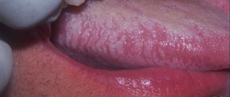
Gum diseases
Some form of gum disease, also called periodontal disease or periodontitis, affects nearly half of adults age 30 and older. Severity ranges from mildly swollen gums to bleeding gums and complete loss of teeth.
Most gum diseases develop in several stages:
- Plaque and tartar build up on the teeth, leading to gingivitis (inflammation of the gums).
- Gingivitis irritates the soft tissue along the gum line, which gradually leads to periodontitis.
- Periodontitis occurs when the gums separate from the teeth, forming pockets. This can lead to further gum infection, requiring treatment with antibiotics, surgery, or tooth extraction.
Treatment:
Like tooth decay, gum disease in the gingivitis stage is highly treatable if detected early. If it can't be corrected, your dentist will likely suggest scaling, a professional deep cleaning to remove all plaque from your mouth. As noted, antibiotics may also be prescribed. If periodontitis is advanced, surgery may be required.
“Periodontal disease is indeed a common problem. If such diseases are left untreated, sooner or later this will lead to the formation of deep and painful periodontal pockets, and ultimately to tooth loss. In addition, the microflora that lives in the mouth is very aggressive. Once it enters the bloodstream, it can spread and complicate the course of chronic diseases of the joints, heart, lungs, and brain, causing disruption in their functioning. The relationship between the presence of pathogenic microflora in the oral cavity and diseases such as Alzheimer's and Parkinson's disease has been proven. Getting dental plaque or the contents of periodontal pockets into the lungs when coughing can worsen the prognosis of any acute respiratory viral infection, as well as aggravate the course of COVID-19. Therefore, it is so important to monitor the condition of the periodontium, undergo preventive examinations every 6 months, and have a professional. oral hygiene, remove dental plaque, both hard and soft. When diagnosing gum disease, it is imperative to carry out timely treatment. For prevention, it is important to use simple care methods at home - brushing your teeth with high-quality toothpaste - 2 times a day, and also use dental floss, irrigators, and special rinses for additional hygiene.”
Kalashnikova Oksana Yurievna
dentist, implantologist-orthopedist, gnathologist, maxillofacial surgeon, specialist in aesthetic dentistry, candidate of medical sciences, doctor of the highest category, doctor of integrative interdisciplinary anti-aging medicine Work experience: 30 years
Material and methods
This prospective, non-randomized cohort study included 20 menopausal, 10 premenopausal (45 to 50 years) and 10 postmenopausal (51 to 59 years) women. The study took place at the Department of Therapeutic Dentistry of the Institute of Dentistry of the First Moscow State Medical University named after. THEM. Sechenov. All study participants received full information about the purpose and objectives of this study and gave written informed consent to participate.
To determine the composition of the oral microflora, gingival fluid was collected using sterile paper strips 0.3–0.8 mm in size and placed in a test tube for further PCR diagnostics in the laboratory.
Additionally, the pH of mixed saliva was determined to identify the relationship with the qualitative composition of the oral microflora. The collection of mixed saliva was carried out in a graduated test tube on an empty stomach, indicator paper was introduced into the test tube for 5 s, then compared with the attached color scale and the pH value was recorded.
As part of a study of the dental status of pre- and postmenopausal women, the prevalence and intensity of periodontal tissue damage was studied using the periodontal index (PI). The periodontal condition of each tooth was determined by a score from 0 to 8, taking into account the degree of gum inflammation, tooth mobility, and the depth of the periodontal pocket.
Infectious diseases
An infection of the oral cavity leads to the development of a disease in which the mucous membranes become inflamed. The pathological process can involve the teeth and gums. Symptoms of infections in the mouth are often hidden, but sooner or later they appear.
Online training in Anti-Age medicine Learn the intricacies of anti-aging medicine from anywhere in the world.
For the convenience of doctors, we have created the Anti-Age Expert online training platform: Lectures from our educational programs are consistently posted here, and are accessible 24/7. Doctors can study the materials as many times as necessary, ask questions and discuss interesting clinical cases with colleagues in special chats. Find out more
Causes of infectious diseases of the oral mucosa:
- Uncontrolled medication use.
Treatment of any disease should be under the supervision of a doctor. Improper use of antibiotics, antibacterial and other agents leads to consequences, including diseases of the oral cavity.
- HIV and AIDS.
Infectious diseases of the mucous membranes of the oral cavity often occur against their background.
- The occurrence of infections in the oral cavity
may be associated with a weakened immune system.
- Poor nutrition.
If the mucous membrane is exposed to aggressive food, it injures it. Thus, the mucous membrane becomes more vulnerable and susceptible to infection.
In addition, smoking and alcohol provoke oral diseases. Those whose bodies are dehydrated or experience hormonal imbalances also face similar problems.
- Stomatitis.
There are many bacteria living in the mouth, and due to a decrease in immunity, they are activated. Thus, an infectious disease develops. One of the most common is stomatitis. It usually develops in people who brush their teeth incorrectly or not thoroughly enough. The disease can also occur due to tonsillitis or diseases of the gastrointestinal tract. There are several types of stomatitis:
- Catarrhal.
It is manifested by swelling of the oral mucosa. As catarrhal stomatitis progresses, plaque appears on the tongue.
- Ulcerative.
In this case, the lymph nodes become enlarged. This stomatitis is manifested by dizziness and weakness. May occur in people with stomach ulcers or enteritis.
- Aphthous stomatitis.
Leads to damage to the oral mucosa, on the surface of which erosions form. Aphthous stomatitis is associated with an imbalance in the gastrointestinal tract. Signs of aphthous stomatitis: swelling of the oral mucosa, malaise, erosion in the mouth.
- Diseases caused by viruses.
The best known oral infection is herpes, clinically known as herpes simplex virus type 1 (HSV-1). It is often found in children between 6 months and 5 years of age.
Once HSV-1 enters a child's body, he will carry the virus throughout his life. It is estimated that 50-80% of adults live with herpes simplex, either dormant or active.
The herpes virus manifests itself as rashes around the mouth. As the disease progresses, ulcers form in the mouth: they are localized on the inside of the lips and cheeks. Herpes can also infect tissues near the teeth.
After the first attack of oral herpes, the body produces antibodies to fight the virus and its effects. This way, your subsequent outbreaks of HSV-1 may not be as severe or the virus may remain dormant.
Otherwise, taking antiviral drugs may help.
- Candidiasis.
This is a manifestation of a fungal infection. Candida fungus microorganisms live in the mouth and are activated under the influence of unfavorable factors. Candidiasis occurs in people with weakened immune systems. To avoid illness, it is necessary to strengthen the immune system and avoid hypothermia. There are several types of candidiasis:
- Pseudomembranous.
It occurs in an acute form. As the pathology progresses, the mucous membrane of the cheeks begins to dry out, and the same thing happens with the lips and tongue. A coating of curd consistency forms on the tongue. Pseudomembranous candidiasis causes discomfort when chewing and itching. The disease can occur in people with a weakened immune system, as well as against the background of blood pathologies. Other causes of pseudomembranous candidiasis are diabetes mellitus and hypovitaminosis.
- Atrophic candidiasis.
In this case, the mucous membrane of the mouth becomes red and dry. The chronic form of the disease develops in those who use dentures for a long time.
- Hyperplastic candidiasis
may be acute or chronic. In chronic cases, plaque forms on the mucous membranes, including the tongue. When you try to remove it, the mucous membrane becomes more inflamed. Brushing your teeth may cause bleeding.
- A disease that occurs due to HIV.
Secondary immunodeficiency is characterized by the active proliferation of pathogenic flora in the oral cavity. Early diagnosis will allow faster initiation of treatment and improve the prognosis of immunodeficiency.
It is important to note that diseases of the oral cavity that occur against the background of HIV often become chronic.
Lek Microflora floor of the mouth dental plaque
9
Lecture. Microflora of the oral cavity, dental plaque, dental plaque. Pathogenicity factors of cariogenic streptococci, veillonella, fusobacteria. Causative agents of purulent-inflammatory processes in
dentistry. Dental sepsis. Anaerobic infection
maxillofacial region.
The microbial flora of the oral cavity is a collection of various bacteria, fungi, protozoa and viruses that can enter into a symbiotic relationship with humans as a biological species.
The dominant place, both in the diversity of species living in the oral cavity and in quantity, is occupied by bacteria. The number of bacterial species in this ecological niche ranges from 120 to 200. The content of microorganisms in saliva (oral fluid) ranges from 4 million to 5 billion per ml, in dental plaque (plaque) - from 10 to 1000 billion per g of material. They are widely represented by gram+ and gram– microorganisms, both coccoid bacteria and rod-shaped and convoluted forms.
Obligate anaerobic (fusobacteria, bacteroides) and microaerophilic (aerotolerant) flora (streptococci, actinomycetes, corynebacteria) of the oral cavity account for 80 to 96% of microorganisms. The remaining part consists of facultative anaerobic species - staphylococci, streptococci, enterobacteria, and aerobic neisseria, Haemophilus influenzae, non-fermenting gram bacilli (acinetobacter, moraxella, bordetella).
A significant place in the microbiocenosis of the oral cavity is occupied by convoluted forms: vibrios, campylobacters, spirillum and spirochetes. They are mainly obligate anaerobes and less often microaerophiles.
The number of anaerobic species of microorganisms is 106–1011 CFU/ml or g of material. The ratio of anaerobes to aerobes is approximately 100:1. Almost without exception, all types of anaerobic bacteria isolated during pathology are representatives of the normal microflora of the oral cavity.
Brief characteristics of the most clinically significant genera and species of obligate anaerobic
bacteria.
Genus Peptostreptococcus
– gram+ non-spore forming cocci, round or slightly elongated, often forming short and long chains. They have low saccharolytic activity, but decompose peptone and amino acids. They are contained in large quantities in dental plaque, released from carious cavities, root canals during pulpitis and periodontitis, from periodontal pockets, purulent exudate during acute and chronic inflammatory processes of the maxillofacial area. Important species: P.anaerobius, P.micros, P.asaccharolyticus.
Genus Veillonella
– gram-anaerobic cocci, in smears they are located in groups and clusters. Lactate, pyruvate, and acetic acid, which form streptococci and lactobacilli from carbohydrates, ferment well. They are found in saliva, on the mucous membrane of the mouth and gums, in the deep layers of dental plaque. They are often isolated during various pathological processes: from purulent exudate and the contents of periodontal pockets.
Genus Actinomyces
– gram+ rods with a tendency to form branching filamentous elements that form the basis of dental plaque. Due to adhesion, other symbionts of dental plaque, for example, cocci, are fixed on them. Actinomycetes are important in the etiology of caries, periodontitis, nonspecific inflammatory processes and actinomycosis. When fermenting carbohydrates, actinomycetes form lactate, acetic, formic and succinic acids, which have a strong cariogenic effect. Their dental plaques are isolated normally and in pathology, from carious cavities, periodontal pockets, and are contained in purulent materials in actinomycosis, chronic inflammatory processes of soft tissues and osteomyelitis of the maxillofacial region. Main species: A.israellii, A.naeslundii, A.viscosus, A.odontolyticus.
Genus Fusobacterium
– gram sticks of elongated spindle-shaped form with pointed ends. Produce butyric and other acids. Normally they inhabit the oral mucosa and dental plaque. In pathology, they are isolated from purulent exudate, the contents of the periodontal pocket, in ulcerative necrotic stomatitis and Vincent's angina in association with spirochetes. Main species: F.nucleatum, F.necroforum, F.mortiferrum.
Genera Bacteroides, Porphyromonas and Prevotella
– small gram rods and coccobacteria. They do not have spores or flagella. Produce fatty acids. Normally they are detected on the oral mucosa and as part of dental plaque. They are isolated in various inflammatory processes of the maxillofacial area, often in association with anaerobic cocci, fusobacteria, streptococci and staphylococci. Main species: B.fragilis, Porph.gingivalis, Prev.melaninogenica.
Genus Clostridium
– gram+ spore-forming rods, some are mobile, biochemically active. Normally they are part of the intestinal microbiocenosis. They are not constantly detected in the oral cavity. Isolated in patients with purulent wounds of the maxillofacial area. If the wound surface is contaminated and there is extensive tissue trauma, a gas anaerobic infection may develop. Main species: C.perfringens, C.septicum, etc.
Facultative anaerobic microflora. Staphylococci are isolated from inflammatory foci in 10-15% of cases: Staphylococcus aureus, less often S.epidermidis, S.saprophyticus, S.hominis, S.warneri, S.xylosus; in 15% streptococci: S.pyogenes, S.faecalis, S.viridans.
Microaerophilic streptococci play a huge role in the microbiocenosis of the oral cavity. The frequency of their isolation from purulent exudate or the contents of periodontal pockets is 10-35%; they constitute a significant part of the material of dental plaque and other biotopes of the oral cavity.
The metabolic features of one of them - S.mutans - release a significant amount of lactic acid, leading to decalcification of tooth enamel due to the low pH of the environment. In combination with high adhesive properties to tooth enamel, it can be considered as one of the leading factors in the development of caries. S.sanguis also play an equally important role.
Microaerophilic streptococci S.sanguis, S.salivarius, S.milleri, S.mitis, S.intermedius are often found in pathological material from periodontitis, abscesses, phlegmon and osteomyelitis of the maxillofacial area, the contents of periodontal pockets and purulent discharge.
Aerobic non-fermenting gram bacteria of the genera: Acinetobacter, Eikenella, Moraxella, Pseudomonas, Neisseria also take part in the development of purulent-inflammatory processes. In the development of purulent periodontitis, the role of gram bacteria Actinobacillus actinomycetemcommitans has been proven.
In immunodeficiency states, purulent-inflammatory processes in the maxillofacial area are associated with gram-enterobacteria of the genera Escherichia, Enterobacter, Proteus, Providencia and bacilli (B.licheniformis, B.coagulans, B.cereus).
Biotopes of the oral cavity:
- Oral mucosa
- Ducts of the salivary glands containing saliva
- Gingival fluid and gingival groove area
- Oral fluid
- Dental plaque
The microbiocenosis of each biotope differs due to differences in physical and chemical characteristics, pH of the environment, viscosity, temperature, the presence of organic compounds and food residues, and partial pressure of gases.
Oral mucosa
– the most extensive and diverse biotope. On the surface of the mucous membrane, predominantly gram-anaerobic and facultative anaerobic flora, as well as microaerophilic streptococci, grow.
In the sublingual region, on the inner surface of the cheeks, in the folds and crypts of the oral cavity, anaerobes predominate: veillonella, peptostreptococci, lactobacilli, and streptococci (S.mitis). The dorsum of the tongue is colonized by S. salivarius.
Streptococci, corynebacteria, neisseria, hemophila, pseudomonads, yeast-like fungi and nocardia are found on the mucus of the hard and soft palate, palatine arches, and tonsils.
There are from 200 to 20,000 microbial cells per 1 cm2 of CO.
Salivary gland ducts and saliva
practically sterile due to the high bactericidal activity of enzymes, lysozyme, secretory immunoglobulins and other factors.
Gingival fluid and gingival groove
. The biotope is dominated by filamentous and convoluted obligate anaerobic species of bacteria: fusobacteria, leptotrichia, actinomycetes, spirillum, anaerobibrio, campylobacter and spirochetes. This is the main habitat of anaerobic bacteroids, porphyromonas, prevotella. Protozoa, yeast-like fungi and mycoplasmas are also found here. The number of bacteria in the gingival fluid in a healthy person is no more than 105 mt/ml, and with the development of gingivitis or periodontitis up to 108 mt/ml.
Oral fluid.
The basis of oral fluid is saliva, which receives microbes from the mucous membrane, gingival groove, folds and dental plaque. This biotope contains Veillonella, microaerophilic streptococci (S.salivarius), facultative anaerobic streptococci, aerococci and mycoplasmas. The number of microorganisms in healthy people is 106–1010 mt/ml.
Dental plaque
– the most complex and multicomponent biotope formed on the surface of the tooth. Almost all representatives of the microbial flora of the oral cavity are determined in the composition of dental plaque. Their number varies significantly among different people and at different periods of their lives.
In the formation of a biotope, the determining role of the macroorganism and environmental factors (diet, lifestyle, occupational hazards, etc.) is undoubted. Quantitative and qualitative disturbances in the composition of the symbionts of a given biotope, disturbances in their interaction with the macroorganism play a decisive role in the emergence of such important nosological forms as dental caries and periodontitis.
To study the composition of dental plaque, a technique is used to take the material with a probe or metal spatula, followed by weighing on an analytical balance. After this, mechanical grinding of the plaque or its disintegration by ultrasound and quantitative inoculation on optimal nutrient media and cultivation under both aerobic and anaerobic conditions are carried out. The number of bacteria is expressed in colony-forming units per g of material (CFU/g).
According to modern ideas, on the surface of the tooth enamel there are:
1 – cuticle, which is a reduced enamel epithelium,
2 – pellicle – an organic polymer film formed when enamel comes into contact with saliva,
3 – dental plaque or dental plaque.
Dental plaque consists mainly of microbes with a small inclusion of structureless substance of organic nature.
In the formation of dental plaque, several main mechanisms can be distinguished:
- Adhesion of epithelial cells invaded by bacteria to the enamel, followed by the growth of microcolonies,
- Precipitation of extracellular glycans produced by S. mutans and S. sanguis
- Precipitation of salivary glycoproteins forming a pellicle with subsequent specific adhesion of bacteria to it,
- Agglutination of bacteria with antibodies followed by fixation on the enamel surface. The bacteria in dental plaque are coated with IgA and IgG.
Dental plaque begins to form within 1–2 hours after brushing the teeth, and in the dynamics of its formation, significant changes in the nature of the microbiocenosis occur. The general trend is a change in the composition of the flora from the dominance of aerobic and facultative anaerobic forms, mainly cocci, to obligate anaerobic gram-negative rods and convoluted forms.
1 phase
formation of dental plaque – the first 2-4 hours after thorough brushing of teeth. It mainly consists of cocci (streptococci, neisseria and staphylococci) and short rods (lactobacillus). This is the so-called "early" dental plaque.
2 phase
– up to 4-5 days. It is characterized by a decrease in the proportion of Gram+ cocci and the prevalence of gram-variable filamentous forms - leptotrichia and fusobacteria.
3 phase
– from 6-7 days onwards. The dental plaque takes on its final form in terms of the composition of symbionts, although quantitative changes in it occur constantly. The number of aerobic species - Neisseria, Rotium, and facultative anaerobic streptococci - sharply decreases. The dominant species are gram-obligate anaerobic bacteria - bacteroides, fusobacteria, veillonella and gram+ actinomycetes, microaerophilic streptococci and peptostreptococci.
The total number of bacteria in a dental plaque increases from 90-100 CFU/g in the 1st phase of formation to 1-10 million CFU/g in the 2nd phase and to tens and hundreds of billions per 1 g in the 3rd phase of formation.
Bacteria have unequal adhesion to different tooth surfaces. The adhesion process is influenced by mechanical factors associated with the chewing process, physicochemical conditions, etc. Therefore, on different surfaces of teeth, in pits and fissures, the composition of the microflora differs even within the same tooth.
Dental plaque also forms on the surface of fillings, and its composition is somewhat different and depends on the nature and quality of the filling material. The microbial flora on cements and amalgams is most richly represented. An average level of colonization is typical for macrocomposite filling materials. On microcomposite materials, dental plaque is poorly formed due to the low affinity of bacteria.
It has been established that after eating food, especially one rich in carbohydrates, a sharp increase in the enzymatic activity of bacteria occurs in the oral fluid - a “metabolic explosion”. At the same time, the process of glycolysis is activated, which leads to a sharp shift in the pH of the environment to the acidic side due to the release of acidic catabolites - acetic, lactic, formic, pyruvic and other acids.
In turn, this leads to the release of calcium ions from the hard tissues of the tooth (demineralization), as well as a decrease in the phosphate content during the process of phosphorylation in bacteria. In addition, dental plaque bacteria accumulate excess carbohydrates in the form of reserve polysaccharides - dextrans and levans.
Etiology and pathogenesis of inflammatory diseases of the maxillofacial area
In the balance of all components of the microbiocenosis, the violation of which leads to the development of the infectious process, regulatory (immune and non-immune) mechanisms play a determining role. This is confirmed by the polyetiological nature of opportunistic infections. As a rule, associations of several types are isolated from the inflammatory focus.
At the same time, the properties of the microorganisms themselves may change and under certain conditions they may exhibit their pathogenic properties. Opportunistic bacteria have genes that encode the formation of the most important phenotypic signs of pathogenicity. However, their implementation of this information is significantly reduced or blocked due to the constant influence of the regulatory systems of the human body and other components of the normal microbiocenosis.
Signs of pathogenicity of UPM
- Colonization
– the ability of bacteria to colonize a specific biotope or ecological niche of an organism. This is possible if they have
- Adhesion factors
ensuring adhesion to the surface of tissues or tooth enamel. Adhesion is realized through several basic mechanisms:
- Through cell wall molecules such as galactose-binding lectins of actinomycetes or lipoteichoic acids of streptococci, interacting with fibronectin, a protein in blood plasma and tissue fluid,
- Through specific bacterial villi - pili or fimbriae, as well as surface vesicles in Porphyromonas gingivalis,
- Due to hemagglutinins and coaggregation factors with other bacteria, which are identified in oral streptococci, actinomycetes, fusobacteria and bacteroides,
- Due to the adhesive properties of the capsule in streptococci and bacteroides.
- Factors of protection
which allow bacteria to “protect themselves” from bactericins that produce antagonist microbes and resist the action of numerous protective factors of the human body.
Protection factors include: 1) the polysaccharide capsule
of bacteroides, streptococci and other bacteria, which determines resistance to phagocytosis, the action of antibodies and some antibacterial drugs;
2) enzyme systems
of bacteria that break down IgG and IgM, C3 and C5 components of complement, transferrin, hemopexin, haptoglobin, etc. - Invasiveness
– the ability of bacteria to leave the biotope with subsequent local spread in tissues (contamination) or spread throughout the body by blood and lymph flow (dissimination). Invasiveness is the trigger point in the pathogenesis of an infectious disease. In its implementation, the main role is played by aggression enzymes - hyaluronidase, chondroitin sulfatase, lecithinase, heparinase, protease, DNAase, neuraminidase, alkaline phosphatase, catalase, peroxidase, β-lactamase, etc.
- Toxigenicity
– the ability of bacteria to form exo- and endotoxins, as well as toxic metabolic products – indole, ammonia, formic acid, hydrogen sulfide, etc. Hemolytic and hemotoxic properties are expressed in streptococci, prevotella, bacteroides, clostridia. Clostridia exotoxins have necrotic, histolytic, neurotoxic and other properties.
Inflammatory diseases caused by opportunistic microbes affect any tissue of the maxillofacial area: mucous membrane, fatty tissue, muscles and fascia, ligaments and bones. For the development of a disease, it is necessary that conditions arise for the microflora to go beyond the limits of its inherent ecological niche or biotope in the body.
They can be local, purely mechanical, or general, associated with disruption of regulation and protection throughout the body.
To local conditions
include: trauma to the oral mucosa, tooth extraction, blood stasis in the capillaries, tissue necrosis, decreased partial pressure of oxygen, deficiency of various local systems and protective factors.
General terms
: somatic diseases and conditions leading to a lack of nonspecific resistance or immunological reactivity of the body as a whole.
Etiology of purulent-inflammatory diseases
maxillofacial area and neck
In the vast majority of cases, purulent-inflammatory processes are caused by resident microflora of the oral cavity. The development of these diseases is preceded by the activation of chronic odontogenic foci (chronic periodontitis, periodontitis, periocoronitis, lymphadenitis, etc.), acute processes (acute periodontitis, lymphadenitis, etc.). Penetration of infection into soft tissues and bone can occur in various ways: lymphogenous, hematogenous, along the length, contact, etc.
In 90-100% of cases, the focus of inflammation is determined from 2 to 7 types of different microorganisms, while in more than half of the patients a combination of obligate and facultative anaerobic species is determined, in 30% of associations only anaerobes are detected.
About 65% of the strains were represented by non-spore-forming anaerobic bacteria, predominantly gram-bacillus (bacteroides and fusobacteria) and gram-+ cocci (peptostreptococci and peptococci); actinomycetes and veillonella were less frequently identified. Facultative anaerobic and aerobic bacteria account for an average of 35% of all isolated strains. Of these, the most frequently identified are staphylococci (17.4%), streptococci, enterobacteria and bacilli.
A special place in the development of odontogenic inflammation belongs to anaerobic bacteria of the bacteroid group. In purulent foci there are more than 10 types of these microorganisms, which belong to three genera Bacteroides, Porphyromonas, Prevotella. The main species identified are: P.melaninogenicus, P.assaccarolyticus, P.capillosus, P.oralis. These species form pigment and produce a variety of fatty acids.
Bacteroides are often isolated from purulent foci and in 1/3 of patients they predominate in the microbial association. Bacteria of the genus Peptostreptococcus predominate in 20%, staphylococci - 15% of associations. Other microorganisms are leading in 3-8% of cases. The concentration of microorganisms in purulent exudate is lower with limited foci and higher with diffuse ones, involving two or more cellular spaces.
Microflora in acute odontogenic purulent-inflammatory diseases
is mixed with a clearly defined anaerobic accent, which must be taken into account when conducting antibacterial therapy.
In chronic inflammatory diseases, both nonspecific (chronic lymphadenitis, chronic osteomyelitis, etc.) and actinomycosis, the microflora of the inflammatory focus differs little: obligate anaerobes make up about 60%, microaerophilic streptococci up to 6%, in other cases facultative and aerobic microorganisms. The dominant microflora in these patients are bacteroides, peptostreptococci, staphylococci and, less commonly, bacilli. Anaerobic actinomycetes (A.israelii, A.naeslundii, etc.) are isolated in 8-10% of patients.
Oral cancer
Oral cancer most often affects the tongue, tonsils, gums and oropharynx. Since its various types often do not cause obvious signs and symptoms in the early stages, regular dental examinations are the most important method of detecting them.
Your dentist may also test you for oral cancer during your exam, especially if you have any of these symptoms:
- Persistent pain in the mouth or lip;
- Red or white spot in the mouth;
- Loose teeth;
- Painful swallowing, constant pain in the mouth or ears.
Treatment:
Depending on the type and stage of oral cancer, treatment may include a combination of surgery, radiation therapy and chemotherapy.
Prevention
Maintaining oral health is not difficult if you follow simple recommendations:
- Eat a balanced diet.
The diet should contain a sufficient amount of protein foods, as well as foods containing amino acids, trace elements and vitamins. Carbohydrate foods, on the contrary, are best consumed in small quantities.
- Careful oral hygiene.
It is important, for example, to use dental floss and brush your teeth with toothpaste or gel twice a day.
- Use of vitamins and nutritional supplements (as prescribed by a doctor).
This will make up for deficiencies in the body that affect oral health.
- Regular visits to the dentist.
You should visit your dentist for a checkup at least once a year. Even if there are no complaints.
Brief conclusions
- The most common oral diseases are tooth decay, gum disease, infectious diseases and oral cancer.
- Caries and gum disease are highly treatable in the early stages.
- The causes of infectious diseases of the oral cavity vary - from viruses to poor diet and uncontrolled use of medications.
- For prevention, it is important to review “life hygiene” and regularly visit the dentist.
Anti-Aging Medicine Seminars Gain knowledge based on evidence-based medicine first-hand from the world's leading experts. As part of the Anti-Age Expert Modular School, in-person two-day seminars are held every month, where the intricacies of anti-age medicine are revealed to doctors of more than 25 specialties
Find out more