What it is
Officially, painful cracks on the tongue and along the edge of the tongue are called glossalgia. This is a fairly common neurosomatic disease.
Most common symptoms:
- dryness in the tongue area;
- itching and burning;
- swelling;
- soreness.
The insidiousness of glossalgia is that it does not go away on its own; without appropriate treatment it is impossible to get rid of a crack in the tongue.
At an early stage of the disease, the doctor may not notice pronounced cracks in the tongue, since they are microscopic and mainly manifest themselves as burning and pain. If you do not respond to this symptom in a timely manner, over time a crack may form at the tip or in the middle of the tongue, longitudinally, as well as cracks under the tongue and on the sides. In this case, the doctor also notes swelling and swelling of the tongue, atrophy of the salivary glands and filiform papillae on the surface of the tongue. The sooner you start treatment, the easier it is to get rid of glossalgia, so if you have cracks in your tongue, you should immediately consult a doctor.
Signs of pathology
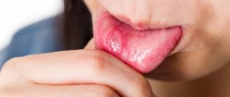
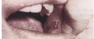
Sores on the tongue can appear in different places, and the process of their formation goes through several stages:
- In the first stage, swelling and small blisters appear on the tongue.
- Next, bubbles appear.
- The wounds are covered with a layer of white or yellow plaque, and a red border appears.
There are no age preferences for sores on the tongue; they can appear in children and adults. The frequency of their formation varies from several times throughout life, while in others they may appear with enviable regularity.
Usually the sores go away quickly, but if the wound on the tongue does not heal, then you should visit a doctor and find out the cause.
Congenital fissures
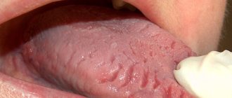
It happens that a patient has cracks in the tongue since birth or childhood, which practically do not cause him any concern. Such cracks are usually transverse and are caused by the development and formation of the tongue.
Some patients have a so-called folded tongue, a congenital feature of tongue development. It is usually considered normal and does not require any treatment. The exception is red cracks in the tongue, which form at the bottom of the folds and cause pain to the patient. This happens with insufficient oral hygiene, so those with a folded tongue must carefully clean not only their teeth and gums, but also their tongue from plaque. If the owner of a folded tongue has developed cracks, treatment should be prescribed by a competent doctor, taking into account the design features of the patient’s tongue. To prevent the formation of folds in the depths, more attention should be paid to the sanitation of the oral cavity.
Glossitis
This pathology is a type of stomatitis. Manifests itself as an infectious inflammation of the mucous membrane of the oral cavity and tongue. The disease most often develops against the background of a variety of reasons that were listed earlier, and is manifested by a variety of symptoms.
Without therapy, the situation will only get worse, so it is better to visit a doctor for advice.
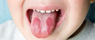
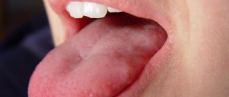
How do they look
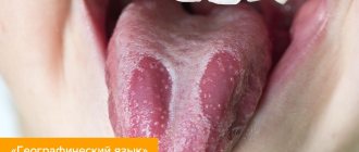
Small cracks in the tongue can be completely invisible to the naked eye; only an experienced doctor can examine them using special instruments. The larger ones are clearly visible to the naked eye; they resemble longitudinal or transverse grooves on the tongue. Deep cracks look like crevices; sometimes the tongue appears to be cracked or split into two parts. If you notice such a problem, consult a doctor, even if nothing is bothering you at the moment. If an infection gets into the crack, the course of the disease may be complicated by the inflammatory process.
Reasons for appearance
Reasons for education:
- lack of nutrients, iron and vitamin PP in the patient’s body;
- anemia;
- lack of B vitamins;
- allergic reaction to toothpaste or medications;
- chronic fatigue and constant nervous tension;
- mechanical damage to the tongue (nervous biting, chewing);
- disruption of capillary blood flow in the tongue;
- the patient has diseases such as gastritis, enterocolitis, hepatitis, cholecystitis;
- wearing uncomfortable dentures.
The main reason why the tongue is cracked is mechanical injuries due to a lack of vitamins and microelements in the patient’s body. In this case, it is enough to bite your tongue while chewing food so that a painful crack forms at its tip.
Another fairly common reason why a patient’s tongue is cracked is a malfunction in the patient’s central nervous system, which is caused by chronic fatigue, frequent stress, and lack of sleep. This is a reason to suspect a problem with the hypothalamus.
It happens that patients are perplexed: why there are cracks on the tongue, there seems to be no visible reason for their appearance. In this case, the answer to the question of why cracks appear can only be given by an experienced doctor, after a thorough diagnosis. It is necessary to do a detailed blood test to determine which microelements and vitamins are missing in the patient’s body. Quite often, correction of the nutritional system helps get rid of the disease.
Many people are probably interested in the question of what cracks mean. After all, the tongue, as is known, signals the presence of many diseases and pathological conditions of the human body. Cracks in the tongue are a sign of a lack of vitamins and microelements in the body. Quite often, cracks are caused by worms, as they deplete the human body, causing anemia and iron deficiency. They also indicate a diseased liver, since with this disease the human body does not receive many microelements important for health.
Cracks in the tongue on the side can be a sign of thyroid disease; in this case, it swells and is injured at the edges by the patient’s teeth.
Results and discussion
Analysis of local criteria for assessing the healing of tongue wounds showed that all patients of group 2 who received “traditional” local treatment, a day after surgery, indicated aching severe pain in the oral cavity and inability to swallow. 16 (88.9%) patients in this group experienced an increase in body temperature, on average to 37.8 °C, within 2-3 days after surgery. All patients were found to have severe swelling and hemorrhage of the tongue stump, as well as moderate hyperemia of the suture line. In 15 (83.3%) patients, a clinical blood test revealed leukocytosis (on average up to 11.5·109/l), and an acceleration of ESR (on average up to 21.5 mm/h) was noted in all.
3 days after surgery, severe pain and inability to swallow were noted by 7 patients of group 2 (38.9%). All patients had low-grade fever. Severe swelling of the tongue tissues and hyperemia of the suture line were detected in 4 (22.2%) patients. In 16 (88.9%) cases, partial divergence of the sutures in the middle and distal fragments of the wound was noted. All patients had no wound discharge; the stump of the tongue was covered with fibrinous plaque.
On the 5th day after surgery, 4 patients in this group (22.2%) still had low-grade body temperature. A clinical blood test revealed leukocytosis up to 11·109/l in 14 (77.8%) patients and acceleration of ESR up to 20 mm/h in 16 (88.9%) people. 7 (38.9%) patients complained of minor pain in the wound, and 14 (77.8%) patients complained of difficulty swallowing water. All patients had partial suture divergence, with 4 (22.2%) of them having an area of divergence of more than 70%. The edges of the wound in 5 (27.8%) patients were covered with purulent-necrotic masses, as well as purulent wound discharge. Sluggish granulation of wound surfaces was detected in 7 (38.9%) patients.
On the 9th day after surgery, minor pain in the wound was noted by 7 (38.9%) patients of group 2, swallowing was free in all patients. In 4 (22.2%) there was slight hyperemia around the preserved suture material, in 5 (27.8%) patients there was a serous nature of the wound discharge. All patients had active granulation, in some areas even with the growth of hypergranulation, and partial epithelization of the wound surface. On the 9th day, the existing sutures were removed.
On days 12–14 after surgery in patients of the study group, complete epithelization of the wound surface was observed in only 3 (16.7%) patients. In 15 (83.3%) there was an area in the center of the wound that was not covered by epithelium. Complete epithelization of the wound surface in this group with the formation of a dense, linear scar was completed by the 19th day after surgery.
A comparative analysis of the nature of healing of tongue wounds in patients who received a 2% solution of acid-soluble chitosan in the postoperative period revealed that on the 5th day after treatment, 13 (72.2%) patients of the 1st group had partial suture dehiscence on average and distal fragment of the wound and slight swelling of the tongue tissue. In 5 (27.8%) patients in this group, wound healing occurred without suture dehiscence. The surfaces of the wound and tongue stump were covered with a fibrinous film, after removal of which formed granulations were visible.
On the 9th day after surgery, 13 (72.2%) patients in the group with identified wound dehiscence had an active granulation process and partial epithelization of the wound surface. In 5 (27.8%) patients with primary wound healing, the sutures were removed. Complete epithelization of the wound surface in all patients of group 1 was completed by the 14th day after surgery. All of them developed a linear, dense scar.
Thus, in patients of this group, a favorable picture of reparation of damaged oral tissues was clinically observed and in earlier stages of healing than in patients of group 2.
A study of the microbiocenosis of mixed saliva in 36 patients with tongue carcinoma before surgery established the predominance of streptococci and staphylococci (100 and 87.5%, respectively), less often - bacteria of the Enterobacteriaceae
(37.5%),
Bacteroides
(31.3%),
Sarcina
and
Candida
(25%),
Lactobacillus
(18.8%),
Stomatococcus
(13.3%),
Veillonella
(12.5%),
Corynebacterium
and
Fusobacterium
(6.3%).
The number of isolated microorganisms was as follows: Streptococcus
spp.
— 7.4±0.13 lg CFU/ml, Fusobacterium
spp.
— 6.3±0.23 lg CFU/ml, Corynebacterium
spp.
— 6.1±0.26 lg CFU/ml, Staphylococcus
spp.
— 5.9±0.08 lg CFU/ml, Candida
spp.
— 5.6±0.78 lg CFU/ml, Bacteroides
spp.
— 5.3±0.07 lg CFU/ml, Lactobacillus
spp.
— 5.2±0.09 lg CFU/ml, Stomatococcus
spp.
— 5.1±0.20 lg CFU/ml, Sarcina
spp.
— 4.8±0.25 lg CFU/ml, Veillonella
spp.
- 4.5±0.11 lg CFU/ml, family Enterobacteriaceae
- 3.9±0.24 lg. The pathogenic potential of the isolated microbiota in patients with tongue carcinoma was more pronounced than in healthy people during the initial examination. 66.7% of staphylococcal strains had hemolytic and lecithinase activity, and 43.8% of streptococci had hemolytic activity. In 62.5% of patients, associations of 3-4 cultures of microorganisms were detected, and in 38.5% - of 5-6 cultures.
Thus, the identified dysbiotic state of the oral microbiota of patients with tongue carcinoma before surgery is regarded as grade III dysbiosis, requiring correction in order to prevent purulent complications of the upcoming surgical intervention on oral tissues.
On the 10th day of using acid-soluble chitosan (group 1), a repeated study showed positive dynamics of changes in the microbiocenosis of the oral cavity (Fig. 1).
Rice.
1. Frequency of isolation of microorganisms in mixed saliva in patients with tongue tumors before and after treatment. The frequency of isolation of pathogenic and opportunistic microbiota decreased and representatives of normal microbiota began to be isolated to a greater extent. Thus, the prevalence of staphylococci decreased by more than 4 times, and yeast fungi of the genus Candida
and micrococci decreased by 2 times; Enterobacteriaceae, bacteroides, stomatococci, corynebacteria, veillonella, and fusobacteria were not isolated. At the same time, the frequency of detection of representatives of normal microbiota, lactobacilli (60%) increased 3 times; peptostreptococci began to be isolated in 90% of cases, peptococci, non-pathogenic candida, epidermal staphylococci, and actinomycetes began to be isolated in 20-30% of cases. Quantitatively (Fig. 2)
Rice. 2. The number of microorganisms in the mixed saliva of patients with a tongue tumor before and after treatment. There was also a decrease in the level of representatives of pathogenic and opportunistic microbiota - 2 times staphylococci, 1.5 times streptococci, candida, micrococci. Against this background, the amount of normal microbiota - peptostreptococci, peptococci, etc. - increased.
The microbiocenosis of saliva in patients of group 2 after the use of traditional antiseptic drugs was also restored to normocinosis, but much more slowly than after the use of chitosan irrigations. The quantitative and qualitative composition of normobiota and non-pathogenic representatives of the microbiota is lower than in patients of group 1.
A study of the level of lysozyme activity in mixed saliva of healthy people showed (Fig. 3),
Rice. 3. Lysozyme content in mixed saliva of healthy people and patients with tongue tumors before and after surgical treatment. that the amount of lysozyme averaged 47.4 ± 10.1 μg/ml.
The lysozyme level of all primary patients with tongue carcinoma before surgical treatment averaged 25.4±8.5 μg/ml.
After tumor removal, an increase in the level of lysozyme was observed: in group 2 - up to 78.125±18.7 μg/ml; in group 1, the amount of lysozyme increased 8 times and averaged 202.87±56.85 μg/ml.
Treatment
The main question that worries patients with glossalgia is how to cure cracks in the tongue. In fact, everything is not so difficult. The treatment method depends on the cause of glossalgia.
- First, it is necessary to eliminate the dental causes of tongue cracks - correct an incorrect bite, replace defective fillings, and adjust the prosthesis. Next, the patient should be carefully examined by a therapist, neurologist, and, if necessary, by an endocrinologist and gastroenterologist. And only after this should treatment for glossalgia be prescribed.
- Typically, treatment for fissures consists of drug therapy and physiotherapeutic procedures. Treatment with medications is to improve blood circulation in the tissues of the oral cavity, as well as to improve general cerebral circulation. For this purpose, drugs such as trental, cavinton, nicotinic acid, and vitamin C are prescribed.
- The tone of the autonomic nervous system should also be improved; for this purpose, sedatives are prescribed - tincture of valerian, motherwort, persen.
- Additionally, the doctor may prescribe procedures such as hirudotherapy or electrical neurostimulation using a TENS device. A good effect is achieved by using electrophoresis, as well as iontophoresis of novocaine.
- If the tongue is severely painful, the doctor may prescribe local painkillers - lidocaine, dicaine.
- If the patient has cracks in the tongue, plaque and burning, it is necessary to adjust the diet. During treatment, you should completely exclude from the diet foods that irritate the surface of the tongue - such as sour juices, vegetables, pickles and marinades. You should not consume large amounts of spices, especially pepper and salt. It is advisable to quit smoking.
- After each meal, be sure to brush your teeth and rinse your mouth with salted water or a decoction of medicinal herbs. Oak bark, sage, propolis, and honey have a healing effect.
Use of Medicines
If home remedies do not help get rid of sores on the tongue, then you cannot do without medications. Most often, doctors prescribe:
- Antiseptic drugs that destroy bacteria, reduce the sensitivity of the mucous membranes of the tongue and oral cavity, thanks to analgesic components. This category includes: “Inhalipt”, “Gexoral”, “Strepsils”.
- Products with disinfecting properties can be used: ethyl alcohol, alcohol solution of iodine, hydrogen peroxide.
- Dental gels and ointments that not only disinfect, but also relieve pain. These include: “Kamistad”, “Cholisal”, “Solcoseryl”.
- Rinsing the mouth with a solution of “Furacilin”, “Chlorhexidine”, “Chlorophyllipt”. If you don’t have these drugs on hand, you can prepare a rinse solution from iodine, salt and soda.
All of the listed remedies must be used only with the permission of a doctor and with extreme caution so as not to cause even greater harm to the mucous membrane of the tongue. Medicines should be applied to wounds purposefully, trying not to affect adjacent healthy areas.