Occlusal curves are lines passing in transverse or longitudinal section through the molars of the same name on the right and left chewing side of the jaw. In dentistry, two types of occlusal planes are used, which have functional significance. Together, they are analyzed during the diagnosis of the patient and are of significant importance in making a diagnosis. Also, these zones allow you to determine the need for treatment and select the appropriate correction technique.
On a note!
It is advisable to determine the position of occlusal parameters after the age of 13, when the process of eruption of chewing units is completed. However, already in childhood it is possible to determine the likelihood of anatomical defects in a row for a particular patient.
Occlusal curves
Lesson No. 9
Topic: Particular anatomy of permanent dentition teeth. The structure of the dentition. Dental arches, shape of dental arches.
Learning objective:
Assessment of knowledge acquired at specialized departments. Study of the structure of the dentition. Shapes of dental arches. Dental formulas.
The duration of the lesson is 2 hours.
| Lesson stages | Equipment | Visual aids | Time |
| 1. Teacher briefing | 5 minutes. | ||
| 2. Control of initial knowledge | Control questions. | 20 minutes. | |
| 3. Discussion of the topic | Slides, presentations | 45 min. | |
| 4. Monitoring the results of assimilation. | Questions to monitor the results of mastering the material. | 20 minutes. | |
| 5. Assignment for the next lesson. | 5 minutes. |
Questions studied previously and necessary to test knowledge:
1. What are teeth and what groups of teeth are distinguished in humans?
2. List the main parts and surfaces of the tooth.
3. Indicate the main differences between teeth in temporary and permanent dentition?
4. List the hard tissues of the tooth and indicate their location and function.
The structure of the dentition. Dental arches.
The teeth located in the jaws form dental arches (dentition).
Dental arch
– a line drawn through the cutting edges and occlusal surfaces of the teeth near the vestibular contour.
The upper dental arch has the shape of a semi-ellipse, the lower – a parabola. The upper dental arch is slightly wider than the lower one, so the chewing surfaces of the upper teeth are located anterior and external to the lower ones.
In addition to dental arches, dentistry distinguishes alveolar and apical arches.
Alveolar arch
– a line running along the edge of the alveolar process near the necks of the teeth on the vestibular side.
Apical arch
- a line drawn along the tops of the roots of the teeth.
On the upper jaw, the crowns of the teeth are inclined towards the vestibular side, so the widest arch on the upper jaw is the dental one, and the narrowest is the apical one. The teeth of the lower jaw are inclined lingually, so the widest arch is apical, and the narrowest is occlusal.
Occlusal curves
The occlusal surfaces of the chewing teeth are not located in the same plane, but form the so-called sagittal occlusal curves.
Sagittal occlusal curve (curve of Spee)
– a line passing through the buccal cusp of the first premolar and the distal buccal cusp of the last molar.
On the upper jaw the sagittal occlusal curve is convex, and on the lower jaw it is concave. Due to the presence of the curve of Spee, when lowering and moving the lower jaw forward, contact is maintained between the chewing teeth (the so-called Bonville three-point contact
).
Therefore, this curve is also called compensation
.
The plane passing through the incisal point on the lower jaw and touching the occlusal curves on the right and left is called occlusal.
In addition to the sagittal curve, there is also a transversal occlusal curve
. It is formed by tilting the upper molars to the buccal side, and the lower molars to the lingual side.
Transverse occlusal curve (Wilson curve)
– a line passing through the occlusal surfaces of the chewing teeth of the right and left sides in the transverse direction.
The Wilson curve ensures contact of the dentition during transversal movements of the lower jaw. In the area of the first premolars there is no transverse occlusal curve.
Due to the presence of interdental contacts, the pressure during chewing is distributed to the periodontium of adjacent teeth. At the same time, the load on individual teeth is reduced. For optimal perception of chewing pressure by the jaw bones, according to the direction of the load, there are certain abutments (buttresses)
– bone thickenings through which the force of chewing pressure is transmitted to the cranial vault.
There are four buttresses on the upper jaw: frontonasal, alveolar-zygomatic, pterygopalatine
and
palatine
.
There are two buttresses defined on the lower jaw : alveolar
and
ascending
.
Dental formula
The order of the teeth is recorded in the form of a dental formula, in which individual teeth or groups of teeth are designated by numbers or letters.
In the clinic, the complete temporary occlusion formula is written in Roman numerals, which correspond to the serial number of the tooth in each half of the jaw.
| V IV III II I | I II III IV V |
| V IV III II I | I II III IV V |
The group dental formula of a child means that in each half of the upper and lower jaws there are 2 incisors, 1 canine, 0 premolars and 2 molars:
| 2 0 1 2 | 2 1 0 2 |
| 2 0 1 2 | 2 1 0 2 |
In the clinic, the full formula of the teeth of the permanent bite is marked in the same way as the temporary one, but in Arabic numerals:
| 8 7 6 5 4 3 2 1 | 1 2 3 4 5 6 7 8 |
| 8 7 6 5 4 3 2 1 | 1 2 3 4 5 6 7 8 |
The group formula of adult teeth shows the number of teeth in each group on the halves of the jaws:
| 3 2 1 2 | 2 1 2 3 |
| 3 2 1 2 | 2 1 2 3 |
The World Health Organization (WHO) has proposed the following notation for the dental formula: numbers indicate each tooth and each half of the upper and lower jaw, and the numerical value increases clockwise.
Formula of permanent dentition teeth (WHO):
| 8 7 6 5 4 3 2 1 | 1 2 3 4 5 6 7 8 |
| 8 7 6 5 4 3 2 1 | 1 2 3 4 5 6 7 8 |
When recording the tooth formula in this way, an icon is not placed to mark one or another half of the jaw, but a number corresponding to one or another half of the jaw is placed. So, for example, to write down the formula for the second molar of the lower jaw on the left, the designation 37 is used (3 - the left half of the lower jaw, 7 - the second molar).
Temporary occlusion teeth formula (WHO):
| 5 4 3 2 1 | 1 2 3 4 5 |
| 5 4 3 2 1 | 1 2 3 4 5 |
Questions to control the assimilation of the material:
1. What is a dental arch? What shape do dental arches have?
2. What is the sagittal occlusal curve (curve of Spee)?
3. What is the transversal occlusal curve (Wilson curve)?
4. List the main buttresses and indicate their role.
5. What is a dental formula? Indicate the types of dental formulas and principles of their use?
Literature
- Phantom course of therapeutic dentistry. Atlas. Yu.M. Maksimovsky, textbook. Benefit. –M. “Medicine”, 2005. – 328 p.
- Propaedeutic dentistry. M.M. Pozharitskaya, T.G. Simakova, M., “Medicine”, 2004. – 304 p.
- Orthopedic Dentistry Ed. V.N. Trezubov.-7th ed., revised. And additional – St. Petersburg: Foliant, 2006. – 592 p.
- Surgical dentistry Robustova
- Phantom course of therapeutic dentistry. A.I. Nikolaev, L.M. Tsepov “Medpress-inform” M. 2009., 430 p.
6. Propaedeutic dentistry. Textbook. Edited by Professor E.A. Bazikyan. M. "GEOTAR-Media", 2010.
- Lecture material.
©2015 arhivinfo.ru All rights belong to the authors of the posted materials.
1 … 10 11 12 13 14 15 16 17 … 33
Why is there a need for a sagittal occlusion curve?
With the mandibular movement forward and downward, the articular head slides along the slope of the articular tubercle. Accordingly, the lower jaw (its back part) descends and forms a gap in the area of the lateral teeth between the rows of teeth. Contact of the cusps of the lateral teeth is possible only if the chewing surfaces of the teeth are located along the sagittal straight line. Following this teaching, the occlusal straight line is called the compensation curve.
The radius is determined to be 5.8-21.2 centimeters, the average value is 6.5-8.5 cm. The deepest point of the occlusal curve is the mesial cusp of the lower first molar.
The article describes the Spee curve in detail.
Factors ensuring the stability of dentition. Age characteristics.
The dentition is a single whole, both morphologically and functionally. The unity of the dentition is ensured by interdental contacts, alveolar process and periodontium. A significant role in the stability of the dentition is played by the nature of the location of the teeth, the direction of their crowns and roots.
With age, contact points are erased and contact pads are formed instead. The erasure of contact points is indirect evidence of the physiological mobility of teeth. Also, with the loss of teeth, the lower jaw, as it approaches the upper jaw, protrudes forward, creating the appearance of progeny (senile progeny).
Vertical, transversal movements of the lower jaw
This depends on the angle of bend of the articular tubercle. During
lateral movements opening the upper and lower jaws in
the molar area on the non-working side is provided
non-working joint path. It depends on the angle of bending of the articular
tubercle and the angle of inclination of the mesial wall of the glenoid fossa on
non-working side.
Incisal path
The incisive path when moving the lower jaw forward and in
side is made up of the front guide component of it
movements and ensures the opening of the posterior teeth during these
movements. Group work guiding function
ensures opening of teeth on the non-working side during
labor movements.
Question 22.
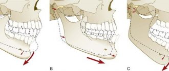
Transversal movements of the lower jaw . Lateral movements of the mandible result from unilateral contraction of the lateral pterygoid muscle. When moving to the right, the left lateral pterygoid muscle contracts, and when moving to the left, the right muscle contracts.
In this case, the articular head on one side rotates around an axis running almost vertically through the articular process of the lower jaw. At the same time, the head of the other side, together with the disc, slides along the articular surface of the tubercle. When the lower jaw moves to the right, on the left side the articular head moves down and forward, and on the right side it rotates around a vertical axis.
On the side of the contracted muscle, the articular head moves downward and forward and somewhat outward. Its path is at an angle to the sagittal line of the articular path. This angle was first described by Benet and for this reason named after him (the angle of the lateral articular path), on average it is 17°. On the opposite side, the ascending ramus of the mandible moves outward, thus becoming at an angle to its original position.
Transverse movements are characterized by certain changes in the occlusal contacts of the teeth. As the lower jaw shifts to the right and left, the teeth describe curves that intersect at an obtuse angle. The farther the tooth is from the articular head, the blunter the angle.
Of significant interest are changes in the relationships of chewing teeth during lateral excursions of the jaw. When lateral movements of the jaw are made, it is customary to distinguish between two sides: working and balancing. On the working side, the teeth are set against each other with cusps of the same name, and on the balancing side with opposite cusps, i.e., the lower buccal cusps are set opposite the palatal cusps.
Transversal movement is therefore not a simple, but a complex phenomenon. As a result of the complex action of the masticatory muscles, both heads can simultaneously move forward or backward, but it never happens that one moves forward while the position of the other remains unchanged in the articular fossa. Therefore, the imaginary center around which the head on the balancing side moves is never actually located in the head on the working side, but is always located between both heads or outside the heads, i.e., there is, according to some authors, a functional rather than an anatomical center .
These are the changes in the position of the articular head during transversal movement of the lower jaw in the joint. During transversal movements, changes also occur in the relationship between the dentition: the lower jaw alternately moves in one direction or the other. The result is curved lines that intersect to form angles. The imaginary angle formed when the central incisors move is called the Gothic angle, or the angle of the transversal incisal path.
It is on average 120°. At the same time, due to the movement of the lower jaw towards the working side, changes occur in the relationship of the chewing teeth . On the balancing side there is a closure of opposite cusps (the lower buccal cusps close with the upper palatine ones), and on the working side there is a closure of the homonomous cusps (buccal - with the buccal and lingual - with the palatine).
Question 23.
Date added: 2015-06-04; ; Copyright infringement?;
Purpose, structure and functioning of the dental system
Understanding the complex process, which is called the biomechanics of the masticatory apparatus of the dentofacial system, contributes to the timely detection of pathology in the development of muscles, articular structures, closure of teeth and the condition of the periodontium (tooth - Greek odontos, Latin dente - hence the formation: odontology - the science that deals with the description of odontos , periodontitis is a disease of periodontal tissues).
It is on a healthy periodontium - the complex of tissues surrounding the tooth, which is a single component of the temporomandibular joints - that its normal functioning depends. It follows from this that the biomechanical functions of the periodontium are determined by the anatomical and physiological features of its structure and are closely related to the work of other elements.
The laws of biomechanics of the dental system are successfully applied in orthopedics at the stages of design and creation of various prostheses, as well as some auxiliary devices.
Devices that reproduce the movements of the lower jaw include:
- Occluder . A device for assisting the patient, allowing to simulate and make the correct adjustment of the orthopedic structure.
- Facial bow . This device allows you to make an impression as accurately as possible for further correction of the bite.
- Articulator . They come in different types: universal, medium (simplified). This device is used for the manufacture and fitting of removable and fixed dentures and bridges, crowns and caps.
Photo:
Occluder
Facebow
Articulator
It is worth noting that the articulator is an extremely important device that helps to correctly make an exclusive fit of various prostheses. After all, it is articulation, in dentistry perceived as a multi-vector movement of the lower jaw (lat. mandibula) relative to the upper one, which occurs during compression and stretching of the masticatory muscles, which decisively determines intelligible and articulate pronunciation.
If some pathology associated with low frequency develops, then speech, chewing food, laughter, and swallowing are immediately impaired.
The table of movements of the lower jaw in a concise form sets out the basic provisions and determining factors of the dominant theories of articulation, the authors of which are Hanau, Gysi, Monson. Despite some discrepancies in the interpretation of the processes, their authority is indisputable, and their role in the development of orthopedics is beyond doubt.
| Articulatory theories of dentition construction | Basic provisions | Determining factors |
| Gysi's theory | The inclination of the articular path determines the displacement vector of the mandibula, which is influenced by the size and shape of the articular tubercle | Accurate determination of the articular path. Recording the incisal path. Determination of the sagittal compensation curve. Determination of the transversal compensation curve of the line. |
| Monson's theory | Complex vector movements of the LF are determined not by the articular paths, but by the surfaces of the dental cusps, which give direction to the movements | |
| Hanau's theory | The theory is similar to Gysi's theory, which analyzes the entire system of articulation. In particular, she highlights the differences between the position of the prosthesis in the articulator and in the mouth due to a decrease in the elasticity of muscle tissue | Inclination of the articular path Depth of the compensation curve Inclination of the reference plane Inclination of the upper incisors Cusp height |
| Balancing theory | Takes into account:
| |
| Spherical theory | Provides:
|
In addition, correct and healthy breathing and aesthetic emotions (expression) are impossible if the muscles that push the lower jaw forward are subject to obstruction (spasm, remission).
Complete chewing of food occurs only if the teeth of the upper and lower jaws come into correct contact - occlusion. Therefore, it is the closure of the dentition that is the defining characteristic of chewing movements.
All connecting elements of the LF move as a result of the synchronous interdependent action of the temporomandibular joint (TMJ), muscular chewing tissues and teeth. Their actions are organized, coordinated and controlled by the central nervous system.
Displacements of a spontaneous and reflex nature are entirely subordinate to the neuromuscular system and can be reproduced consistently.
The initial voluntary movements include the process of biting food and directing it into the mouth. And the following chewing and swallowing are reflexive-unconscious actions.
Due to the tasks that are defined for the jaw, its complex structure is determined.
First of all, it is the only movable bone of the facial skull, which vaguely resembles a horseshoe.
This structure is due not only to its defining purpose as a responsible component of the chewing process, but also to its development, which originates from the first gill arch.
Mandible structure:
- Body.
- The edge of the body where the cells for teeth (alveoli) are located is the alveolar process.
- Chin hole. It serves as a communicator for nerves and blood vessels.
- Corner.
- Head.
- Mandibular canal and foramen.
- Branches.
- Articular and coronoid processes.
The bone formations would remain constantly in a static position if it were not for the muscle tissue connecting them.
The muscles that move the lower jaw are called mastication.
Moreover, each muscle structure, or rather their groups, produces certain movements:
- The medial pterygoid, masseter and temporal muscles raise the jaw.
- The digastric, mylohyoid, and geniohyoid are involved in the process of descent.
- Lateral movements are possible thanks to the lateral pterygoid muscles.
Anomalies of the dentition in the transversal plane
In the first, the disc together with the head slides along the surface of the articular tubercle. In the second phase, the sliding of the head is joined by its articulated movement around its own transverse axis. The distance that the head of the mandible travels when it moves forward is called the sagittal articular path. It is on average 7-10 mm. The angle formed by the intersection of the line of the sagittal articular path with the occlusal plane is called the angle of the sagittal articular path (Fig. 20). Depending on the degree of protraction of the lower jaw, this angle changes. According to Gizy, it averages 33°.
45
Fig.20.
Angles of the sagittal joint and incisal paths (diagram) a - angle of the sagittal articular path, b - angle of the sagittal incisal path of natural teeth and artificial teeth in a removable denture (c)
With an orthognathic bite, the protrusion of the lower jaw is accompanied by sliding of the lower incisors along the palatal surface of the upper ones. The path made by the lower incisors when moving the lower jaw forward is called the sagittal incisal path. The angle formed by the intersection of the line of the sagittal incisal path with the occlusal plane is called the angle of the sagittal incisal path (Fig. 20 b, c). According to Gysi, it is on average 40 - 50° .
When the lower jaw is advanced to the position of anterior occlusion, contact of the dentition is possible only at three points. One of them is located on the front teeth, and two are located on the distal cusps of the second or third molars. This phenomenon was first described by Bonneville and was called Bonneville's three-point contact.
Catalog:
ld ld -> Anxiety and depressive disorders and quality of life in elderly patients with coronary heart disease complicated by chronic heart failure, possibilities of correction 14.00.05 internal diseases ld -> Turtles to the very bottom. Prerequisites for personal genius ld -> Intracavitary photodynamic therapy for bladder cancer and prostate adenoma 14.00. 40. Urology ld -> Extrapineal melatonin in the aging process 14.00. 53 gerontology and geriatrics ld -> The relationship between burnout syndrome and socio-psychological personality characteristics in extreme conditions of professional socialization ld -> 5. Dermatovenereology ld -> Abstract topics on pathophysiology
Share with your friends:
On the side of the contracted muscle, the articular head moves downward, forward and somewhat outward. Its path during this movement is at an angle to the line of the sagittal articular path. This angle was first described by Benet and for this reason is named after him. Otherwise it is called the angle of the lateral articular path. It is on average 17°. On the opposite side, the ascending ramus of the lower jaw moves outward, thus becoming at an angle to its original position (Fig. 34).
Transversal movements are characterized by certain changes in the occlusal contacts of the teeth. Since the lower jaw shifts to the right and left, the teeth describe curves intersecting at an obtuse angle. The farther the tooth is from the articular head, the blunter the angle. The most obtuse angle is obtained from the intersection of the curves formed by the movement of the central incisors. This angle is called the angle of the transversal incisal path or the Gothic angle (Fig.
What else is remarkable about the Spee curve?
Also, according to Spee, a researcher and practitioner, the occlusal curve is completely connected with the articular path, since it, together with the curve of the row of teeth, is formed by one radius, and accordingly, the teeth and the articular head slide along one circle forward and the steeper the inclination of the articular path, the more uniform the occlusal curve is concave. Such assumptions made by the scientist met with many objections. Other scientists argue that the occlusal curve cannot be called a segment of a circle. In its continuation, the sagittal curve of Spee often passes below or above the articular path, and its center is not in the orbit.
Transversal movements of the lower jaw.
35). It determines the range of lateral movements of the incisors and is equal to 100-110°.
Of significant interest are changes in the relationships of the chewing teeth during lateral excursions of the jaw (Fig. 36). When lateral movements of the jaw are made, it is customary to distinguish between two sides: working and balancing. On the working side, the teeth are set opposite each other with cusps of the same name, and on the balancing side they are opposite, i.e. e. the buccal lower cusps are set opposite the palatal cusps.
Until now, when studying the movements of the lower jaw, the latter were artificially decomposed into their component elements (lowering, moving forward, to the sides). This was done for methodological reasons. In reality, mandibular excursions are very complex because they are a combination of different movements. Chewing movements are of greatest practical interest for orthopedic dentistry. Knowing them can facilitate the manufacture of dentures and artificial teeth. When chewing food, the lower jaw undergoes a cycle of movements. Gisi presented the cyclical movements of the lower jaw in the form of a diagram (Fig. 37). The initial moment of movement is the position of the central occlusion. Then four phases follow continuously one after another. In the first phase, the jaw lowers and moves forward. In the second phase, the jaw moves to the side (lateral movement). In the third phase, the teeth close on the working side with cusps of the same name, and on the balancing side with opposite cusps. In the fourth phase, the teeth return to the position of central occlusion and the chewing cycle repeats. After chewing is completed, the jaw is set to a position of physiological rest.
There is no doubt about the statement that on the working side there is a closure with the same tubercles. A different relationship of the lateral teeth would not ensure grinding of food. As for the balancing side, here it is possible either the formation of contact between opposite tubercles, or their absence. The latter is confirmed by research by A. Ya. Katz and A. K. Nedergin. This appears to depend on the severity of the transversal occlusal curves.
During lateral movements of the lower jaw, the trajectories of tooth movement form an angle open posteriorly. It is called the Gothic angle, or the angle of the transeveral incisal path. Its size depends on the position of the teeth. The farther the tooth is from the articular head, the greater the angle. The largest angle is observed at the central incisors (100-110°) (Fig. 14).
The division of the movement of the lower jaw into its component elements - vertical and lateral, forward movement - is conditional and is done for methodological reasons. But it helps to understand the nature of these movements when performing different functions.
are of greatest practical interest . Their significance facilitates the manufacture of artificial teeth for dentures and the design of artificial dentition. When chewing food, the lower jaw undergoes a cycle of movements, accompanied by the appearance of rapid sliding contacts of the teeth on the working side. Maximum chewing forces develop in the position of central occlusion, when the movement of the lower jaw stops momentarily before the start of the next chewing cycle. In the first phase, the jaw lowers and moves forward. In the second phase, the jaw moves to the side (lateral movement). In the third phase, the teeth close on the working side with like-named tubercles, and on the balancing side with opposite ones. However, there may be no contact between the teeth on the balancing side, which apparently depends on the severity of the transversal occlusal curves. In the fourth phase, the teeth return to the position of central occlusion and the chewing cycle repeats (Fig. 15).
• questions for exam questions
Part I
Rice. 15. Complex of chewing movements of the lower jaw (according to Gisi): a - cycle of chewing movements ((-IV phases of the chewing cycle).