Osteomyelitis of the jaw is a dangerous inflammatory disease that is caused by infection. A purulent-necrotic process develops on the tooth bone - most often on the lower jaw. Next, the inflammation affects the bone tissue of the head and spreads to soft tissues - gums, salivary glands, skin, chewing and facial muscles. In severe cases, pus forms and necrosis of bone tissue occurs.
If the process turns into purulent inflammation of the soft tissues of the face, the patient suffers not only from local inflammation of the bone, but also from general intoxication.
Inflammatory processes in the mouth do not always develop into osteomyelitis. This disease most often affects people with weakened immune systems.
General information
Osteomyelitis of the mandible is more common, more severe and has more complications. With chronic osteomyelitis of the lower jaw, the bone is affected deeper, which can lead to jaw fractures. Inflammation affects a dangerous area - the jaw processes. Acute osteomyelitis of the lower jaw easily turns into subacute and then chronic.
Osteomyelitis of the upper jaw develops more rapidly and can lead to inflammation of the maxillary sinuses. However, since the bone tissue here is less dense, abscesses and phlegmons develop less often with osteomyelitis of the upper jaw, and the disease itself is easier.
The entry point for infection is a diseased tooth, and the causative agents of inflammation are Staphylococcus aureus and Staphylococcus alba, pneumococci, Escherichia coli and typhoid bacilli. This microflora is usually located in areas of chronic infection of the tissues surrounding the tooth.
Inflammation in osteomyelitis does not occur suddenly; initially, a chronic infection develops in the gum for a long time - for example, in periodontitis or periodontitis. Then, if the outflow of waste products from the infection is disrupted, the microflora from the lesion spreads to the surrounding tissue.
Osteomyelitis of other parts of the skeletal system is caused by only one type of pathogen - staphylococcus (streptococcus), which penetrates the tissue through the bloodstream. Since osteomyelitis of the jaw can be caused by different pathogens, the course of the disease has much more distinctive features than with ordinary osteomyelitis.
Provoking factors
For the development of osteomyelitis, the presence of only reasons is not enough. The infection begins to actively “attack” the bone tissue of the jaw under the influence of provoking factors. The main condition for the occurrence of a pathological process is immunodeficiency. A weakened immune system is unable to resist bacteria, which leads to tissue damage.
Other factors that reduce immunity can also provoke osteomyelitis:
- bad habits (alcoholism, smoking);
- diabetes;
- immunodeficiency diseases;
- poor nutrition, diet, fasting;
- oncological diseases.
In the absence of treatment or incomplete therapy, chronic osteomyelitis of the jaw develops. The chronic form of the disease is difficult to treat and provokes serious complications.
Classification
Osteomyelitis of the jaws (photo) is classified according to several parameters.
Osteomyelitis may be caused by:
- Infectious
a) Single-gene. It is a consequence of dental disease - caries, stomatitis. The most common type of pathology accounts for 75% of all cases of osteomyelitis.
c) Non-odontogenic (hematogenous). The pathogen enters the bloodstream from other foci of infection. May be a consequence of chronic tonsillitis, diphtheria, scarlet fever.
- Non-infectious (traumatic). The result of mechanical damage to the jaw or a wound through which infection enters. Osteomyelitis can occur under the influence of a growing malignant tumor or become a complication after dental surgery. For example, if the nerve tissue is not carefully removed from the tooth socket.
According to the course of the disease, osteomyelitis of the jaw occurs:
- Spicy. It lasts 7–14 days, then passes into the subacute stage - when a fistula forms at the site of inflammation and exudate flows out from the site of inflammation.
- Chronic. Lasts from a week to several months. It ends with the rejection of dead bone areas through the fistulous tract. Treatment at this stage is mandatory.
- Subacute. The transitional form between acute and chronic disease lasts 4–8 days. The painful signs subside, but the infection is actively spreading.
According to the location of the pathology:
- Osteomyelitis of the upper jaw.
- Osteomyelitis of the lower jaw.
By coverage:
- Limited. The inflammation covers the alveolar process or the area of 2–4 teeth of the jaw.
- Diffuse. The pathology covers most or all of the jaw.
According to clinical and radiological forms, chronic odontogenic osteomyelitis is:
- productive. During the healing process, it does not form sequesters (dead bone areas);
- destructive. Upon recovery, it forms sequesters.
- destructive-productive.
Complications
If you see a doctor in time, make a diagnosis and choose the right treatment, the prognosis will be favorable.
If you ignore all the conditions, complications will arise:
- Meningitis
- Abscess of the brain and lung.
- Orbital phlegmon.
- Sinusitis.
- Vein thrombophlebitis.
- Sepsis.
- Mediastinitis.
Pathological data require immediate assistance to prevent death.
The chronic process, with its long course, affects the soft tissue and bone areas of the maxillofacial area, and is accompanied by:
- traumatization;
- changes in the TMJ;
- the formation of adhesions in the joints and scars on the masticatory muscles;
Such disorders limit chewing movements or lead to immobility.
Causes of osteomyelitis of the jaw
Odontogenic osteomyelitis is caused by advanced diseases of the oral cavity. The infection penetrates into the bone tissue through the affected pulp or tooth root. Among the most common gates of infection are:
- caries;
- pulpitis;
- periodontitis;
- pericoronitis;
- alveolitis;
- granuloma or dental cyst.
With hematogenous osteomyelitis, infection through the bloodstream can come from abscesses on the neck or face (boils, carbuncles) or result from:
- purulent otitis;
- tonsillitis;
- umbilical cord infections in infants;
- inflammatory foci in diphtheria and scarlet fever.
With traumatic osteomyelitis, the infection from the external environment enters the open wound, then into the bone tissue. This may happen:
- with a gunshot wound;
- jaw fracture;
- damage to the nasal mucosa.
If a person has good immunity, his body successfully resists the invasion of pathogenic bacteria and in 60% of cases can prevent the development of the disease. If the patient suffers from chronic ailments - diseases of the blood, liver, kidneys, endocrine system, arthritis and polyarthritis, he will most likely develop osteomyelitis of the jaw.
Signs of inflammation.
As soon as an infection gets into the gum, an inflammatory process begins in it, it swells and increases in volume. After some time, pathogenic bacteria spread to nearby soft tissues, as a result of which the cheeks and lip begin to swell. There may also be an increase in body temperature, a deterioration in general condition, and the occurrence of pain in the ears and temples. All these signs indicate a problem that requires immediate correction. There are several stages of periostitis:
Acute serous form.
Symptoms appear 1-3 days after the gums become infected. There is redness and swelling of the mucous membrane, mostly imperceptible changes occur.
Acute purulent.
All previous manifestations of the disease remain, and in addition to them, the patient begins to be bothered by acute pain, due to which the movement of the jaw is limited. Pus accumulates in periodontal tissues, which can open on its own, i.e. rupture and leak out. When biting a damaged tooth, unpleasant sensations appear, and it also becomes mobile.
Acute diffuse.
Loss of appetite, deterioration in health, lethargy. If the abscess has not opened, it provokes the formation of severe edema at the site of damage to the periosteum. Flux in the acute diffuse stage is accompanied by throbbing, unbearable pain that can radiate (give) to the head: temples, ears, neck.
Chronic form.
Sluggish course of the disease with periodic exacerbations. The patient has slight facial asymmetry and inflammation of the lymph nodes. On the x-ray you can see thickening of the bone tissue, due to sharply changed dimensions of the periosteum.
Symptoms of osteomyelitis of the jaw
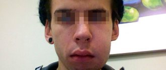
At the initial stage, osteomyelitis of the jaw has no characteristic signs. The person feels unwell, as with most inflammatory diseases. Unaware of the real cause of poor health, the patient may assume another disease and self-medicate. Not only will this not help, but it will also delay the moment of recovery. Therefore, if you feel unwell, it is important to consult a doctor.
External symptoms of maxillary osteomyelitis:
- General weakness, sweating, headache. The person is depressed, he sleeps poorly, refuses to eat.
- Depending on the type of infection and the patient’s immunity, the body temperature rises to 38 or higher or remains normal. If there is no temperature, then the body is not fighting the infection.
- In the acute form of odontogenic osteomyelitis, the tooth affected by the infection hurts. The pain intensifies with pressure and is not relieved by taking painkillers. The tissue around the tooth is swollen and has a reddish tint. Not only the diseased tooth moves, but also those nearby.
- Sometimes an abscess develops on the periosteum of the tooth root. Inflammation affects neighboring teeth, and acute pain shoots into the ear, temple and eye area.
- With osteomyelitis of the lower jaw, the lower lip, mouth and chin become numb. When inflammation invades the jaw tissue, pain spreads throughout the face and neck. The submandibular and cervical lymph nodes become enlarged and painful.
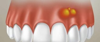
- A periodontal pocket filled with pus forms in the space between the tooth and gum.
Symptoms of acute osteomyelitis of the jaw:
- it hurts to chew;
- at the site where the infection develops, the skin turns pale and becomes covered with plaque;
- the sclera of the eyes turn yellow;
- blood pressure surges;
- with osteomyelitis of the lower jaw, part of the lower lip becomes numb and stops moving, this is due to the fact that the source of inflammation compresses the alveolar nerve.
In subacute osteomyelitis, the inflammatory process continues and sequestration is formed. The teeth in the affected jaw retain and even increase mobility.
With chronic osteomyelitis, the patient feels satisfactory. During remission, the pain subsides. However, the following warning signs remain:
- lack of appetite;
- poor sleep;
- soreness of the facial skin;
- lymph nodes are enlarged;
- fistulas in the mouth do not heal, pus is regularly discharged from them;
- the mucous membranes of the mouth are swollen;
- The teeth on the affected jaw are mobile, and their mobility increases over time.
During an exacerbation, the patient again feels pain spread throughout the jaw and cannot always accurately indicate the location of its localization.
Infants and young children can also develop osteomyelitis of the jaw (usually the upper jaw). The disease develops against the background of sepsis or becomes a complication of ARVI. The symptoms of osteomyelitis in children are pronounced; the disease progresses rapidly and can lead to severe complications, including pneumonia and meningitis. Therefore, it is important to consult a doctor as soon as possible and begin treatment if your baby has the following symptoms:
- high temperature that cannot be reduced with antipyretic drugs;
- cheeks, eyes, lips swell, facial asymmetry appears, it is difficult for the child to open his eyes, and the swelling of the nose makes it difficult to breathe. If you do not see a doctor, the swelling spreads to the neck;
- lymph nodes enlarge;
- purulent foci are noticeable on the gums, and over time infiltrates and fistulas open;
- pain in the eye area.
Features of the treatment of acute purulent periostitis
In the case of acute purulent periostitis, the patient is required to be prescribed painkillers, antibiotics and physiotherapy. In addition, the dentist makes an incision in the gum, removes all pus and installs drainage, for which the tip of a rubber strip is inserted into the cavity located at the site of the incision. This prevents the wound from healing very quickly and allows the fluid formed at the site of inflammation to flow out of it for some time without accumulating.
Acute periostitis is usually provoked by a diseased tooth, which must be removed if its crown is severely damaged or there is obstruction of the root canals. In addition, the tooth that is the source of infection is removed if it has become mobile, if its functionality has been lost, or if the conservative treatment method has proven ineffective.
If acute periostitis is caused by inflammation of the tonsils or throat, then it is necessary to eliminate the causes that caused the development of the infection and restore immunity. The most effective approach to treating this form of periostitis is complex, combining surgical treatment, physiotherapy and drug therapy.
Diagnostics
Osteomyelitis cannot go away on its own; without medical help, the patient’s condition will worsen.
Periodontitis, cysts and tumors of the jaw, as well as lesions of oral tissue due to syphilis, tuberculosis, and fungal skin infections have similar symptoms. Therefore, self-diagnosis is highly not recommended.
When visiting the clinic you must:
- Undergo an initial examination of the jaw by a dentist. With osteomyelitis, tapping and palpating the affected areas will be painful.
- Take an x-ray. In the acute form, it is not very informative, but in subacute and chronic pathology, areas of change in bone density and areas of dead bone tissue will be noticeable.
- Take a general urine and blood test, a biochemical blood test, and bacteriological culture to identify the type of pathogenic microorganisms.
- Get a CT scan.
Treatment of gumboil with folk remedies
In order not to harm the body, folk remedies for treating flux can only be used for temporary pain relief or as a supplement during the recovery period.
- Infusions of oak bark, sage, St. John's wort, and calendula are used to rinse the mouth (strictly warm solutions).
- Chamomile flower tea is taken orally and is also used as a mouth rinse.
- Honey or honey propolis products are used to lubricate the gums.
In case of treating periostitis at home, you should consult a doctor. In any case, you should not postpone a surgical visit to the dentist when the first symptoms of gumboil appear on the gums or replace medical therapy with self-medication. When treating flux only with folk remedies, inflammation can spread further or develop into chronic periostitis.
With a timely and comprehensive approach to the treatment of periostitis, the disease is successfully cured. However, full recovery will require the help of a qualified dentist, medical procedures and medications.
Treatment of osteomyelitis of the jaw
Treatment of osteomyelitis of the jaw is complex and consists of surgery, drug treatment and restorative therapy, which promotes tissue regeneration.
During surgery:
- the surgeon removes the affected tooth;
- treats fistulas and boils in the oral cavity;
- drains pus;
- removes dead (sequestered) areas of bone;
- fills hollow areas with bone materials;
- strengthens mobile teeth with splints.
At the same time, drug treatment is carried out:
- antibiotics;
- immunostimulants;
- vitamin complexes.
To consolidate the result, you must undergo a course of physical procedures:
- UHF;
- magnetic therapy;
- ultrasound therapy.
During the treatment process, the patient is prescribed plenty of fluids and a gentle diet of light, crushed and nutritious foods. It is necessary to carefully monitor oral hygiene.
Desensitization therapy
The treatment complex is supplemented with antihistamines:
- diphenhydramine;
- chloropyramine;
- fenkarol;
- diazolin.
Medicines help eliminate intoxication, activate the anti-inflammatory functions of antibiotics, minimize the permeability of the vascular wall, and fight swelling. To increase the effect, drugs containing calcium and glucose are prescribed in combination.
This treatment protocol is used for patients with diabetes, since this approach minimizes the negative impact of concomitant disorders in the foci of infection and facilitates the recovery process.
General restorative therapy
As part of a general strengthening course, the patient is prescribed vitamin complexes (groups B, C), adaptogenic drugs, and stimulants.
In severe forms of the disease, extracorporeal detoxification is performed. For this purpose, hemosorption, plasmapherosis or lymphosorption are prescribed. Courses of autohemotherapy (administration of one’s own venous blood intramuscularly) have a positive effect.
Physiotherapy
Such treatment is allowed only after acute inflammatory symptoms have reduced, so manipulations are prescribed a few days after the start of therapy. The methods may be as follows:
- hardware treatment with UV rays;
- electrophoresis using anti-inflammatory and antibacterial drugs;
- massage;
- oxygenation;
- laser irradiation of the site of the extracted tooth.
Regardless of the course of the disease, the treatment protocol is determined only by the doctor, taking into account the physiological characteristics of the body.
Forecast and prevention of osteomyelitis of the jaw
If the patient sought medical help on time, followed the doctor’s recommendations and received the correct treatment in full, osteomyelitis will be successfully cured. Incorrect treatment in combination with a weakened immune system can lead to the following complications:
- sepsis;
- acute purulent inflammation;
- general intoxication;
- the formation of multiple abscesses;
- meningitis;
- inflammation of the facial nerves;
- bone deformities;
- purulent sinusitis and destruction of the walls of the maxillary sinuses;
- infection of the orbit and formation of phlegmon;
- jaw fractures.
To prevent osteomyelitis of the jaw, it is important to:
- Have dental checkups every year.
- Properly brush your teeth, maintain oral hygiene, and use interdental floss and rinses.
- Treat teeth and gums in a timely manner.
- Protect your jaw from injury.
- Strengthen immunity.
- Do not start treatment for infectious diseases, including tonsillitis and ARVI.
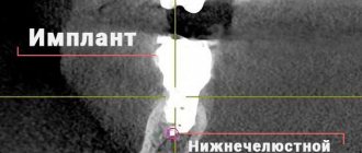
Causes of complications after tooth extraction
General disorders of the condition usually occur against the background of the patient’s nervous overstrain, which are associated with fears, often formed in childhood. But technologies are changing, and now treatment by dentists is not accompanied by pain or any other unpleasant sensations. An individual method of anesthesia is selected for each patient to ensure that tooth extraction is easy and painless.
Local complications after tooth extraction can be associated with several factors. The most common of them are:
- anatomical features (cause a root or crown fracture, injury to adjacent teeth);
- careless work of the dentist (improper use of forceps leads to fractures of the tooth or its root, an incompletely separated tooth ligament injures the gum during removal, and when the forceps or elevator slide, damage to soft tissues is caused);
- lack of x-ray control (causes tooth remains to remain in the gum);
- severe carious lesions (provokes fractures of the root or dental crown);
- failure to follow the doctor’s recommendations (excessive or poor hygiene after tooth extraction causes alveolitis or insufficient opening of the mouth during surgery can cause injury to the gums or soft tissues).
Anatomical features that provoke complications after tooth extraction
- The roots are thin and long.
- Thick alveolar walls, which are difficult for the doctor to work with.
- High level of root curvature.
- Hypercementosis.
- Dense partitions between roots.
- If there are many roots, the degree of their divergence is taken into account.
Knowing the causes of complications after tooth extraction, you can prevent their occurrence. But if it was not possible to avoid complications after the operation, it is necessary to immediately proceed to treatment; delaying seeking medical help leads to pathologies.