Leukoplakia refers to the presence of small areas of altered mucosa. This nonspecific process can be found in any organ where there is a mucous membrane, but most often it occurs in the oral cavity and on the genitals.
We can talk about the problem as a precancerous condition, since it has the potential to develop into a malignant tumor.
With this nonspecific process, the appearance of an area of dense cells of the surface epithelium is observed. In a normal state, there is a regular process of death and exfoliation of the upper layers of the skin and mucous membrane. As the pathology develops, it disrupts it, thus the cells that have died do not disappear and form a multilayered whitish island.
Uterus and reproductive organs
The female reproductive system is represented by the mammary glands and pelvic organs.
The main function of these organs is procreation. The gonads produce regulatory substances that affect the development of the reproductive system and other organs. The most important structures are the ovaries, in which the maturation of female germ cells occurs. During ovulation, the reproductive cell leaves the ovarian follicle and enters the fallopian tube. The fusion of male and female reproductive cells in this organ leads to the formation of the rudiment of a new organism. The remaining stages of embryo development occur in the lumen of the uterus. The uterus is a hollow muscular organ located next to the rectum, vagina, ovaries and bladder. The uterine cavity is connected to the external environment through the cervical canal and the vaginal opening. The fallopian tubes allow the fertilized egg to migrate to the uterus. Attachment of the embryo to the inner layer (endometrium) of the organ is necessary for the formation of embryonic organs.
The main part of the cervix is the narrow cervical canal, which connects the vaginal opening to the uterine cavity. This anatomical structure is necessary for the transport of sperm into the fallopian tubes. The mucous membrane of the cervical canal contains a large number of glands that secrete a special fluid. Gynecologists include the functions of cervical mucus as protection against pathogenic microorganisms and ensuring the transportation of sperm. Smooth muscle and elastic fibers of the cervix provide expansion of the cervical canal during childbirth.
Causes
The exact causes of leukoplakia are unknown. Gynecologists associate the etiology of this disease with pathological external and endogenous influences. Thus, a significant increase in estrogen production against the background of progesterone deficiency can trigger the development of the disease. Hereditary factors play an important role in the formation of leukoplakia: certain genetic mutations can cause epithelial changes.
Possible reasons:
- Diseases of the endocrine system. The endocrine glands regulate the development of all organs, including the structures of the reproductive system. The greatest risk of developing cervical leukoplakia is associated with a disorder of the pituitary gland and ovaries. Diseases of the thyroid gland and adrenal glands can also be complicated by damage to the cervix.
- Chronic infections and inflammatory diseases of the pelvic cavity. In particular, human papillomavirus infection directly affects the condition of the cervical epithelium.
- Sexually transmitted infections including chlamydia, gonorrhea and herpes. Sexually transmitted diseases affect the condition of the external and internal genital organs.
- Mechanical damage to the cervical epithelium that occurs during trauma, diagnostic and therapeutic procedures. Artificial termination of pregnancy leads to tissue trauma.
- Mutation of the gene responsible for suppressing the growth of tumor cells. According to research, a mutation in the p53 gene, which controls cell development, leads to impaired tissue growth.
Normally, the outer lining of the cervix is represented by stratified non-keratinizing epithelium. Certain diseases can affect the internal structure of epithelial cells. Changes in the regulation of epithelial cells lead to keratinization and thickening of the outer layer of the membrane. It is assumed that one of the mechanisms of cell changes is the expression of a mutant gene.
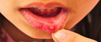
Leukoplakia is a hyperkeratosis of the oral mucosa (ORM), accompanied by inflammation of the stroma and usually occurring in response to chronic exogenous and endogenous irritations [12, 22, 29].
Leukoplakia refers to one of the types of keratoses, characterized by a chronic course and affecting the oral cavity and the red border of the lips [2] under the influence of endogenous and exogenous factors.
Leukoplakia is usually diagnosed in middle age, and the prevalence of the disease increases over the years. Idiopathic leukoplakia of the oral cavity accounts for 10% of cases, and leukoplakia caused by exo- and endogenous factors accounts for 90% [49]. Men are more often affected [44].
Etiology and pathogenesis.
Despite many works devoted to leukoplakia, the question of its etiology still remains open [20, 23, 33, 43]. It has been established that the appearance of areas of leukoplakia on the oral cavity is provoked by weak but long-acting irritants [36]: hot or spicy food, strong alcoholic drinks, tobacco and betel, meteorological (cold, wind) and occupational factors (aniline paints and varnishes, pitch, coal resins, phenol, some benzene compounds, formaldehyde, products of dry distillation of coal, gasoline vapors, bromine, etc.) [7].
The main exogenous causes of impaired keratinization of the oral mucosa include consumption of alcohol, hot, hot and spicy foods, as well as smoking [4, 45].
Recently, a relationship has been identified between the presence of a viral infection (namely human papillomavirus - HPV), the possible onset of the development of leukoplakia and its subsequent malignancy [2, 18, 35].
Chronic trauma to the oral cavity by sharp edges of decayed teeth and improperly manufactured dentures can lead to limited hyperkeratosis and the formation of leukoplakic plaques. The galvanic current that occurs when dissimilar metals are present in the alloy of the prosthesis can also contribute to the disruption of keratinization of the mucous membrane [5, 16]. When leukoplakia is localized on the red border of the lips, chronic injury from a cigarette, pipe, or mouthpiece is important [16, 46].
Leukoplakia is an occupational disease of people working in chemical production and in hot shops [40].
Endogenous factors also have a huge impact on the occurrence and clinical course of leukoplakia.
A relationship has been established between the occurrence of leukoplakia and stress, hormonal disorders [10], the presence of pathology of the gastrointestinal tract (GIT) [9, 15], lack of vitamins A, E, B12 [24, 27, 38, 47, 48], as well as genetic predisposition [13, 14, 19, 32].
There is a connection between the occurrence and development of leukoplakia and chronic candidiasis infection [31, 54].
Some patients are diagnosed with endocrine diseases, in particular diabetes types 1 and 2. The causes of leukoplakia are indicated by fluctuations in the level of the ratio of sex steroid hormones associated with age-related involution, which disrupts the processes of metabolism and protein synthesis and, in turn, reduces the resistance of tissues to exposure to unfavorable factors, but a direct correlation between these factors and the occurrence of leukoplakia was not observed [10, 15].
The relationship between the presence of leukoplakia and HPV remains controversial, although many patients with leukoplakia have HPV type 16 [2, 39, 42, 51].
Classification of leukoplakia SOP
According to the modern WHO classification, 10th revision (1999), a distinction is made between homogeneous and inhomogeneous leukoplakia. Non-homogeneous leukoplakia, in turn, is divided into erythroplakia, nodular, spotted and verrucous leukoplakia [19, 22, 28, 34, 50]. Evidence has accumulated that inhomogeneous leukoplakia, especially macular leukoplakia, is epithelial dysplasia in 50% of cases and is characterized by a high incidence of malignancy [8].
In 2005, WHO adopted another classification of head and neck diseases, in which the concept of “epithelial precancer” appears [52], which, in turn, includes erythroplakia and leukoplakia SOP. According to this, leukoplakia is histologically divided into focal epithelial hyperplasia (without atypia), low, moderate and high grade dysplasia. For the last 3 types of leukoplakia, the concept of “squamous intraepithelial neoplasia” (Squamous Intraepithelial Neoplasia - SIN) from 1 to 3 degrees of severity was introduced. The main feature of the SIN classification is a clear display of each stage of malignant transformation of the epithelium of the oral cavity [53].
It should be noted that this classification is primarily pathomorphological and complements the clinical diagnosis itself.
Histological structure of unchanged epithelium of the oral cavity
The unchanged, non-keratinized stratified squamous epithelium of the oral cavity includes a basement membrane, a basal layer, represented by 1 row of cells having a cylindrical or cubic shape; Next comes the parabasal cell layer, consisting of 1-2 rows of cells located above the basal ones and having no connection with the basement membrane, then the spinous layer, which contains 5-6 rows of large, irregularly shaped cells with processes, then flattening of the cells and their turning flat. On the hard palate, dorsum of the tongue and gums in the keratinized epithelium, adjacent to the spinous layer is a granular layer, consisting of elongated cells with keratinosomes and keratohyaline granules, as well as a stratum corneum with rows of keratinized cells, in which nuclei and cytoplasmic organelles are not visible. Cytologically, from the basal to the superficial keratin layer of the epithelium, a progressive decrease in the nuclear-cytoplasmic ratio and accumulation of intracellular keratin is noted [3].
Clinical and morphological characteristics of leukoplakia
The clinical picture of leukoplakia depends both on the form of the disease and the factor causing it, as well as on the location. Leukoplakia is a chronic disease; patients often cannot clarify the moment of its onset. Clinically, leukoplakia is manifested by hyperplastic processes in stratified squamous epithelium under the influence of various damaging agents and is expressed in the formation of superficial hyperkeratosis. The parabasal, basal and superficial layers of the epithelium change. Clinically, leukoplakia has several forms; histologically it can be characterized by various nosological processes - from benign hyperkeratosis to invasive squamous cell carcinoma. Therefore, all excised tissue must be sent for pathomorphological examination, which should include histological and immunohistochemical (IHC) examination.
IHC research is a method for identifying the specific antigenic properties of malignant tumors. IHC methods are used to localize a particular cellular or tissue component (antigen) in situ
by binding it to labeled antibodies and are an integral part of modern cancer diagnostics, providing detection in tissues of various cells, markers of proliferation, cell maturity, and acanthosis factors.
Subsequently, specific antibodies and reagents are used to visualize antigens.
Leukoplakia is a strictly clinical term; it is not currently used for morphological description. With leukoplakia, 4 types of changes can be detected: focal epithelial hyperplasia; squamous intraepithelial neoplasia grade 1 (SINI), squamous intraepithelial neoplasia grade 2 (SINII), and squamous intraepithelial neoplasia grade 3 (SINIII) [17].
With focal epithelial hyperplasia, an increase in the thickness of the epithelium is noted due to an increase in one of the components of the basal, spinous (acanthosis) or superficial (hyper- and parakeratosis) layers. It may be accompanied by inflammatory and reactive cytological changes.
In grade I squamous intraepithelial neoplasia, the epithelium contains cells with slightly altered cytological characteristics; changes are limited to the lower third of the epithelium; at the same time, normal maturation of epithelial cells in the upper 2/3 of the epithelium is maintained. Mitoses of a normal configuration limited to the basal layer may be detected, and hyperkeratosis is also possible.
With squamous intraepithelial neoplasia of grade II, altered cells with atypical cytological characteristics are observed already in the lower and middle third of the epithelium. Large nucleoli are detected in the nuclei; thus, cellular atypia is more pronounced. Differentiation of cells and tissues is preserved in the upper third of the mucous membrane, mitoses are observed in the intermediate and parabasal layers.
In grade 3 squamous intraepithelial neoplasia (SIN III), mitotic activity is detected in actively proliferating cells; they occupy more than 2/3 of the epithelial layer. However, they do not have the cytological atypia characteristic of carcinoma in situ
. Cell differentiation is preserved in the superficial layers. Hyperkeratosis may be present. As neoplastic changes in the epithelium increase, inflammatory phenomena intensify in the underlying dermis, accompanied by infiltration of lymphocytes.
Clinically, the process usually begins with the so-called preleukoplakic stage, which is characterized by inflammation of the oral cavity. This stage is rarely observed on the red border of the lips [12].
After this, keratinization occurs, and the so-called flat, or simple, form of leukoplakia develops, which is a uniform keratinization of a limited area of the mucous membrane, which is the result of epithelial hyperplasia and is accompanied by chronic inflammation of the stroma. Most often, this form of leukoplakia is localized on the mucous membrane of the cheeks, along the line of closure of the teeth and in the corners of the mouth, on the tongue. The affected areas of the mucosa may have different shapes; Thus, keratinization of a limited area of the mucous membrane of the cheek is possible in the form of stripes with clear boundaries and irregular outlines, which cannot be removed by scraping.
There are almost no subjective sensations with flat leukoplakia. Patients may notice a cosmetic defect in the form of a whitish area of the mucous membrane or roughness. The disease is characterized by varying degrees of thickening of the epithelial layer and involvement of the lamina propria of the oral cavity in the chronic inflammatory process; Clinically, the inflammatory reaction has the appearance of chronic inflammation or does not manifest itself at all. An important feature of this form of leukoplakia is that the areas of the oral cavity that have undergone keratinization do not rise above the surrounding tissues.
Pathomorphologically, this form of leukoplakia is described as focal epithelial hyperplasia, in which manifestations of hyperkeratosis alternate with parakeratosis. If keratinization is of the nature of parakeratosis, the phenomena of acanthosis are most pronounced. The lamina propria of the oral cavity in the affected area has an inflammatory infiltrate of mononuclear cells and plasma cells, which alternates with foci of fibrosis and sclerosis. The connective tissue is plethoric, edematous, loosened. The parabasal layer of the epithelium is prominent. The thickness of the spinous layer is 40-50 rows of cells, acanthosis is noted.
Thus, parakeratosis in flat leukoplakia is more often combined with epithelial hyperplasia, in which the basement membrane remains well defined as a line separating the thickened epithelium from the underlying stroma.
The flat form of leukoplakia can proceed for years without changes or can develop into a verrucous form.
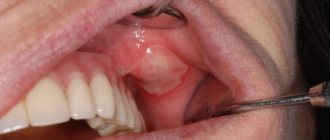
Verrucous leukoplakia is a special form of leukoplakia. Its main clinical sign is an area of pronounced keratinization that rises significantly above the level of the SOR and sharply differs in color from it. Foci of hyperkeratosis have a bright milky white color.
There are plaque and warty forms of verrucous leukoplakia [30]. In the plaque form, limited milky-white, sometimes straw-yellow, rounded formations with clear contours are identified, rising above the surrounding mucous membrane. Verrucous leukoplakia with warty growths has the most pronounced tendency to malignant transformation [1, 8, 21, 26].
The warty variety of verrucous leukoplakia is characterized by dense grayish-white bumpy formations, rising 2-3 mm above the level of the mucous membrane; it has a greater potential for malignancy than plaque. A number of foreign and domestic authors note that it is verrucous leukoplakia that in 70% of cases transforms into squamous cell carcinoma [6, 8, 11, 25, 37].
Histologically, the epithelial cover of the mucous membrane is sharply thickened due to the proliferation of the horny and granular layers. In the cytoplasm of the cells of the granular layer, the amount of keratohyalin increases. The most superficial cells have the appearance of thin scales with dense homogeneous eosinophilic protoplasm and degenerating nuclei. Thin connective tissue papillae protrude deeply into the thickness of the integumentary epithelium. Acanthosis is pronounced; in the underlying, edematous and loosened connective tissue, chronic inflammation, hyperemia, and cellular infiltration are determined. With verrucous leukoplakia, pronounced hyperkeratosis is found, rarely combined with small foci of parakeratosis; sometimes the stratum pellucida is involved in the pathological process; the granular layer consists of 4-5 rows of cells with well-defined granulomas, the spinous layer - of 8-12 rows of cells. It is possible to identify atypical cells. In some cases, acanthosis is accompanied by elongation and expansion of epithelial processes. Changes in the cells of the spinous layer are observed. The cells are of different sizes and shapes, have large hyperchromatic nuclei containing on average 2-4 large nucleoli.
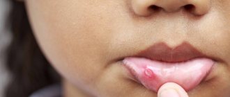
The erosive form of leukoplakia is the most malignant [41]. With the erosive form of leukoplakia, cracks and erosions appear, which are inevitably subject to mechanical and thermal stress.
Erosion occurs as a result of injury to foci of simple and verrucous leukoplakia by mechanical and thermal factors. As a rule, this is observed on the mucous membrane of the tongue, cheeks, and red border of the lips. With increased keratinization and the appearance of infiltration at the base of bleeding and non-healing erosion, we can talk about the malignancy of the process.
Histological examination shows that the mucous membrane is thickened, the spinous layer and intercellular spaces are expanded. Strands of epithelium protrude deeply into the thickness of the connective tissue layer, delimiting elongated, thin papillae - acanthosis. The epithelium is loosened in places; Erosion forms in places where epithelium is lost. Chronic inflammation is observed in the lamina propria of the mucous membrane, intercellular infiltration with an admixture of plasma cells and mononuclear cells is pronounced.
If squamous cell carcinoma is suspected, the histological picture is characterized by the appearance of oval-shaped areas of hyperkeratosis with so-called “cancer pearls”. Complexes of atypical squamous epithelial cells with invasive growth into the lamina propria of the mucous membrane are identified. Atypical cells have cytological signs of malignancy, including a high nuclear-cytoplasmic ratio, prominent 1 or more nucleoli, nuclear hyperchromasia and pleomorphism, and pathological mitoses.
Thus, the pathologist's report will not contain a clinical diagnosis, but diagnoses (SIN), such as focal epithelial hyperplasia, squamous epithelial neoplasia of varying degrees (from 1st to 3rd), the presence or absence of atypical cells, hyperkeratosis, verrucous leukoplakia or squamous cell carcinoma [17].
Based on the above, leukoplakia is a clinical term. The pathomorphological report contains a description in accordance with the 2005 WHO classification: the degree of damage to the mucous membrane is described depending on the depth. The presence of hyperkeratosis and hyperplasia should be considered as confirmation of the clinical diagnosis of leukoplakia. The leading clinical sign of verrucous leukoplakia is foci of hyperkeratosis that rise strongly above the surrounding mucosa, and with ulcerative-erosive leukoplakia - foci of erosion.
If frequent mitoses and a cluster of atypical cells with several large nuclei are described, we can talk about carcinoma in situ
. Leukoplakia is considered a precancerous disease. Its verrucous and erosive-ulcerative forms are most prone to malignancy, therefore, during surgical treatment, pathomorphological examination, including IHC, is mandatory.
Risk factors
In addition to the direct causes of cervical leukoplakia, gynecologists recognize the importance of certain forms of predisposition to the disease associated with heredity, individual history and a woman’s lifestyle.
Known risk factors:
- unprotected sexual intercourse;
- menstrual irregularities;
- drinking alcoholic beverages and smoking;
- radiation therapy of the pelvic organs;
- insufficient intake of vitamins and microelements from food;
- metabolic diseases;
- uncontrolled use of hormonal drugs;
- detection of leukoplakia or cervical carcinoma in a close relative.
Eliminating some of the risk factors listed above is an effective method of preventing the disease.
Leading specialists in the treatment of vulvar leukoplakia in the Southern Federal District
Ermolaeva Elvira Kadirovna is a well-known and recognized specialist in the North Caucasus in the treatment of leukoplakia of the vulva, vagina and cervix, diagnosis and treatment of kraurosis, lichen sclerosus, Keir's erythroplasia and other diseases of the vulva. One of the authors of the method of regenerating microinjections for the treatment of leukoplakia. Desperate people turn to her and women exhausted by suffering. Experienced gynecologist, physiotherapist-health resort specialist, ultrasound doctor.
Ermolaev Oleg Yurievich Candidate of Medical Sciences, gynecologist-endocrinologist with 25 years of experience and successful experience in the treatment of dysplasia, kraurosis and leukoplakia of the vulva Able to see relationships that elude others
About the doctors of the Clinic in detail...
| INTERNATIONAL RECOGNITION of the reputation and achievements of the Women's Health Resort Clinic in the development and implementation of effective and safe treatment methods and the quality of medical services provided is the AWARDING of the Women's Health Resort Clinic in Pyatigorsk with the SIQS International QUALITY CERTIFICATE in the field of medicine and healthcare. International Socratic Committee, Oxford, UK and Swiss Institute for Quality Standards, Zurich, SWITZERLAND. |
The resort women's health clinic is open 7 days a week and on holidays:
Monday - Friday from 8.00 to 20.00, Saturday, Sunday, holidays from 8.00 to 17.00.
Treatment of vulvar leukoplakia in Pyatigorsk by appointment no later than 3 days in advance by multi-channel phone number 8 (calls within Russia are free), or (for foreign calls).
| ONLINE information about the treatment of vulvar leukoplakia in Pyatigorsk can be found at REGISTER ONLINE for leukoplakia treatment here. REGISTER online for leukoplakia treatment here. You can buy a COURSE for treatment by phone or here. |
Booking a course
The doctors of the Women's Health Resort Clinic have gained EXTENSIVE EXPERIENCE in treating vulvar leukoplakia, vaginal leukoplakia, cervical leukoplakia using natural medicines and resort factors.
We accept women from all cities of Russia, near and far abroad.
The spa clinic for women's health facilitates the accommodation and accommodation of women, women with children and couples during examination and treatment.
ACCOMMODATION in Pyatigorsk is NOT INCLUDED IN THE PRICE of the treatment course and is paid separately.
About living conditions and transfer from Mineralnye Vody airport and Pyatigorsk railway station in detail in the article “Accommodation”.
If you need to book accommodation, please coordinate your arrival date no later than 7 days in advance.
We are at your complete disposal if you have any doubts or wishes.
Classification
There are two main types of cervical leukoplakia, differing in morphological and functional features. Determining the specific type of disease is possible only with the help of histological examination. Different morphological forms of leukoplakia differ in the risk of malignant degeneration.
Forms of the disease:
- Simple leukoplakia, characterized by coarsening of the outer layer of the epithelium and excessive cell division. Significant morphological changes in the cells of the basal layer are not detected.
- Proliferative leukoplakia. This form of the disease is characterized by a violation of cell specialization and the appearance of specific morphological changes. Proliferative leukoplakia is considered a precancerous condition.
Determining the form of the pathology is necessary to select treatment.
Types of leukoplakia
How to treat leukoplakia depends on its type. The disease is dangerous because pathological processes in the epithelium can become malignant and provoke oncological tumors.
Today, medicine distinguishes three categories of leukoplakia.
- Simple. The formations are located flush with the epithelium. They are usually white in color and the changes are benign.
- Proliferative. It is expressed in dense growths rising above the epithelium. There are warty and scaly forms.
- Erosive. Characterized by cracking and bleeding lesions. This form is the most dangerous because the structures contain atypical cells.
Symptoms
In most cases, the disease does not manifest itself symptomatically. In itself, the coarsening of the outer epithelium of the cervix does not lead to dysfunction of the organ, so the pathology can remain undiagnosed for a long time. Typically, leukoplakia becomes an incidental finding during a gynecological examination.
Possible symptoms:
- discomfort during sexual intercourse;
- pain during menstruation;
- bloody issues;
- excess vaginal discharge.
The appearance of frequent uterine bleeding not associated with menstruation and pain may indicate the transformation of leukoplakia into a malignant tumor.
What it is
It is believed that leukoplakia occurs against the background of chronic irritation of the oral mucosa and is a kind of protective reaction of the body. This is a local reaction and is not transmitted through contact with a sick person.
The disease is not so harmless, since it most often occurs shortly before the development of oral cancer. Many people mistakenly believe that leukoplakia itself is cancer, however, this opinion is incorrect. This disease affects the epithelial covering of the oral cavity in response to constant external irritants; vitamin deficiency, a decrease in the level of immunity and the presence of chronic foci of inflammation in the oral mucosa serve as an additional impetus for its development.
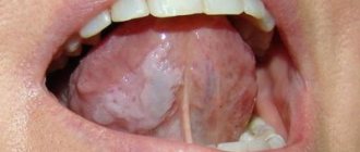
Leukoplakia of the oral cavity is a significant lesion of the mucous membrane of the lips, tongue, cheeks, upper and lower palate, manifested by keratinization (hardening) of the epithelial cover to varying degrees. Doctors usually classify leukoplakia as a disease that precedes the development of a tumor.
The greatest risk of developing this disease are men in the age group from 30 to 70 years, who abuse alcohol, smoke and wear dentures. Leukoplakia does not occur overnight, and its development can last for years.
Diagnostics
If risk factors for the disease are detected, you must make an appointment with a gynecologist. During the consultation, the doctor will ask the woman about her complaints and examine her medical history. Then an initial examination of the genital organs is carried out, including palpation examination and the use of mirrors to assess the condition of the cervix. If suspicious changes are detected, the gynecologist prescribes instrumental and laboratory tests.
Additional diagnostic methods:
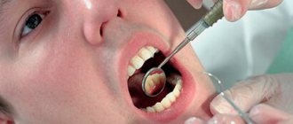
- Colposcopy is a method of visual examination of the cervix. The doctor asks the patient to sit in the gynecological chair. A colposcope equipped with optics and a light source is carefully inserted into the cervix through the vaginal opening. The gynecologist studies in detail the condition of the epithelium of the organ. Treatment of suspicious areas with iodine solution makes it possible to detect pathological changes in tissues. If necessary, the optics are replaced by a video camera, allowing the specialist to see a clear image of the organ tissue on the monitor. Colposcopy is a reliable and safe examination that does not require anesthesia.
- Scraping of cervical epithelial cells. During a colposcopy, the doctor inserts a special instrument into the organ cavity to collect cells from the suspicious area. Cytological examination of the obtained material in the laboratory makes it possible to evaluate morphological changes and determine the form of leukoplakia. Cytology is a reliable method for excluding malignant tissue degeneration.
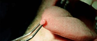
- Cervical biopsy. If the epithelium is severely coarsened, the doctor cannot obtain cells from the lower layers of leukoplakia by scraping. In this case, a knife biopsy is the optimal diagnostic procedure. Using a special instrument, the specialist excises the required amount of tissue. Cytological examination of the biopsy is also necessary to exclude the growth of malignant cells. The procedure is performed under local anesthesia. If necessary, the gynecologist prescribes complete curettage of the epithelium of the cervical canal.
- Microbiological examination of the material. Tissue obtained by scraping or smear can be used to rule out infection. Special nutrient media give a specialist the opportunity to identify the causative agent of the disease.
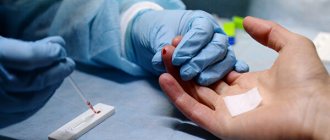
- Blood analysis. In the treatment room, a nurse collects venous blood from a patient. Laboratory examination of the material is carried out to detect signs of inflammation and infection. Serological tests aimed at searching for specific immunoglobulins are necessary to exclude sexually transmitted diseases. If an HPV infection is suspected, sections of the DNA of the virus are searched using polymerase chain reaction (PCR).
The scope of diagnosis depends on the results of colposcopy and general examination. The main goal is to eliminate the risk of malignant tissue degeneration.
Treatment
The treatment method depends on the form of leukoplakia and the patient’s individual medical history. Due to the high risk of transformation of the pathology into carcinoma, it is recommended to remove the focus of the changed tissue. In some cases, the gynecologist prescribes drug therapy. Prescribed medications may include antibiotics and anti-inflammatory medications. Treatment of the root cause of the disease helps prevent relapse.
Pathology removal methods:
- Cryosurgery is the use of cold to destroy the affected epithelium. During the procedure, the patient is in a gynecological chair. After inserting the dilator into the cervix, the surgeon uses a special probe, the tip of which is cooled with liquid nitrogen. Destruction of leukoplakia takes 5-10 minutes. Cryosurgery is not accompanied by tissue trauma or severe pain.
- Radio wave destruction. A special device inserted into the cervix affects the tissue. This is a safe and fast treatment method with a low risk of complications.
- Other methods of minimally invasive removal of the disease, including chemical cauterization and argon plasma coagulation. The duration of rehabilitation after such interventions does not exceed 2 months.
If the gynecologist detects signs of malignant degeneration of leukoplakia, radical methods of surgical treatment are prescribed. The doctor needs to remove not only the pathological focus, but also the cells surrounding it. This is important to prevent abnormal cells from migrating into neighboring tissues.
Other methods of surgical treatment:
- Conization of the cervix. Using a special instrument, the specialist applies radio waves to the organ and removes the affected part of the cervix. This is a low-traumatic procedure that allows you to preserve reproductive function.
- Amputation of the cervix - removal of the affected segment of the organ. The disadvantage of this method is the high degree of tissue trauma, but the operation allows you to preserve the integrity of the reproductive system.
During treatment of the disease, the doctor asks the patient to abstain from sexual intercourse and taking hormonal contraceptives.
Prevention and prognosis
In most cases, the prognosis is favorable. The absence of human papillomavirus infection and dysplasia indicates a low risk of malignant tissue degeneration. Surgical intervention not only eliminates the risk of organ malignancy, but also preserves reproductive function. After treatment, the gynecologist regularly conducts examinations to eliminate the risk of relapse.
Simple medical recommendations can reduce the likelihood of leukoplakia and other diseases of the reproductive system. Prevention methods are aimed at changing lifestyle and eliminating negative external factors.
Basic methods of prevention:
- use of hormonal drugs only under the supervision of a physician;
- regular gynecological examinations;
- keeping a calendar of the menstrual cycle and contacting a doctor if disorders occur;
- refusal of alcoholic beverages and cigarettes;
- timely treatment of chronic diseases of the genital and endocrine organs;
- using a latex condom;
- sexual intercourse only with a trusted partner (HPV infection can be transmitted even when using a condom).
A consultation with a gynecologist or oncologist will help a woman learn more about the risk factors for leukoplakia and treatment methods for this disease. If necessary, the doctor immediately conducts an examination and excludes the presence of suspicious changes in the cervix.
Reasonable restrictions for leukoplakia
For leukoplakia of the vulva, vagina and cervix, it is extremely UNADVANTABLE to take hot general baths, stay in the sun for a long time and sunbathe (regardless of the activity of the Sun, the presence/absence and type of bathing suit).
HEAT TREATMENTS and insolation (sunbathing in natural conditions and solariums) increases the production of estrogens (female sex hormones), the excess of which stimulates oncological processes.
- Reviews about the treatment of vulvar leukoplakia in our Clinic
- About the Clinic
- Clinic team
- Prevention of female diseases
- What is homeopathy?
- How to prepare for an appointment with a gynecologist?
Our specialized clinic for the treatment of leukoplakia is open 7 days a week and on holidays:
Monday - Friday from 8.00 to 20.00, Saturday, Sunday, holidays from 8.00 to 17.00.
Treatment of vulvar leukoplakia in Pyatigorsk by appointment no later than 3 days in advance by multi-channel phone number 8 (calls within Russia are free), or (for foreign calls).
| ONLINE about the treatment of leukoplakia in Pyatigorsk at BOOK ONLINE for leukoplakia treatment here. BOOK online for leukoplakia treatment here. Buy coursework for treatment by phone or here. |
Booking
Subsections
- Program No. 1. Treatment of inflammatory and infectious diseases of the pelvis
- Program No. 2. Treatment of urinary incontinence, prolapse and prolapse of the uterus and vagina
- Program No. 3. Treatment of female infertility of endocrine (hormonal) and mixed origin
- Program No. 4. Treatment of erosion, endometriosis, leukoplakia, dysplasia, polyps, cervical cysts (uterine cysts)
- Program No. 5. Treatment of uterine fibroids
- Program No. 6. Treatment of cervicitis and endocervicitis
- Program No. 7. Postpartum rehabilitation and wumbling
- Program No. 8. Treatment of chronic cystitis
- Program No. 9. Treatment of severe menopause
- Program No. 10. Treatment of mastopathy
- Program No. 11. Treatment of endometriosis
- Program No. 12. Treatment of hydrosalpinx
- Program No. 14. Comprehensive treatment of polycystic ovary syndrome
- Program No. 15. Preconception preparation for IVF, ICSI
- Program No. 16. How to remove belly fat
- Program No. 17. Treatment of kraurosis
- Program No. 18. Treatment of vulvar leukoplakia