1.What is hemophilia and its causes
Hemophilia
is a blood disorder in which the blood does not clot properly. Typically, this occurs because a person with hemophilia lacks a certain clotting factor. This means it can be difficult to stop bleeding. Moreover, this can apply to both ordinary bleeding from wounds and falls, and bleeding during some operations. It also happens that some people with hemophilia begin to bleed internally for no apparent reason.
There are two main types of hemophilia:
- Hemophilia A
is caused by the absence of active blood clotting factor VIII. According to statistics, approximately 1 in 5,000 male infants are born with type A hemophilia. Blood clotting factor VIII is a plasma protein. The greater the deficiency, the more severe the symptoms of hemophilia. - Hemophilia B
(Christmas disease) is caused by a lack of active blood clotting factor IX in the body. This form of hemophilia is less common and is diagnosed in about 1 in 30,000 male infants.
Hemophilia is usually inherited and almost always affects men. In very rare cases, a person can develop hemophilia without a family history. This is called acquired hemophilia, and it occurs in both men and women.
Causes of hemophilia
Hemophilia, both type A and B, occurs due to a defect in a pair of chromosomes that affects the presence of a certain blood clotting factor in the body and how it works. In the case of acquired hemophilia, the blood clotting factor stops working as it should because it begins to be attacked by antibodies that the body itself produces.
A must read! Help with treatment and hospitalization!
Blood clotting
Many people begin to fear blood clots after, when donating blood from a vein, the nurse reports that it is too viscous. Those who have too thin blood that successfully fills the test tube hope that they are protected from thrombosis. But viscosity and coagulability are two completely different concepts, and one does not always determine the other.
How viscous you are
Increased blood viscosity, due to which it becomes less fluid, most often occurs due to the predominance of its formed elements over liquid ones. This happens due to too strict adherence to the recommendations “do not eat 12 hours before taking the test” and because of the decision to add one more restriction to this – not to drink. Just to be sure. The end result is worse - and the blood flows poorly into the test tube, and some indicators (for example, hemoglobin, hematocrit, total number of red blood cells, white blood cells and platelets) turn out to be artificially high. Therefore, it is important to remember: before taking blood tests, you should not limit yourself to fluids.
Another common cause of increased viscosity is increased levels of red blood cells and hemoglobin, characteristic of smokers. After all, the more smoke and less air a person inhales, the greater the concentration of oxygen carriers required. Their compensatory increase is formed. Therefore, visually, the blood of smokers often appears more viscous.
According to hematologists, true diseases (thrombocytosis, erythrocytosis, etc.) associated with increased blood viscosity account for a small number of all cases of “viscous blood”. And this is clearly visible from a routine general blood test - the doctor will immediately notice that the number of red blood cells or platelets is too high.
Normally, the content of red blood cells is 3.7-5.1, platelets - 180-320.
Viscosity and coagulability - what's the difference?
The most important indicator is blood clotting. Unfortunately, it can be difficult to obtain accurate information about coagulation, even despite the level of development of medicine. On the one hand, obvious diseases with clotting disorders, such as hemophilia, have long been known. On the other hand, there is a lot of hidden pathology, which may not show itself for a long time, but once manifested, it can quickly lead to serious consequences.
Only in recent decades have researchers learned to identify these problems using high-tech genetic tests. Considering that, according to statistics, more than 1-3% of the world's population has a congenital pathology of the blood coagulation system, it is likely that in the future these tests will be carried out in the maternity hospital for every newborn. And absolutely for those who need to be prescribed certain medications that can increase the risk of blood clots.
Protection or danger?
For example, this is very important to do before prescribing hormonal contraceptives. Their ability to enhance blood clotting and occasionally lead to thrombosis (as it turns out, this almost always occurs in women who have a hidden clotting disorder) has been known for several decades. Due to the widespread use of this method of protection against unwanted pregnancy these days, the incidence of thrombosis in young, apparently healthy women has increased. At the same time, when prescribing COCs, gynecologists rarely focus their patients’ attention on this danger. They can be understood - the complication is infrequent in general, and I don’t want to once again intimidate patients, as if pushing them into an unwanted pregnancy. Meanwhile, the most advanced Western colleagues have already included a full blood clotting test in the mandatory examination algorithm before prescribing hormonal contraceptives.
What will the analysis show?
What tests need to be done to check blood clotting? The most common and familiar analysis to many, a coagulogram, may not answer all questions, especially in the prevention of thrombosis.
However, a classic coagulogram is the first stage of a screening examination of the coagulation system. If it reveals abnormalities, the next step will be more detailed studies of hemostasis - thromboelastography or thromboelastometry. A separate story is the determination of D-dimer , Leiden mutation and other genetic clotting disorders - tests that reveal a tendency to form blood clots in the future. What is necessary and in what cases?
The most common standard coagulogram today includes five components: PTI (prothrombin index); INR (International Normalized Ratio. Reflects the ratio of the patient’s blood clotting time to the blood clotting time of a healthy patient; APTT (activated partial thromboplastin time. Estimates the time it takes for a blood clot to form after special reagents are added to the plasma), FIBROGEN and PLATELET LEVEL.
At the same time, APTT is informative only in people undergoing treatment with heparin, and INR is important only for people constantly taking blood-thinning drugs from the neodicoumarin group (warfarin).
It turns out that two out of five indicators are not so important for screening. The total number of platelets is also not always indicative, because with most coagulopathies it is not their number that changes, but primarily the functional activity.
Therefore, the most informative method, which allows one to evaluate several stages of blood coagulation at once, is thromboelastography. This is a kind of detailed observation of the formation of a blood clot, and its subsequent dissolution (lysis) with the construction of graphs of each of the stages. Thromboelastometry is another version of this study, which is considered even more informative. Unfortunately, the instruments for conducting these studies are expensive and require special training of personnel, so not every laboratory can offer thromboelastography services.
Another important indicator is D-DIMER (this is a breakdown product of fibrin, a small fragment of protein present in the blood after the destruction of a blood clot).
It is actively used to determine the risk of blood clots. Those with even slightly elevated D-dimer are at much greater risk of developing blood clots than others. It is necessary to monitor D-dimer in case of venous diseases (thrombophlebitis), after surgical interventions and upon discharge from the hospital if you have been bedridden for a long time. Monitoring D-dimer levels is useful during pregnancy and when taking hormonal contraceptives (the risk of blood clots in the presence of the Leiden mutation while taking birth control pills increases almost 9 times). And now during COVID-19 and several weeks after recovery.
Today, in addition to D-dimer testing, genetic tests for congenital bleeding disorders are emerging. The most common of these is the Leiden mutation, which occurs in 2–6% of Europeans. The presence of a defective gene increases the likelihood of venous blood clots 6–8 times, and the risk of heart attack and stroke increases significantly. But other mutations, of which there are more than ten today, are no less dangerous. At the same time, timely prevention of thrombosis (mainly the constant use of anticoagulants, the exclusion of certain foods and medications, wearing compression stockings when flying and working on your feet, etc.) reduces the risk of dangerous complications tenfold.
Flickering problem
If everything is in order with the coagulation system, a possible cause of increased coagulation may be arrhythmia, namely atrial fibrillation. According to statistics, 2% of the planet's population suffers from paroxysmal (occurring in attacks) or permanent form of atrial fibrillation. This problem usually appears after thirty. The basis for the disorder is the appearance of pathological turbulence of the electrical impulse in the atria, which gives the myocardium extraordinary electrical stimuli. In medical language, this is called the “re-entry” or re-entry mechanism. As a result, the atria become a churn that churns blood into clots like milk into butter. The situation worsens even more in the case where the person initially had the same clotting disorders. Blood clots can form within 48 hours after the onset of an attack and “fly away” into the arteries of the brain, causing an ischemic stroke, into the intestines, leading to mesenteric thrombosis, into the arteries of the extremities, provoking their acute ischemia.
Von Willebrand factor and COVID -19.
Severe COVID-19 may be associated with elevated levels of one of the blood coagulation factors, von Willebrand factor. Anna Aksenova, a senior researcher at the Laboratory of Amyloid Biology at St. Petersburg State University . Her scientific article was published in the journal Ecological Genetics. It has already been proven that the SARS-Cov-2 virus can have a direct damaging effect on the inner wall of blood vessels. In response to damage, the body strives to “patch” the hole as quickly as possible, and the leading role in this is played by the von Willebrand factor, which is involved in the activation of platelets and, in fact, triggers the process of local thrombus formation. In the course of research, it turned out that some people are characterized by an increased concentration of this factor in their cells, so, as a rule, it is higher in people with blood group II. An individual characteristic of the body is also possible. As a result, in response to massive microdamage to blood vessels, massive microthrombosis occurs, which causes the appearance of larger and more dangerous blood clots.
Genetic mutations of the coagulation system identified during tests:
MUTATION OF COAGULATION FACTOR V OF BLOOD CLOTTING (LEIDEN FACTOR)
PLASMINOGEN ACTIVATOR INHIBITOR 1
MUTATION OF COAGULATION FACTOR II (MUTATION OF PROTHROMBIN)
MUTATION OF METHYLENETETRAHYDROPOLATE REDUCTASE (MTHFR C677T)
MUTATION OF COAGULATION FACTOR VII OF BLOOD CLOTTING (F7 ARG353GLN)
POLYMORPHISM OF THE METHIONINE SYNTHASE REDUCTASE GENE (MTRR A66G)
FIBRINOGEN MUTATION, BETA (FGB G-455A)
MUTATION OF THE PROMOTER OF THE COAGULATION FACTOR GENE FVII (-312 INS 10BP)
INSERTATION/DELETION OF ALU ELEMENT IN THE ANGIOTENSIN CONVERTING ENZYME GENE (ALU INS/DEL)
PLATELET GLYCOPROTEIN 1B ALPHA SUBUNIT MUTATION
PLATELET ADP RECEPTOR MUTATION (P2RY12 H1/H2)
MUTATION A1298C OF METHYLENETETRAFOLATE REDUCTASE GENE
D-dimer is significantly elevated in most patients with moderate to severe COVID-19. Therefore, all patients receive therapeutic doses of anticoagulants.
The Leiden mutation is the most common hidden bleeding disorder, occurring in 2-6% of Europeans.
Published: January 19, 2022
2. Symptoms of the disease
Symptoms of hemophilia may include:
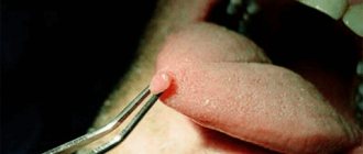
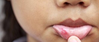
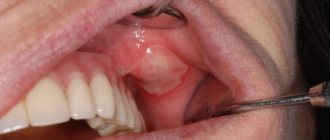
- Bleeding in a joint or muscle area that causes pain and swelling;
- Abnormal bleeding after wounds or surgery;
- Bruising;
- Frequent nosebleeds;
- Blood in the urine;
- Bleeding after dental surgery.
Some people with mild hemophilia may not experience all of these symptoms, especially as the person gets older. However, in infants it is usually possible to diagnose hemophilia based on some signs. So, signs of hemophilia in infancy may be an unusual reaction to the most common vaccination - intramuscular bleeding and serious bruising. Or, for example, bleeding that begins after cutting the umbilical cord and does not stop for a long time (but this happens very rarely).
Visit our Therapy page
Forecast
Acute forms of idiopathic purpura disappear within a few months and it often happens that the disease does not recur. In the chronic form, it is possible to induce remission, but the disease often recurs.
When using modern medications and following all recommendations for lifestyle changes and employment, thrombocytopenic purpura has a favorable prognosis.
Untreated hemophilia Lack of treatment often leads to joint pathology ( hemophilic arthropathy ), which then requires the use of crutches and wheelchairs or special orthopedic treatment. During treatment, life expectancy is practically no different from healthy individuals.
3.Diagnostics and treatment
Diagnosis of hemophilia
If your doctor thinks you or your child may have a blood clotting problem, a blood test will be done. The tests will help check your body's clotting factor, the type of hemophilia, and the severity of the disease. The severity of the problem depends on how much clotting factor the body produces and how often and under what conditions bleeding occurs.
Depending on this, there are several types of hemophilia:
- Mild hemophilia
. The blood clotting factor level is at least 5% of normal. This type of disease is not always noticed, especially if the person has not had heavy bleeding after a major injury or surgery. - Moderate hemophilia
- the level of blood clotting factor is from 1% to 5% of normal. Bleeding usually begins after a fall, sprain, or severe muscle strain. - Severe hemophilia
is diagnosed when blood clotting factor levels are below 1% of normal. People with severe hemophilia often bleed up to several times a week, without any obvious reason.
If you have a family history of hemophilia and you are planning a pregnancy, ask your doctor about special tests that can show whether you are a carrier of the disease (only women can be carriers).
Treatment of hemophilia
Hemophilia can be treated by replacing missing clotting factors
. During this therapy, clotting factor concentrate is injected into a vein. This replacement therapy can prevent or treat bleeding.
You may need to take special medications to treat hemophilia. Sometimes such drugs are prescribed before a certain procedure, which may be accompanied by blood loss - surgery or, for example, dental treatment at the dentist. In some cases, it is also necessary to take pain medications to help manage pain due to joint damage.
By following all your doctor's recommendations for treating hemophilia, you will be able to lead a normal life with this disease. As a rule, modern clinics have the necessary resources to help patients with hemophilia.
About our clinic Chistye Prudy metro station Medintercom page!
In children
Poor blood clotting in a child is often associated with immune thrombocytopenic purpura . Acute purpura develops between the ages of 2 and 9 years. This is an immune-related disease characterized by a constant (or periodic) decrease in platelets of less than 100 thousand. The disorder in children occurs 1-3 weeks after a viral infection. Such pathological reactivity can start not only under the influence of a viral infection, but also after taking medications, vaccination, exposure to temperatures (both low and high), surgical interventions or emotional stress. Against the background of normal health, the child develops a petechial rash (on the mucous membranes and skin), bruises, repeated nosebleeds and bleeding gums. In severe cases, there may be brain hemorrhages and stomach bleeding. Since antigens gradually leave the blood, in most people the disease goes away on its own after 2 months.
Vitamin K deficiency coagulopathy occurs in children in the first months of life and newborns . With Vit-K deficiency, the activity of several factors decreases: prothrombin , proconvertin , Christmas factor and Prower factor . hypocoagulation develops , accompanied by hemorrhagic syndrome.
Causes of Vit-K deficiency in a newborn:
- taking anticoagulants during pregnancy;
- antibiotics;
- anticonvulsants;
- severe damage to the pregnant woman’s liver and intestines;
- the presence of fetoplacental insufficiency;
- gestosis and preeclampsia .
The manifestations of this coagulopathy in newborns are not very specific - skin syndrome, increased bleeding during blood sampling and bleeding from the umbilical wound. With a lack of Vit-K, the duration of bleeding and platelet levels are within normal limits. Many authors recommend prophylactic administration of Vit K to all children immediately after birth - 2-3 administrations for the first 1.5 months, and in some cases weekly administration continues up to 3 months.
In later life, Vit-K deficiency coagulopathy is caused by breastfeeding only. At the same time, 78% of children develop massive intracranial hemorrhages. Much less frequently, the cause of decreased coagulability in children is disseminated intravascular coagulation syndrome in severe sepsis, congenital metabolic changes and hereditary coagulopathies.
4.What can be done at home for hemophilia?
To prevent bleeding and improve your well-being, patients with hemophilia can recommend the following:
- Ask your doctor about how to manage bleeding if you have hemophilia;
- Maintain a healthy weight. Additional stress on joints due to excess weight can cause bleeding in hemophilia;
- Choose forms of physical activity with caution. It is better to give preference to swimming and other sports that do not put unnecessary stress on the joints;
- Consult your doctor before taking any medications. And do not take aspirin, ibuprofen or other non-steroidal anti-inflammatory drugs because they affect blood clotting.
- Organize your living space to avoid injuries and accidents as much as possible.
Pathogenesis
The pathogenesis of hemorrhagic syndrome includes:
- damage to vascular endothelial cells by inflammatory mediators or endotoxin ;
- activation of protein C , which inhibits FV and FVIII and suppresses the synthesis of plasminogen activator inhibitor (the latter promotes the transition of plasminogen to plasmin, which breaks down the fibrin of the blood clot);
- activation of fibrinolysis, which plays a role in the development of non-stop bleeding and depletion of f I, II, V, VIII, XIII;
- accumulation in the blood of metabolites that have an anticoagulant effect.
In the pathogenesis of uremic thrombocytopathy, platelet deficiency is important, which is associated with the action of toxic plasma metabolites. In addition, patients with uremia undergo an extracorporeal circulation procedure, in which platelet dysfunction occurs due to their interaction with the tubes and membranes of the device. In this case, platelets are activated and release granules (platelet degranulation). Platelet dysfunction causes bleeding so severe that platelet transfusion is required.
Drug-induced thrombocytopenia is associated with the interaction of the drug (or its metabolite) and a platelet membrane glycoprotein. As a result of this interaction, an immunogenic complex - a glycoprotein-drug . Altered platelets are removed from the bloodstream by RPE cells. With drug-induced thrombocytopenia, the level of IgG and platelet-bound antibodies to the drug increase. Idiopathic purpura based on the production of antibodies against viral antigens. Platelets are damaged by adsorbing a viral antigen or a virus-antibody immune complex on their membrane.
Disorders of vascular-platelet hemostasis
Disturbances in this part of hemostasis most often manifest themselves as increased bleeding, a tendency to form hematomas (bruises) with the slightest contact, or even spontaneously, for no apparent reason. In some situations, on the contrary, there is a tendency to excessively easy thrombosis.
There are factors that stimulate the formation of a primary thrombus and those that disrupt it. Stimulants include the inflammatory process, because inflammation increases the content of biologically active substances in the blood. We can say that there is a readiness for the formation of a blood clot; it is only a matter of local damage to the vessel. Therefore, in severe infectious diseases, blockage of blood vessels may occur. There is an increased readiness for thrombosis during pregnancy, as well as in some hereditary diseases (thrombophilia). Among food products, table vinegar (marinades) and coffee increase platelet activity.
The process of formation of a primary thrombus is disrupted when the number of platelets decreases (thrombocytopenia) and when there is a qualitative deficiency of platelets (thrombocytopathy). Thrombocytopathy can occur when taking certain medications. First of all, these are anti-inflammatory drugs: aspirin, analgin, brufen, some antibiotics. Thrombocytopathy also develops in kidney diseases. Spices and strong alcohol can also reduce the usefulness of platelets.
The blood coagulation system is actually several interconnected reactions occurring in the form of a cascade, or chain reaction. At each stage of this process, the proenzyme (the inactive form of the enzyme) is activated. Thirteen of these proteins (clotting factors) make up the coagulation system. They are usually designated by Roman numerals from I to XIII.
List of sources
- Fatkullin I.F., Zubairov D.M. Hereditary and acquired defects of the hemostatic system in obstetric and gynecological practice. M., 2002. P. 64.
- Galstyan G.M., Sukhanova G.A. Introduction to hemostasis, modern blood products and their effect on coagulation // Medical Council. 2013. from 11-13.
- Barkagan Z.S. Diagnosis and controlled therapy of hemostasis disorders / Z.S. Barkagan, A.P. Momot. – M.: Newdiamed, 2001. – 296 p.
- Barinov S.V., Dolgikh V.T., Medyannikova I.V. Hemocoagulation disorders in pregnant women with gestosis. Journal of Obstetrics and Women's Diseases. – 2013; 62 (6): 5–12.
- Degtyarev D.N., Karpova A.L., Mebelova I.I. and others. Draft clinical recommendations for the diagnosis and treatment of hemorrhagic disease of newborns // Neonatology. 2015. No. 2. P. 75–86.
Reasons for the development of pathology
The mechanism and type of inheritance of hemophilia have been studied in sufficient detail. Genes that provoke insufficient production of blood clotting factors are linked to the X chromosome. The pathology is inherited recessively through the female line. Hereditary pathology occurs exclusively in boys.
The sons of a healthy man and a woman who is a carrier of the pathological gene are equally likely to be born without signs of hemophilia or with them. A man suffering from bleeding disorders will be able to conceive healthy children with a woman who is not a carrier of the altered gene.
Medicine knows of isolated cases of hemophilia in women. Their mothers were carriers of a mutated gene, and their fathers suffered from insufficient production of blood clotting factors. The cause of hemophilia in such cases is a combination of recessive and dominant genes.
Diagnosis of pathology
Diagnostic procedures are performed by doctors of several specializations: neonatologist, pediatrician, geneticist and hematologist. For concomitant pathologies, consultation with a gastroenterologist, orthopedist, otolaryngologist and neurologist may be required.
At-risk couples should visit a doctor before conceiving a child. Molecular genetic research of biomaterials from future parents will allow us to take into account the risk of having a child with hemophilia. After conception, prenatal screenings may be performed. Their results will confirm or refute the fact that the child inherited hemophilia.
Neonatal tests performed in the first days of a baby’s life are no less effective. A coagulogram provides the neonatologist with comprehensive information about the production of coagulation factors by the newborn’s body.
In case of hemarthrosis, the child is prescribed an X-ray examination of the joints. Ultrasound diagnostics is performed when signs of internal bleeding and retroperitoneal hematomas are detected.
Questions and answers
Is it possible to be completely cured of hemophilia?
Drug therapy and blood or plasma transfusion procedures relieve acute pathology syndromes. Sometimes patients are prescribed long-term use of medications selected based on medical history. But completely eliminating the symptoms of hemophilia remains impossible.
Does hemophilia pose a threat to a child's life?
If parents seek medical advice in a timely manner, the baby will be out of danger. A quick correct diagnosis and initiation of treatment will allow the child not to limit himself in physical activity and games.
Can a boy with hemophilia pass it on to his sons?
The risk of having children with hemophilia from a father with bleeding problems is minimal. The disease will be inherited by sons only if their mother is one of the carriers of the altered gene.
Summarizing
In recent years, the complexity of the coagulation system has gradually become less mysterious. The discovery of all essential components of the system, the development of mathematical models and the use of new experimental approaches made it possible to lift the veil of secrecy. The structure of the coagulation cascade is being deciphered, and now, as we saw above, for almost every significant part of the system, the role it plays in the regulation of the entire process has been identified or proposed.
Figure 7 shows the most recent attempt to reconsider the structure of the coagulation system. This is the same diagram as in Fig. 1, where parts of the system responsible for different tasks are highlighted with multi-colored shading, as discussed above. Not everything in this scheme is securely established. For example, our theoretical prediction that activation of factor VII by factor Xa allows clotting to respond in a threshold manner to flow rate remains as yet untested experimentally.
Figure 7. Modular structure of the coagulation system: the role of individual coagulation reactions in the functioning of the system.
[1]
It is quite possible that this picture is not yet completely complete. However, progress in this field in recent years gives hope that in the foreseeable future, the remaining unsolved regions of the coagulation circuitry will gain meaningful physiological function. And then it will be possible to talk about the birth of a new concept of blood coagulation, which replaced the old cascade model, which faithfully served medicine for many decades.
The article was written with the participation of A.N. Balandina and F.I. Ataullakhanova and was originally published in Priroda [10].