What is sinusitis?
Sinusitis is an inflammation of the maxillary sinuses. It usually develops as a complication of a runny nose. The most characteristic signs:
- temperature rises to 37 - 37.5 C;
- nose is stuffed;
- yellow-green nasal discharge appears;
- the bridge of the nose, cheeks near the wings of the nose and temples hurt;
- headache worsens in the evening;
- the sense of smell disappears;
- the voice becomes nasal;
- feeling of general weakness.
When the maxillary sinuses are inflamed, a person quickly gets tired, it is difficult for him to concentrate, and his memory weakens. It is very difficult to work with sinusitis, so it is better to take sick leave.
Sinusitis can be acute or chronic.
Anatomy of the sinuses
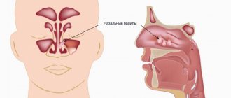
There are 4 types of sinuses in the human skull:
- frontal (in the forehead area);
- maxillary, also known as maxillary (in the area of the cheeks under the eyes);
- ethmoid sinuses, or cells of the ethmoid labyrinth (in the area between the nose and eye);
- wedge-shaped (in the very middle of the skull, behind the eyeballs).
The sinuses are peculiar voids that reduce the weight of the skull, participate in the formation and sonority of the voice, in the process of smell, and also serve as shock absorbers for injuries to the facial skeleton.
They also perform a protective function: when foreign particles and bacteria enter the nasal cavity, the nasal mucosa is irritated, sneezing begins, and the particles are evacuated from the body along with mucus. But if the body’s defenses are weakened, bacteria can enter the sinuses from the nasal passages and cause severe inflammation, which is what we observe during the development of acute manifestations of the disease.
Causes of sinusitis
The disease occurs more often in the cold season in people with weakened immune systems, but this is not the only reason. Inflammation of the maxillary sinus can develop due to:
- complications of a runny nose;
- curvature of the nasal septum;
- damage to the nasopharynx;
- adenoiditis;
- infections and bacteria;
- allergies;
- too dry indoor air;
- work in hazardous industries;
- poor condition of the upper teeth;
- trauma to the mucous membrane of the maxillary sinus.
How does dental sinusitis develop?
The paranasal sinuses (sinus paranasales) consist of a system of several cavities in the near-nasal space.
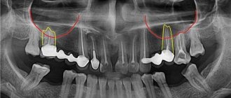
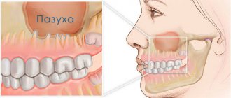
In the case of a cold, less ventilated sinuses are especially susceptible to developing sinusitis. The maxillary sinuses (sinus maxillaris, maxillary) are relatively well ventilated. However, the floor of the maxillary sinus is separated only by a narrow bony plate from the molars of the upper jaw. Due to this anatomy, the development of dental (odontogenic) sinusitis is quite common. The main cause of odontogenic sinusitis may be inflammation that forms in the area of the roots of the teeth and easily spreads to the mucous membrane of the maxillary sinus. Common pathogens include bacteria such as:
- Streptococcus pneumonia - streptococcus;
- Haemophilus influenza – hemophilus influenzae;
- Moraxella catarrhalis is a protobacterium of Moraxella.
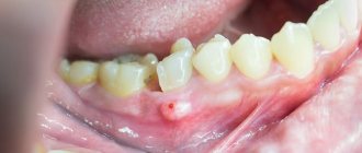
Odontogenic maxillary sinusitis can also form due to tooth extraction (extraction). If one of the maxillary molars is removed, with damage to the bone grafting of the upper jaw, bacteria from the oral cavity can enter the sinus. In this case, they talk about the formation of an unnatural connection between the oral cavity and the paranasal sinus - an oroantral fistula. This trigger factor is considered one of the common causes of “dental” sinusitis.
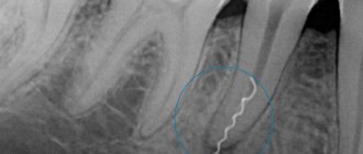
A third dental cause of sinusitis involves inflamed roots that go unnoticed. As a result, cysts are formed that “grow” into the sinus cavity.
Diagnosis of sinusitis
Treatment of sinusitis will be effective if the patient contacts an ENT specialist in time, the specialist will diagnose and prescribe treatment. The examination includes a number of procedures:
- examination of the nasal cavity;
- palpation or feeling the area around the nose and under the eyes;
- clinical blood test;
- X-ray of the paranasal (paranasal) sinuses - according to indications;
- computed tomography of the sinuses.
X-rays can detect the accumulation of mucus in the sinuses and swelling of the mucous membrane of the sinuses. With sinusitis, a darkening will be visible in the picture, since the mucus and swelling of the tissues in the cavities do not allow x-rays to pass through.
CT scan of the paranasal sinuses is necessary to identify polyps, cysts and anatomical changes. The examination is painless and lasts no more than 5 minutes.
If you feel heaviness when you tilt your head down, and the discomfort subsides some time after you raise your head, contact an ENT specialist. Self-medication with folk remedies, randomly selected antibiotics and nasal drops causes serious complications. Sinusitis can become chronic and cause more severe diseases: otitis media, meningitis, brain abscess, orbital phlegmon (inflammation of the tissue around the eyeball).
I needed to take care of my teeth!
The cause of the disease can be not only a poorly placed filling. Odontogenic sinusitis can also occur due to individual structural features of the upper jaw. But in the vast majority of cases, the disease is caused by improper dental care. For example, a patient starts caries, and necrotic decay of the pulp (dental nerve) begins. The products of necrosis cause inflammation in the perihilar tissues, which then develops through the membrane of the maxillary sinus.
Article on the topic
Expert advice: How to prevent sinusitis?
If, due to a long-term inflammatory process of the upper “sixes”, the tooth has to be removed, an additional problem may arise – a fistula. Then the only solution is surgery: a flap is cut out from the side of the oral cavity, which is used to cover the tooth socket. If the infection has managed to lead to the development of sinusitis, then it is necessary to operate on the maxillary sinus.
However, sometimes the tooth can still be saved. In such cases, the doctor carefully moves the gum with special instruments, cuts out a piece of the root, filling the void with artificial bone tissue and treating the surgical site with medications. Naturally, therapy is simultaneously prescribed for the mucous membrane of the maxillary sinus - this is the prerogative of the otolaryngologist. But it should be noted that healing is a fairly long process, at least a month to a month and a half.
Treatment of sinusitis
Depending on the patient’s condition and the course of the disease, the doctor prescribes therapy and selects antibacterial, decongestant and anti-inflammatory drugs. Treatment can be conservative or surgical.
Conservative treatment
For mild sinusitis, in addition to drug treatment, according to indications, nasal cavity lavage according to Proetz, or “cuckoo”, is performed. A special solution is injected into the nose of the patient lying on his back. It passes through the nasal cavity, sinuses, after which it is pumped out with a vacuum pump. “Cuckoo” is carried out in a course of 2 to 10 procedures.
For moderate and severe cases of the disease, antibiotics are prescribed in the form of tablets, solution for injection or inhalation.
Surgical treatment (puncture of the maxillary sinus)
In case of severe purulent sinusitis or lack of effect from conservative therapy, a puncture is used. This allows you to quickly remove pus from the sinus, clean it and treat it with an anti-inflammatory drug. Sometimes several similar manipulations are required. Punctures are performed under local anesthesia.
When sinusitis cannot be treated or is in an advanced stage, surgery is performed under general anesthesia. The period of complete recovery after surgery can take up to two weeks.
Implant into a modified sinus - is it possible?
Some time after tooth extraction, the patient may decide to install an implant. If sinus treatment was not carried out in a timely manner after tooth extraction, it remains to treat sinusitis before sinus lift surgery and implant installation.
It is advisable to do this together with an otolaryngologist.
But at the same time, the patient must understand that during a sinus lift, the altered sinus may rupture because it has an altered structure. And if this does happen, the sinus lift may not take place. Therefore, odontogenic sinusitis still needs to be treated during tooth extraction. And prepare the sinus together with the otolaryngologist.
Prevention of sinusitis
To avoid sinusitis you should:
- spend enough time outdoors;
- strengthen immunity;
- Healthy food;
- take vitamins;
- dress according to the weather;
- seek medical help in a timely manner.
If you have discovered the first symptoms of sinusitis and are looking for a qualified specialist who will diagnose and help you cope with the disease, call the phone number listed on the website or leave a request in the feedback form. Medical staff will answer your questions and conduct all necessary examinations within one business day. Let's take care of your health together!
Treatment of addiction to nasal drops
The first stage is awareness of the problem. It is necessary to accept that an addiction to nasal drops or sprays has formed. Addiction worsens the quality of life and threatens serious complications.
The next step is to seek help from a competent ENT doctor. A specialist will help distinguish medicinal rhinitis from seasonal or allergic rhinitis, decide how serious the complications are, and select adequate treatment to restore nasal breathing. The sooner the patient contacts the doctor, the more successfully vascular function will be restored.
Drug addiction sometimes takes years to form, so you can’t get rid of it in a couple of days. Nasal breathing improves gradually. Medicines, physiotherapy methods and surgical interventions are used. To make you feel better, the doctor prescribes intranasal anti-inflammatory drugs containing synthetic glucocorticosteroids. The drugs improve the condition of the mucous membrane, facilitate the discharge of mucus, and relieve the symptoms of inflammation. The therapy is complemented by physiotherapeutic procedures - laser treatment, medicinal extracts (herbal medicine), the use of ultraviolet irradiation, tube quartz treatment, etc.
Odontogenic sinusitis... Who is to blame and what to do?
An unpleasant odor from the nose, a purulent runny nose, a sore tooth in the upper jaw are constant companions of odontogenic sinusitis, or as it is popularly called “dental sinusitis.” Odontogenic sinusitis occurs in 8% of cases of all sinusitis and is a joint problem of the two specialties of otolaryngology and dentistry.
Anatomical prerequisites:
Anatomically, the roots of the 4th, 5th, 6th, 7th and 8th teeth in the upper jaw are in close relationship with the bottom of the maxillary sinus; sometimes they are separated by a very thin plate, and sometimes the roots of these teeth enter the sinus.
Causes:
- Since the root of the tooth can enter the maxillary sinus, during their treatment, perforation of the bottom of the sinus and, accordingly, ingress of filling material is possible; such sinusitis develops as a chronic type and is often fungal in nature.
- The development of an inflammatory process in the maxillary sinus during the removal of teeth in the upper jaw.
- The inflammatory process in the area of the upper small and large molars is a source of infection and can penetrate into the maxillary sinus and cause inflammation in it.
- Impacted teeth can cause inflammation in the maxillary sinus area.
All this leads to disruption of the ventilation and drainage function of the maxillary sinus. As a result of stagnation, ideal conditions are created for the growth and reproduction of bacteria. If this vicious circle is not broken, irreversible changes in the mucous membrane may occur.
Clinical symptoms:
- Increase in body temperature to 38C.
- Difficulty in nasal breathing.
- Purulent runny nose on one side.
- Exacerbation of tooth inflammation.
- Headache, pain in the sinus projection area.
- Unpleasant, foul odor from the nose.
Diagnostics:
Diagnosis of odontogenic sinusitis in modern conditions does not cause difficulties.
Performing an x-ray of the paranasal sinuses will determine the degree of inflammation in the sinus.
A consultation with a dentist will help identify the problem tooth.
If necessary, a CT scan (computed tomography) of the paranasal sinuses is performed to visualize the bottom of the sinus and the roots of the teeth.
Treatment:
Treatment consists of sanitizing the affected maxillary sinus and restoring ventilation in it, as well as treating the causative tooth.
The work of the ENT doctor in this case is to perform a puncture of the affected sinus. This manipulation is performed after local anesthesia with lidocaine. The wall of the maxillary sinus is punctured in the thinnest place, after which the sinus is washed with an antiseptic solution. Depending on what was obtained in the rinsing waters, this procedure may be repeated. In parallel, antibacterial and anti-inflammatory therapy is prescribed to sanitize foci of infection.
The patient is then sent to the dentist for treatment of the causative tooth.
Complications:
In some cases, when the filling material has been in the maxillary sinus for quite a long time or after tooth extraction a communication has formed between the sinus and the mouth (oroantral fistula), surgical treatment is resorted to.
