Today we will talk about the Black classification of caries, known in dentistry.
This scientist devoted a lot of time to researching this disease and, as a result, systematized the knowledge gained and invented his own gradation of this disease, which became popular among medical practitioners.
The most fundamental is the classification of carious cavities, which was invented by Black in 1896. He identified 6 classes of dental damage from this disease. The purpose of introducing this classification was to standardize the methods of preparation and filling of carious cavities. The filling technique directly depended on the type of caries localization.
The discovery of this system was more than a hundred years ago, therefore it is considered not a complete classifier, since carious lesions of the root system and secondary nature are not taken into account.
Despite this, Black's classification of caries is still widely used by dentists. Over time, the system for ranking damage to this disease was modernized, and an additional 6th class was added to its 5 elements. Let's take a closer look at each class separately!
Classification of caries lesions according to the process
In this direction, there are 3 types of dynamics of the course of this disease: fast, slow and stabilized.
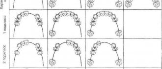
Also, this pathogenic process can be considered by the extent of its localization: caries manifests itself on one tooth, on several elements, or is systemic in nature and affects most of the different teeth in the upper and lower rows.
Classification of caries according to the sequence of occurrence
As in the previous gradation, experts distinguish 3 types of carious lesions.
The first category includes caries that appeared on a tooth for the first time.
The second is a re-injury to a previously filled tooth. In the vast majority of cases, this disease spreads around or under the filling.
The third category includes the so-called recurrent caries lesion. It occurs due to insufficient treatment of the area or a poorly installed filling.
Secondary caries is all new carious lesions that develop near a filling in a previously treated tooth. Secondary caries has all the histological characteristics of a carious lesion.
The reason for its occurrence is a violation of the marginal seal between the filling and the hard tissues of the tooth; microorganisms from the oral cavity penetrate into the resulting gap and optimal conditions are created for the formation of a carious defect along the edge of the filling in the enamel or dentin.
Recurrent caries is the resumption or progression of the pathological process if the carious lesion was not completely removed during previous treatment. Recurrence of caries is often detected under the filling during an X-ray examination or along the edge of the filling.
There are a large number of caries classification systems, almost all of them are repeated. Therefore, for an accurate diagnosis, it is very important for a specialist to correctly determine the depth of the cavity, the nature of the course and the main reason why the carious pathology formed.
The effectiveness of treatment and the absence of relapse processes in the future will depend on the reliability of the diagnosis.
In the world of economic and social competition, it is simply necessary to have a pleasant appearance. Some patients, realizing that beautiful, white, healthy teeth are an element of modern culture, a symbol of youth, health, beauty and success, turned to the dentist with a request to recreate a Hollywood smile, thereby promoting doctors to understand what methods and materials will help them improve the appearance of their patients. Nowadays, therapeutic dentistry, taking into account the importance of aesthetics, is developing a large number of treatment methods and using various types of filling materials.
Knowing the anatomical features of the tooth and the color rendering properties of the material you are working with, you can perform any complex aesthetic restoration.
The purpose of the study is to demonstrate a method for restoring class III cavities (according to Black) with modern materials using minimally invasive preparation techniques.
Material and methods
M. contacted the specialized department of the Department of General and Aesthetic Dentistry of the Moscow State Medical University
23 years old, with complaints of food debris getting and getting stuck between teeth 2.1 and 2.2. When collecting anamnesis, it turned out that the patient did not have an allergic reaction to the drugs and there was no concomitant pathology of the internal organs (cardiovascular system, gastrointestinal tract, endocrine and nervous systems). According to the patient, two years ago she sought help from a dentist with complaints of nightly, prolonged pain from all types of irritants in the area of tooth 2.1. Endodontic treatment was performed. Over the past 2 months, the patient has noted food getting and getting stuck between teeth 2.1. and 2.2.
Upon external examination, the configuration of the face is not changed, regional lymph nodes (submandibular, mental and posterior cervical) are not palpable.
When examining the oral cavity, the mucous membrane is pale pink in color and moderately moist.
When examining tooth 2.1, a carious cavity is noted on the distal contact surface, filled with softened dentin, the filling on the palatal surface is sound, the color is not changed, the marginal fit is not broken (Fig. 1).
Figure 1. Initial view of tooth 2.1 before restoration.
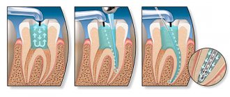
To control the previously performed endodontic treatment, an X-ray examination of tooth 2.1 was carried out, according to which the root canal is obturated to the physiological apex, evenly, in the area of the apical foramen there is an expansion of the periodontal fissure (Fig. 2).
Figure 2. X-ray of tooth 2.1.
Based on the data obtained, a diagnosis was made: tooth 2.1 - condition after complete removal of the pulp, according to ICD-10 - dentin caries K 02.1.
Before starting treatment on the tooth surface 2.1. Soft plaque was removed and the tooth color was determined using the VITA scale. Under infiltration anesthesia Sol. Ultracaini DS forte 4% 0.8 ml formed a class III cavity (according to Black) with an additional platform and fold within the enamel on the palatal surface of tooth 2.1. To control the quality of mechanical treatment of the carious cavity, a caries detector was used (Fig. 3).
Figure 3. Application of a caries detector.
To improve the quality of the restoration, tooth 2.1 was isolated from the oral fluid using an optradam (Fig. 4).
Figure 4. Tooth 2.1 - optradam insulation.
The prepared cavity was treated with a 2% chlorhexidine solution. After adding the VII generation adhesive system, the first layer was a low-modulus light-curing composite material of shade A3, the main volume of the carious cavity was filled with opalescent shade OA3, the last layer was filled with enamel shade A3. Next, the restoration was finished with a brush and polishing paste (Fig. 5).
Figure 5. Finishing of the tooth restoration 2.1.
The final view of the restoration of tooth 2.1 from the palatal and vestibular sides (Fig. 6, 7).
Figure 6. Tooth 2.1, view of the restoration from the palatal surface.
Figure 7. Tooth 2.1, view of the restoration from the vestibular surface.
Thus, this clinical case illustrates that the use of modern composite materials, the correct implementation of hard tissue preparation techniques, and compliance with recommendations for selecting colors taking into account knowledge of tooth anatomy undoubtedly play a key role in recreating an aesthetic restoration.
Topographic classification of caries distribution
In many countries, this classification is most widely used.
It takes into account the depth of the lesion, which is very convenient for the practice of the dentist. There are 4 stages of development of this disease:
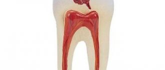
- The appearance of a carious spot. The source of demineralization of the dental element. The process of this harmful phenomenon can last either slowly or quickly, depending on the individual characteristics of the patient’s body.
- Superficial caries is characterized by local damage to the enamel on the tooth.
- Moderate caries manifests itself in damage to the surface layer of dentin.
- Deep caries clings to the peripulpal dentin and affects the tooth right down to the nerve endings.
Differences between chronic caries and acute
Let's take a closer look at the features of the chronic and acute forms of this disease.
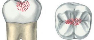
The acute form of caries is characterized by the rapid development of destructive changes in the hard tissues of the tooth, the rapid transition of uncomplicated caries to deep caries.
The affected tissues are soft, slightly pigmented (light yellow, grayish-white), moist, and can be easily removed with an excavator.
Chronic caries is characterized as a slow process (several years).
The spread of the carious process (cavity) is mainly in the planar direction. The altered tissues are hard, pigmented, brown or dark brown in color.
Type of caries according to degree of activity
There are 3 types of caries in this category: compensation, subcompensation and decompensation.
Compensatory caries is characterized by a slow or non-progressive process.
Damage to the surface of the teeth is insignificant and does not cause any discomfort in the patient.
With regular and systematic hygiene procedures, as well as special preventive measures, it is possible to stop the development of the disease at its initial stages.
Subcompensatory caries is characterized by an average rate of progression, at which it can go unnoticed and not cause concern to the patient at all.
Decompensation caries is expressed by intensive development and dynamics of progression, accompanied by such acute pain that it affects both the patient’s ability to work and everyday life.
Because of this, the disease is often called acute caries. It requires immediate treatment procedures, since otherwise the process may spread to third-party teeth with the subsequent addition of pulpitis and periodontitis.
Dr. Greene Vardiman Black
Greene Vardiman Black (1836-1915) is widely recognized as one of the founders of modern dentistry in the United States. Also known as the father of dental surgery. Born near Illinois on August 3, 1836. Parents William and Mary Black. He spent his childhood on a farm and quickly developed an interest in the natural world. At the age of 17 he began to study medicine, in which his older brother helped him. In 1857 he met Dr. JC Speer, who began teaching him practical dentistry.
After the Civil War, in which he served as a scout, he moved to Jacksonville, Illinois. It was here that he began an active career in the growing field of dentistry. He researched many important topics, including the causes of fluorosis and the development of dental caries.
Clinical principles for preparing carious areas
To carry out all the necessary therapeutic manipulations, many specialists rely in their work on Black’s classification of caries.
For any of the above types of tooth damage from caries, it is necessary to carry out full preparation and filling.
The durability of your tooth (or several) depends on the quality of these manipulations.
Experienced dentists, when removing soft carious dentin, can leave its deep pigmented elements to avoid damage to the tooth pulp. After carrying out this work, there should be no affected tissue left on the walls of the cavity.
At all stages of preparation and filling, the dentist sets the main goal - to destroy the carious areas of the affected tooth, disinfect the remaining parts and apply a hermetically sealed structural material that can restore the structure of the tooth and help it fully perform its functions in the future.
Who was Dr. Black and what kind of system did he come up with?
In the dental world, Dr. Black is a cult figure, because it was he who became one of those who initiated the rapid development of dentistry in the United States. Green Vardimar Black was born on a farm in the town of Winchester back in 1836. His first development was a new filling material - gold amalgam, which is still used in dental practice. In 1870, Black introduced the world to his next invention, a foot-powered mechanical drill.
In the dental world, Dr. Black is a cult figure.
Later, the doctor developed an anatomical classification of carious cavities and brought it to a unified terminological standard. It was he who was one of the first to describe the key principles of caries treatment in various clinical cases, and his works to this day remain the main training material for dentists all over the world.