Why is root canal retreatment prescribed and how to avoid it
Treatment of periodontitis does not always end with a solution to the problem, especially when it comes to interaction with an incompetent doctor. Sometimes the dental tissues become inflamed and this causes severe pain. In addition, inflammation is fraught with further complications, which are long and difficult to treat.
The reasons may be different: unprofessional dental care, poor-quality fillings, poorly cleaned root cavities. In any case, the first thing to do when inflammation occurs is to make an appointment with a doctor.
Indications for retreatment
Additional therapy, of course, can be prescribed to you only after a thorough examination. The main indications, which are a strong argument for retreatment, include:
- a fragment of a medical instrument stuck in the canal;
- pain that still does not go away, although a week or more has passed since treatment;
- a canal that is not completely sealed - this is usually indicated by discomfort when eating;
- hidden inflammation shown in the picture;
- infection affecting the periodontal tissues.
Retreatment of tooth canals before prosthetics
In recent years, doctors have been trying to save the patient’s teeth, even if it is the remainder that is hidden inside the gums. There is always the opportunity to place a pin and restore the coronal part. Such a single prosthesis will protect adjacent teeth from the installation of a bridge, which will certainly require preparation of the enamel of adjacent dental units to eliminate the gap in the row. This means that retreatment of the canals saves a person from unwanted interference in healthy tissues.
Stages of root canal retreatment:
- Opening;
- Cleaning to the top using a microscope;
- Disinfection and actual retreatment;
- New filling with modern materials under the control of devices.
The described technique relieves a person of cystic formations, including the possibility of tooth loss.
Features of the procedure for re-treatment of a dental canal
Refilling the canal of a previously treated tooth is a special case in the practice of dentists. Before doing this, it is necessary to remove the old filling, clearing the entire dental canal of the filling material. During such work, the flexible rod may break. This will somewhat complicate the dental treatment process.
Typically, this problem occurs with a very dense filling that is closely adhered to the walls of the canal of the tooth being treated.
Difficulty also arises if it is necessary to refill curved dental canals. When cleaning such channels, you can damage their walls or even break the instrument. Such situations require increased attention from the specialist to ensure high-quality completion of the canal filling. Specialists are required to inform the patient about possible difficulties during treatment before the procedure.
Why is the procedure so effective?
With the right approach, the procedure allows you to save the tooth due to:
- removal of tissue affected by caries;
- no need for anesthesia;
- eliminating the symptoms of diseases that developed under the filling
The use of a microscope in the process significantly improves its quality, since it allows you to correctly assess the features of the anatomical structure and the condition of the root canals. It is worth noting that the cost of canal retreatment with their use is higher, however, the effectiveness of the procedure increases significantly.
Why undergo a dental canal refilling procedure?
There are several reasons why you need to re-treat a dental canal:
- persistence or resumption of pain after filling;
- X-ray shows the presence of inflammation;
- incomplete closure of dental canals.
In addition, complications may result from errors or inaccuracies that arose during treatment. For example, when preparing the canal for treatment, an infection was introduced, access to the base of the canal is impossible, or there are pathological holes in the walls of the tooth or its bottom. In addition, no one is immune from the mistakes of the doctor performing root canal treatment.
When treating a dental canal before filling, the following problems most often occur:
- Dentim gets into the sawmill,
- the middle part of the canal is greatly expanded,
- tool failure,
- the integrity of the root walls is compromised.
When filling a canal, the main problems arise if it is not completely filled, the filling material extends beyond the hole, or there is a longitudinal fracture at the root of the tooth. Medical errors are also possible at this stage - the doctor may incorrectly estimate the length of the canal or not completely clean it. This may contribute to the development of inflammation.
What diseases are they susceptible to?
If problematic root canals are not treated, there are risks of serious consequences. The inflammatory process will not go away on its own, but can spread to nearby tissues and harm the entire body. It can negatively affect the organs located in the upper and lower jaw. There is a danger that a constant source of infection in the oral cavity will provoke the development of the following diseases:
- lymphadenitis;
- cellulitis or abscess of soft facial tissues;
- endocarditis;
- pyelonephritis;
- orthodontic sinusitis;
- sepsis.
How should root canals be filled?
To avoid the need for overtreatment, root fillings must meet certain standards:
- The material should fill the entire cavity of the pulp chamber. If an inexperienced doctor leaves voids during manipulation, space appears for pathogenic microflora to flourish. Or this can happen if the cement is already too old, that is, it was added a long time ago, so it shrinks severely and cracks appear. In conditions of complete tightness, a humid and warm environment, bacteria begin to actively multiply, destroying the tooth, creating pressure in the cavity. Ultimately, infectious agents go beyond the root apex and give rise to the inflammatory process in the periodontium.
- The cement must reach the physiological apex. The root has an anatomical and physiological apex. The first is the visual extreme border of the root, and the second is the end of the canal where it narrows. It is up to this narrowing that the canal must be obturated. If the material does not reach the right place, a “tunnel” remains for the penetration of liquids and microbes, so the cement is washed out faster, and inflammation develops rapidly. Nature does not tolerate emptiness, as Aristotle said. Any “tube” in the body asks it to send more fibers to this place. Ultimately a fibrin sheath is formed around a potentially dangerous hole, which was originally conceived by nature for good - to isolate healthy tissues from the source of possible infection.
In this case, such a restriction plays a cruel joke - it encloses the apex of the tooth and the bacteria in its area in a closed system, where the population of microorganisms continues to grow. As a result, after some time a granuloma appears, it enlarges and becomes a cyst. Both types of formations tend to fester sooner or later, which can lead to root resorption, the development of extensive inflammatory reactions, loosening and even removal of the tooth. If the cement extends beyond the apex, this is another problem that can also be surrounded by a cyst. Some materials can be absorbed by the body on their own, but others require special therapy - a course of electrophoresis procedures. - The filling material should fill not only the main canals, but also their side branches.
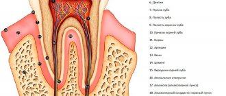
In the root apex area there are often deltoid branches - small tubules extending from the main one. They provide better nutrition and innervation of the pulp normally, but when it becomes infected and removed, they present difficulties for endodontic treatment. It is possible to see and efficiently fill the smallest canals only by using a dental microscope, which not all clinics have. If these areas are left unfilled, the patient may still have pain even after depulpation and the entire complex of therapy, which will require re-treatment. In addition, microscopic infected areas can cause periodontal damage and all its consequences.
Retreatment of tooth canals under a microscope and x-ray
X-rays have long served faithfully in dentists' offices, allowing doctors to see the field of activity and track the steps of root canal treatment in real time. And today doctors are not going to abandon the X-ray examination procedure; this diagnosis will remain in demand for years to come. However, along with objective data, x-rays do not always provide the attending physician with a complete picture of the event.
Disadvantages of X-ray:
- Inaccurate photographs. This is possible when a patient is photographed with treated canals that were filled with non-contrast material. Then the image does not show all the areas that require re-treatment. In addition, x-rays do not show fluid in the canals.
- The procedure is prohibited for pregnant women and small children. Sometimes there is an urgent need to insert a tooth into the canal, but without an x-ray the doctor will have to work almost blindly.
- Irradiation of the patient. Of course, the X-ray machine emits microdoses of gamma rays, but sometimes the dentist is not clear where the canal goes. You have to photograph the tooth root from several sides and make a number of projections. Not wanting to expose the person to unnecessary radiation, the doctor stops the study, but the result may still be unsatisfactory.
X-ray works perfectly in tandem with a microscope. Root canal treatment using magnification greatly simplifies the work of the endodontist. By changing the direction of light and adjusting the sharpness, the doctor distinguishes the direction of even a complex and convoluted canal. Therefore, retreatment takes place as efficiently as possible, right up to the very top.
At the same time, the invasiveness of treatment is significantly reduced. The doctor, looking through a microscope, easily removes nerve tissue from the canals without damaging the walls or touching healthy tissue. It’s no secret: the more natural tissue that was preserved, the more favorable the treatment scenario.
It is incorrect to compare X-rays and microscopes. These are two complementary tools in the hands of an experienced doctor, allowing him to provide quality treatment in the interests of the patient. The microscope will help in the retreatment of tooth canals, in the elimination of carious processes, and is used in excluding other unpleasant diseases of the oral cavity.
If the device is available in the clinic, and doctors do not allow this expensive equipment to sit idle, the clinic clearly demonstrates a high level of treatment. Every person can imagine how much better and more conscientiously a doctor works if the affected area is well illuminated and enlarged. With such careful treatment, the risk of infection and relapse of inflammation is completely eliminated.
At the clinic of Dr. Lopaeva, the task of re-treatment of tooth canals is solved at the highest level. Our highly qualified endodontist dentists cure tooth canals once and for all. During all manipulations, healthy tissues are carefully preserved and strengthened, and damaged tissues are removed without pain or harm to health. Do not hesitate with the procedure; infected tooth tips can provoke serious complications, including the development of tumors.
Dental treatment during pregnancy 2nd trimester
Repeated endodontic treatment of tooth canals for pulp inflammation
Cervical caries on the front teeth: causes and treatment
Treatment of dental caries using the Icon method without preparation
Restoration of tooth enamel, price, drugs
Treat or remove wisdom tooth pulpitis
Retreatment of tooth canals before prosthetics, with granuloma, cyst and aching pain
Contraindications to the procedure
- Even if studies have shown that root canals need to be re-treated, the attending physician may consider the procedure inappropriate or impossible for a number of reasons: if the tooth is clearly not recoverable;
- if periodontal tissue is lost in an amount of about ⅔ of the root length;
- if the general health condition does not allow retreatment (in this case, treatment may be postponed until the condition normalizes or stabilizes);
- in case of unsatisfactory hygienic condition of the oral cavity and the presence of diseases of the mucous membrane in the acute stage (retreatment can also be delayed until the situation improves).
Where to go?
Such a high-tech type of assistance, such as re-treatment of previously filled root canals, can only be offered by a few dental clinics in Moscow. And we are glad that ours is among them. The latest equipment and a team of experienced doctors who know advanced treatment methods help us provide modern services at a high level. The principle of our work: if a tooth can be saved, we need to use all methods to fight for it.
PROCEDURE
After the doctor informs the patient about the upcoming treatment, a step-by-step action plan will be drawn up:
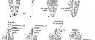
- Opening or unfilling a diseased tooth - complete removal of old filling material from the carious cavity and canals;
- Treatment of inflammation and removal of affected tissue from the apical area, complete disinfection;
- Careful hermetic sealing of all cavities.
Treatment for inflammation depends on its cause. If an x-ray reveals a granuloma or cyst in the area of the root apex, it will be removed after opening the tooth.
The method of removing the inflammatory focus and disinfection after surgery will be chosen by the doctor, in accordance with the financial capabilities of the patient.
WHAT TYPES OF TREATMENT CAN BE OFFERED
Inflamed tissue can be removed using the Doctor smile D5 laser, if the patient is willing to pay the cost of this service. The laser beam completely disinfects the tissues affected by it. Or the operation will be performed using the endosurgical method: removal of the root apex. If this is no longer possible, a hemisection (removal of one of the roots of a multi-rooted tooth) will be performed.
For the purpose of sanitation, the open cavity and roots are treated with disinfectants. In a difficult case: with tortuous and difficult-to-pass canals, they are subjected to intracanal depophoresis, with preparations containing copper and calcium ions.
Then the tooth is carefully filled under the control of radiovisiography (X-ray machine), and only after making sure that there are no cavities left, it is closed with a temporary filling with a medicinal substance. If the tooth can withstand a hermetically sealed filling, no drainage of accumulated fluid is required, and a fistula tract does not appear, then a permanent filling is placed.
RESTORATION OF THE APPEARANCE AND CHEWING FUNCTION OF THE TOOTH
After all therapeutic measures are completed, the crown part of the tooth is restored using an inlay or other methods of microprosthetics. A crown can be placed on a cured tooth and it will last for many years.
If it is too late to see a doctor, the tooth is so damaged that it cannot be restored - it is removed. After removal, it is recommended to immediately proceed with prosthetics, since the absence of even one tooth can lead to further destruction of the rest due to their displacement and loosening.
Our DentaLux-M clinic has European equipment that meets the latest technological advances and qualified medical personnel. In treatment, we use tooth-preserving technologies, because “your” tooth is always better than an implant or removable denture. Prices in our Medical Center are average in the city. You can always make an appointment in advance.

