From this article you will learn
- what is dental granuloma,
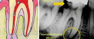
- her appearance in photographs and x-rays,
- How is granuloma on the root of a tooth treated?
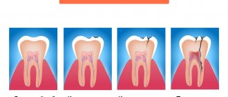
Dental granuloma is an inflammatory formation at the apex of the tooth root, which is a proliferation of granulation tissue. Its development is associated with infection of the root canals and the further development of chronic inflammation at the apex of the tooth root - chronic apical periodontitis. In dentistry, the broader term “apical granuloma” is usually used to refer to this disease, which already implies the location of the source of inflammation directly at the apex of the root (from the word apex - apex).
The granuloma is tightly attached to the root apex, and therefore, when a tooth with a granuloma is removed, the latter usually comes out along with the tooth (Fig. 1-2). It is important to note that granulomas are inflammatory formations only up to 0.5 cm in diameter, but they tend to grow slowly. And if, due to lack of treatment, the granuloma becomes larger than 0.5 cm, then dentists call this inflammatory formation the term “cystogranuloma.” A formation with a size of 1.0 cm is usually called the term “radicular cyst”. Therapy for granulomas is always conservative, but treatment of a cyst may require resection of the tooth root.
What is dental granuloma: photo, diagram
But granuloma differs from cystogranuloma and radicular cyst not only in size. The fact is that a granuloma is not a cavity formation, i.e. inside it there is no cavity filled with pus - like cystogranuloma and radicular cyst. Morphologically, a granuloma is an area of proliferation of inflammatory granulation tissue, surrounded by a fibrous membrane (capsule), due to which the granuloma is tightly attached to the apex of the tooth root.
Outside of periods of exacerbation of the inflammatory process, there is no cavity with pus in the granuloma, i.e. inside the fibrous capsule it is completely filled with granulation tissue. However, during periods of exacerbation, pus forms inside it, the pressure of which on the walls of the capsule from the inside can lead to rupture of the fibrous capsule and the release of pus into the bone tissue and further under the periosteum. The slow growth of granuloma in size leads to its transformation into cystogranuloma (from 0.5 to 1.0 cm), inside of which a cavity is already formed, filled with purulent contents even outside periods of exacerbations.
Treatment of dental granuloma is possible in 98% of cases thanks to the SIEMENS SLA laser
This is confirmed every day by the successful work of the doctors of the Bionic Dentis dental clinic. In the clinical practice of Bionic Dentis, laser treatment is widely used, and now specialists have presented a unique technique - the first in Russia. It was developed in partnership with the German company Sirona Siemens.
Among Russian patients, a disease such as dental granuloma is especially common. The main reason for this is the low level of dental services provided throughout the country. Public clinics still use outdated techniques. With this approach, it is difficult to obtain a positive effect, and the result of improper therapy is the development of dental granuloma.
Causes
The main cause of dental granuloma is infection in the periodontal tissue. More often this happens in cases:
- pulpitis (acute or chronic);
- tooth trauma (cracks - entrance gates for infection);
- inflammation of the soft tissue surrounding the tooth (periodontitis);
- poor quality treatment of pulpitis, dental canals.
Other factors that can trigger the development of the chronic stage of granuloma and cause exacerbation: hypothermia, viral diseases, stress, failure of the immune system.
Tooth granuloma - treatment methods
To treat dental granuloma, various methods are used, which are divided into therapeutic and surgical.
As a rule, the need for surgical intervention arises when the therapeutic course does not produce the desired result.
The list of surgical methods for treating granuloma includes:
- Cystotomy;
- Cystectomy;
- Resection of the apex of the tooth root.
Carrying out operations requires preliminary preparation, and then subsequent rehabilitation and a healing period that lasts a long time. In this case, patients are concerned about pain, discomfort and significant swelling.
However, the main problem of surgical methods is the high likelihood of relapses. In almost 45% of clinical cases, dental granuloma occurs again.
What is the difference between a dental cyst and a granuloma?
Many patients in dental clinics confuse cysts and dental granulomas, while these two pathological processes are significantly different. Seeing the difference is important because the diagnosis affects how the problem is addressed. Thus, treatment of tooth root granuloma is often conservative, but getting rid of the cyst can only be done through surgery - it is excised or removed along with the affected unit. So, how is a granuloma different from a cyst?
- Firstly, by its structure and structure. The first does not have a cavity and is a continuous formation of granulations, while the second is a capsule that is filled with pathological contents;
- Secondly, its size. Cysts are always larger than granulomas: they often reach one centimeter in diameter;
- Thirdly, symptoms. Granuloma is more prone to exacerbation, the development of pain symptoms and exacerbations than a cyst. But if the latter reaches a large size, the unit becomes mobile, which is not observed with granulomas;
- Fourthly, in the condition of the surrounding tissues. With cysts, the mucous membrane does not look inflamed and does not change its color, but the bone tissue, as the process progresses, becomes more pliable and decreases in volume. With granuloma, the mucous membrane reddens and swells, the formation of a fistula is possible, but the bone tissue is almost not affected.
On an x-ray, the cyst has clear contours and boundaries, which cannot be said about the granuloma, which looks like a darkened round shape.
Unique. 100% channel processing. Laser technique
It should be noted that this disease does not occur without a reason. Moreover, he has certain symptoms. You can understand that a patient is developing a granuloma by signs such as the appearance of:
- Abscess on the gum;
- Flux on the gum;
- Feelings of tooth enlargement;
- Pain when eating solid foods;
- Fistula on the gum.
Similar symptoms may also indicate a suppurating dental granuloma. Therefore, the appearance of these disorders requires immediate contact with a dentist. It has been established that purulent lesions quickly spread through the canals and penetrate the bone and other tissues, systematically affecting vital organs - eyes, ears, etc. The result of this is not only severe consequences in the form of a threat to human health, but also to human life.
If you treat this disease in a timely manner, you can get a good result. But the problem is that its symptoms appear only after some time, when it reaches a certain stage. At first, the disease is almost asymptomatic. Many patients may not even realize that they have this dangerous disease. A planned study allows detecting a violation at an early stage. In the panoramic image, its formation is revealed quite clearly. Although the patient may not feel pain or other alarming signals. The only option for detecting the development of the disease is regular medical examination by a dentist.
PREVENTION
Preventive measures must be carried out in a complex manner. They should be aimed at preventing the occurrence of the disease. Preventive actions include the following:
- Constantly maintaining cleanliness of the oral cavity. That is, this is daily, high-quality cleaning and rinsing.
- Treat bleeding gums.
- Scheduled visit (2 times a year) to the dentist.
- Change your toothbrushes regularly to avoid spreading infections in your mouth.
- The slightest pain in a tooth should prompt the patient to immediately go to the hospital. The process cannot be delayed.
- Pay special attention to diseases such as caries, pulpitis and periodontitis, which are common causes of granuloma.
- Use only medicated toothpastes as a preventive measure.
- Regularly rinse your mouth with herbal decoctions.
- Eat food with the maximum content of calcium, trace elements and vitamins.
Treatment not started on time can cause serious complications:
- The appearance of suppuration;
- Due to compression of the nerves, numbness occurs in part of the face;
- Destruction of the jaw leading to a fracture of the lower jaw. The development of the disease leads to fragility and brittleness of the bone. Therefore, a jaw fracture can occur even while chewing solid food;
- Sinusitis formation - this happens in 99% of cases. This disease is also quite severe, accompanied by constant nasal congestion and severe headaches.
Symptoms
A granuloma may not manifest itself for several months or even years until the size of the cystic formation makes itself felt. Symptoms will appear: granuloma of the gums or at the root, the neck of the tooth will show acute pain. This is the first call.
Another sign of a granuloma under a tooth is the ability to feel the lump or ball with your tongue, and feel pain when pressing on it.
The list of symptoms of granuloma at the gum or at the top of the tooth looks like this:
- sharp, dull or aching pain;
- redness and swelling of the gums;
- purulent discharge when pressed;
- darkening of tooth enamel;
- malaise, fever, headache;
- worse from hot food and drinks;
- increased pain during colds (decreased immunity);
- suppuration of granuloma nodules may be accompanied by the development of flux.
Stages of the laser treatment method for dental granuloma
The technique used in clinical practice is particularly complex. Its implementation requires high-tech dental equipment. Moreover, this method shows effectiveness in 98% of cases of treating granuloma.
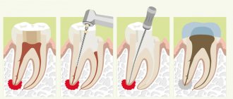
The application of the methodology in practice is carried out in several stages:
- First, a small hole is drilled on the affected tooth. If the front teeth are damaged, then it is done from behind, on the rest - from the side. This is necessary so that the specialist can get to the root canals in the center of the tooth - its pulp.
- Having provided access to the nerve, the specialist removes the contents of the root canal. It may contain an inflamed dental nerve, as well as infected material used to fill the canal, in case of improper treatment.
- The next step is root canal treatment. For this purpose, our clinic’s dentists use a special device, which is a computer-based root canal cleaning system. With the help of special titanium instruments, translational movements - files carefully controlled by microprocessors - clean the root canal. This gives it not only purity, but also evenness.
- Constant monitoring of canal cleaning is performed with a VDW (Germany) apex locator. The main stages of therapy are monitored using a Xios Sirona Siemens radiovisiograph. Such equipment is available in every office of our clinic. Therefore, the research and therapeutic process occurs continuously.
Complete cleansing and unfilling of the root canal requires some time. Usually it is 2-2.5 hours.
After mechanical cleaning and expansion of the canal, the specialist carries out ultrasonic treatment. According to recent research, a large root canal has microscopic branches in which all kinds of bacteria accumulate, causing many dental diseases. Clearing these tiny tubules with files is impossible. For this purpose, our clinic has an ultrasonic root canal treatment system, Sirosonic Sirona, developed by the German company Siemens. It ensures precise penetration of the disinfectant solution into the tubules and pores, sterilizes the root canal and destroys microbes.
After sterilizing the root canal using chemicals and ultrasound, laser treatment can begin. The clinic’s specialists are assisted in this by the Sirolaser laser dental unit. Penetrating through the root canal, the doctor treats the granuloma with a laser. With the use of a unique flexible laser LED, such a procedure becomes possible even in the presence of a curved canal. Therefore, Sirolaser laser equipment is recognized as the best among other analogues.
Having completed the laser treatment, the doctor proceeds to laser sterilization of the root canals and cavities in the bone tissue. This ensures 100 percent disinfection of infected tissues, preventing recurrence of granuloma in the treated tooth.
Following laser sterilization, the drug is installed. For this purpose, a substance is used that stimulates tissue regeneration and disinfection. This drug has a long period of action, 4-6 months.
Then a light-emitting temporary filling or crown, which is extremely durable, is installed on the tooth.
Treatment of dental granuloma is possible in 1 visit. We offer our patients the world's best achievements in dental treatment.
Main types
Depending on the location and certain features, several types of disease are distinguished in dental practice:
- Odontogenic. Localized at the root of the tooth. Usually it does not cause discomfort, there are practically no symptoms. It is recommended to remove the granuloma due to its rapid growth.
- On the gum. This type requires special attention and diagnosis. Often a lump on the gum is of an infectious nature that is not related to the tooth.
- Interroot. In this case, it is located just above the apex of the root. It grows very slowly, but is fraught with a number of consequences. It is recommended to remove the granuloma followed by tooth restoration.
- Apical. According to statistics, it occurs more often than others. It grows at the apex of the root and often leads to purulent inflammation.
3D tooth root canal filling after laser treatment of granuloma
The final stage of therapy and preparation of the tooth for restoration is filling the root canals using permanent materials - alpha gutta-percha. The peculiarity of this biocompatible medicinal drug is that it does not cause an allergic reaction, does not lead to rejection and is an ideal option for obturation of the root canal.
The specialist re-processes the root canal with an antiseptic using an ultrasonic device. Thus, the opening of the entrance for the filling material into the microtubules is achieved.
The alpha gutta-percha pin is first lubricated with a sealant and then inserted into the root canal. Using the BiFill three-dimensional root canal obturation system, gutta-percha is well heated and pressed into the canal. The pressure of a special instrument, a plugger, helps the molten liquid gutta-percha penetrate into the microtubules, instantly solidifying in them. This is how a three-dimensional network or 3d system of a filled tooth canal is formed.
For Russian patients, this method of filling dental canals is new. But in clinics in the USA and European countries, it has become the gold standard when performing obturation of tooth canals. Our specialists also use this technique in 100% of clinical cases. Thanks to this, we are able to prevent recurrences of dental granuloma.
There is also a simpler analogue of filling canals with gutta-percha by using a standard gutta-percha pin. However, its effectiveness is much lower and the results are far from the original technique.
After filling the root canals of a tooth, a filling, inlay or crown is installed on it.
Reasons for the appearance of granuloma on the root of a tooth
In relation to it, this formation can be located in different places, but most often it is found in the area of its apex (or, more simply, the apex). Due to the hidden nature of the development of this pathology, it is not always possible to identify it in a timely manner and begin treatment, and therefore the risk of tooth granuloma degenerating into a cyst and the development of other complications is very high. Experts identify two reasons for its development:
- Spread of the infectious process from the pulp to neighboring tissues with the development of complications of the inflammatory process of the pulp (pulpitis);
- Inflammatory process of tissues around the tooth (periodontitis);
- Traumatic damage to the unit and penetration of an infectious agent;
- Infection during pulp extraction or during endodontic treatment.
As for the factors that initiate acute symptoms of dental granuloma, these include:
- Reduction of internal body temperature to +35°C (hypothermia);
- Past colds;
- Severe stress or physical strain.
Laser treatment of dental granuloma – contact real professionals!
The capital's dental market is distinguished by the variety of services offered. The attention of patients can be attracted by clinics whose specialists are ready to perform laser dental treatment at a price of 3000-5000 rubles. In fact, they use outdated techniques and equipment that are completely unsuitable for providing such services.
Therefore, the choice of a dental clinic should be approached with special responsibility.
Treatment using the original technique is possible only if:
- Fully equipping the clinic with the necessary equipment - a computer system for root canal treatment Gold (wdv), a Sirolaser dental laser, an ultrasound dental system Sirosonic, a device for 3D canal obturation BeeFill (VDW):
- Whether the clinic’s doctors have certificates in training in laser technologies from Sirona;
- Obtaining valid state certificates for doctors in the specialty: therapeutic dentistry.
When you first visit the clinic, you should ask the receptionist to provide all information regarding your treatment. This approach will help you choose a certified clinic where dental granuloma is treated using high-quality original techniques.
Patients with inflammatory diseases of the jaw bone tissue come to our clinic every day. The clinic treats not only patients from Moscow, but also St. Petersburg, as well as other cities of the Russian Federation and CIS countries. The ability to carry out treatment in just 1 visit allows nonresident patients to specifically come to Moscow for treatment for only 1 day.
What causes it to form?
Dentists find it difficult to answer the question of what exactly causes the development of this problem. It has been noted that in 75% of cases, granuloma begins to grow at the site of the extracted tooth. Why is this happening? In the absence of a unit, the void is gradually filled with soft tissue. With insufficient antiseptic treatment, pathogenic microorganisms enter inside, which provoke a kind of cell mutation, as a result of which a nodular formation grows.
The following factors can provoke the growth of a foreign body in the mouth:
- Fungal diseases.
- Tuberculosis, syphilis.
- Inflammation of the pulp.
- Advanced stage of caries.
- Poor quality of dental services.
- Poor oral hygiene.
- Unsuccessful tooth filling.
We were the first in the Russian Federation to master the German method of treating dental granuloma with a laser!
The specialists of the Bionic Dentis clinic were the first in the Russian Federation to master the method of treating dental granulomas with a laser back in 2007. During this time, our dentists and endodontists have gained extensive experience in this area. We can safely say that this is the largest experience in Russia and the CIS countries.
We are ready to demonstrate the results of our successful work with a large collection of images and computed tomography scans of patients before and after treatment.
On this page you can familiarize yourself with the clinical cases of our real patients who were treated by us.
Our statistics: 96% of cases of granuloma are successfully cured using the therapeutic method of laser treatment.
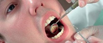
Diagnostics
The doctor will perform an examination of the oral cavity and a series of diagnostic tests, then send you for an x-ray. The presence, size and boundaries of a neoplasm, as well as the shape of periodontitis, can only be determined using an Rg image. An effective component of the West Dental clinic is the presence of a panoramic CBCT Pax-i3D, which can perform OPTG and CT. The photo shows a darkening near the root. There is no cavity in the granuloma; the capsule is surrounded by collapsing tissue.
- Granuloma - up to 0.5 cm;
- Cystogranuloma – from 0.5 to 1 cm.
- Cyst - over 10 mm.
The divisions of the West Dental clinic are equipped with modern equipment, and doctors know innovative methods of treatment, extraction, elimination of granuloma and its consequences after tooth extraction in St. Petersburg. Also, to help our dentists there is a Leica M320 operating microscope.
Treatment of dental granuloma
For more than 10 years, we have collected a large collection of “before” and “after” images of granuloma laser treatment.
ALL IMAGES ARE CLICKABLE
Ozerov Petr Vladimirovich
Chief physician. Dentist, implantologist, orthopedist, surgeon. Laser dentistry specialist
More details
Eremina Anna Arturovna
Dentist therapist
More details
Werner Elena Vladimirovna
Dentist periodontist
More details
What is the danger of granulomatous neoplasms?
Patients who have been diagnosed with dental granuloma under the crown are always interested in what kind of tumor this is. Experienced dentists usually have a photo of a sac-like structure with purulent contents and try to tell their patients why granulomas on the roots of teeth are dangerous. These neoplasms, which develop completely asymptomatic until an advanced stage, threaten the development of the following complications:
- A maxillary abscess is a focus of acute inflammation localized in the soft tissues located in close proximity to the granuloma.
- Jaw osteomyelitis is an infection of the bone structures of the jaw.
- Phlegmon is a purulent lesion of gingival tissue.
In addition to dental complications, patients with granulomatous lesions often experience neural disorders and infectious lesions of internal organs. Their appearance is explained by the active spread of infection throughout the body.
Patient Irina, 37 years old.
A patient came to the dental clinic at her place of residence with complaints of gumboil, pain in the gums, increased body temperature and general malaise. A study was carried out - a panoramic image, which showed a dental granuloma. When contacting various specialists, she was recommended to remove the damaged tooth.
But the patient was not satisfied with this prospect, and she made an appointment with a doctor at the Bionic Dentis clinic. After a thorough examination, a specialist from our clinic suggested laser treatment for granuloma. The patient agreed.
As a result, the treatment was carried out using the Sirolaser laser according to the standard protocol.
After 8 months, the bone tissue was completely restored, all disorders were eliminated. The patient underwent 3D root canal filling and restoration was installed.
By contacting the Bionic Dentis clinic for laser treatment of dental granuloma, you are choosing the world’s best method of therapeutic treatment of this disease.
We achieve complete elimination of granuloma with a 100% guarantee!
You will receive the highest quality treatment that can be found in Moscow, since we work not according to Russian, but according to European standards of endodontics.
As a result of our treatment, the granuloma on the tooth will disappear in 6-12 months, and treatment requires only 1 visit to the clinic. We will save the tooth from removal and save you from the need to install an implant.
Complications
If a pathological process is started, then the first thing to do is remove the unit. But there are also more serious complications:
- Cyst - encapsulation of a formation with pathological bacteria;
- periostitis (flux) - suppuration of the periosteum;
- fistulous tract - formation of purulent discharge through the gingival tissue;
- subcutaneous abscess - occurs when pus enters soft tissue structures.
Suppuration is a dangerous process. If you make lotions from home remedies, many problems will arise. An area of inflammation may lead to resorption of the jaw bone or part of the root system. Suppuration of the formation may be followed by tooth extraction with granuloma, necrosis of surrounding tissues and sepsis. Therefore, it is important to consult a therapist or surgeon in time and find out how to treat dental granuloma. Only highly qualified specialists can carry out diagnostic measures and remove granuloma. West Dental doctors will be happy to help with this.