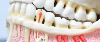
An inflammatory formation affecting the upper part of the tooth root is called granuloma. It is formed during the development of periodontitis. Tooth granuloma is tightly attached to the root, being the initial stage of a granular cyst (cystogranuloma), from which it differs, in fact, only in its slightly smaller size. If the formation is ignored and not treated, it will begin to increase.
Tooth granuloma: symptoms of the disease
Only a doctor can make a diagnosis based on an x-ray, where a darkening will be visible in the root area. Any darkening in the image is a sign of the presence of a cavity, but if a granuloma occurs, it means that periodontitis, which is chronic in nature, begins to develop.
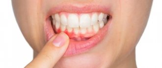
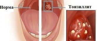
For a long time, the tooth may not bother a person at all. Sometimes pain may occur when biting or when eating hot food: these are the first signs of the development of chronic periodontitis. An exacerbation of the disease is observed during periods of weakening of the body's immunity: then the pain intensifies significantly, which is especially noticeable when biting. During such periods, as a rule, the gums become swollen, and in the area of inflammation it becomes painful to touch.
Types of dental periodontitis
Based on localization, the disease is divided into the following types:
- Regional periodontitis. In this case, tissue damage occurs near the edge of the gums. Often occurs due to injury.
- Apical periodontitis. The tissues are affected near the apical part of the root or at the base. In some cases, it is misdiagnosed as pulpitis.
According to stage, the disease is divided into:
- acute form;
- chronic form.
Acute periodontitis
May be purulent or serous. The first type is especially dangerous, as it can lead to the destruction of periodontal tissue. As a result, the teeth become mobile.
Chronic periodontitis
The chronic form is divided into granulomatous, granulating and fibrous periodontitis. The first two types are the most dangerous, as they are accompanied by pronounced bone resorption.
Why does granuloma appear on the root of a tooth?
At the tops of the roots, such formations can appear for several reasons:
- Poorly performed treatment of pulpitis. If caries is started, the affected cavity gradually becomes quite deep. When microorganisms enter the pulp, its inflammation begins, accompanied by acute pain, which may stop over time, indicating the death of the nerve.
But the development of the disease does not end: bacteria through the root canals extend beyond the boundaries of the affected tooth, as a result of which a focus of inflammation appears near the upper parts of the roots, called periodontitis. The further course of the disease can occur according to several scenarios, one of which is the formation of granuloma.
It is necessary to understand that a tooth with a granuloma does not necessarily have to be affected by caries, since the root can become inflamed near a tooth that has already been treated. If the doctor has not completely removed the tissues affected by caries by placing a filling on top of them, then there is a high risk of developing pulpitis with the subsequent formation of granuloma.
- Filling of root canals performed with violations. A granuloma that appears on the root of a tooth whose canals were filled some time ago indicates the unsatisfactory quality of the procedure performed. Due to inattention, haste or inexperience, the doctor could not fill the canals to the very top. Dentists often refuse to admit their own mistake, because in this case they will have to treat the tooth for free. Such unfortunate specialists can evade until the last minute, pretending that they do not understand the causes of pain, recommending taking a course of antibiotics.
Examples of canine restoration when only the root remains of the tooth
Example 4. The pin in the root of the lower tooth was removed, and the canine itself was restored
In this clinical case, I was restoring the lower canine, and the task was to save the root of the tooth and restore the coronal part of the canine, which was almost destroyed by caries. The anterior wall of the lower canine was made in the form of a filling, and it fell apart from the cutting edge.
As you can see in the photo, an anchor pin was installed inside the tooth - this is a pin that is screwed into the canal to strengthen the tooth. The tooth under the filling and the root of the lower canine were affected by caries. When the carious tissue was removed, only the root and a small part of the wall
:
You and I remember the main topic of our article, which tells us the following - if the tooth root remains, it can be completely restored. And this is not the first time that the unique Cerec technology has come to our aid. Using it and digital scanning, we restored the missing tooth module, which also included the root part:
Next, we restore the lower front tooth using a CEREC inlay and get an excellent result - the lower canine is completely restored:
Example 5. The root of the tooth under the crown of the upper canine has not rotted, and therefore the tooth is 100% restored
In this case, there was a total restoration of the dentition (veneers and crowns) with the replacement of worn-out crowns on the front teeth. One of the upper canines under the crown was seriously affected by caries, but its root was practically healthy - only a small part of the tooth root was damaged, see the following photo:
The missing “crown + root” module and new crowns were manufactured in a laboratory. In general, everything is in a smile, it looks just great:
And the owner of a new beautiful smile herself cannot hide her admiration:
All the main tasks in this total work were successfully solved, but the main thing you should pay attention to is that we managed to save the tooth root here too.
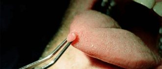
Diagnosis of granuloma on the tooth root
At the early stage of development of the disease, it will not be possible to notice any visual changes. The first signs of pathology become noticeable as the size of the infected area and the amount of pus increase.
To treat the disease with therapeutic methods, it is necessary to examine the affected area in detail: a complete picture of the granuloma must be obtained, which will allow one to detect key signs of the disease that differ from other diseases.
When suppuration occurs, the gums become very red and swollen, and pain appears, which can radiate to the head area in general and the ear in particular. This is the first symptom that should make you wary.
The following methods can be used to make an accurate diagnosis:
- use of classical x-rays;
- the use of radiovisiography or, in simpler terms, computer x-ray.
In the picture, the affected area looks like a dark spot with a clear border in the upper part of the tooth. The size of the spot indicates the following:
- 5-8 mm – the probability of having a dental granuloma is high;
- more than 8 mm – formation in the form of a cyst.
In rare cases, a large granuloma, up to 1.2 cm in size, may occur. Therefore, X-rays may not be enough for diagnosis: it is recommended to perform a biopsy of tissue cells from the area affected by the disease.
In most cases, granuloma is discovered during treatment of other dental diseases: the doctor may pay attention to increased swelling and swelling of the gums. In addition, the bone tissue near the top of the tooth may also bulge.
An increased risk of developing granuloma is observed in patients with crowns and pulpless teeth. Such people are advised to undergo regular examinations in order to promptly identify any changes affecting the gums and teeth.
Material and methods
To identify patterns in the display of CC, including sealed ones, 374 intraoral periapical radiographs, 53 orthopantomograms (OPMG), 47 CT scans before and after endodontic treatment of 194 patients were studied. Under experimental conditions, 127 intraoral periapical radiographs, 26 radiographs using a parallel technique, 32 OPMG, 1434 CBCT sections were performed on skeletonized jaws with teeth.
CBCT data were compared with the results of endodontic treatment, intraoral imaging and OPMG. The influence of the informativeness of radiological techniques on the quality of endodontic treatment was determined, taking into account the time spent on detecting CD of teeth.
The X-ray examination techniques used were applied according to generally accepted rules of survey geometry.
Intraoral radiography was carried out using dental devices Minrey (Finland) and Heliodent DS (Germany), orthopantomography - using Proscan (Finland) and Orthophos XG5 DS Ceph (Germany) devices. Cone beam computed tomographs New Tom 3G (NIM SrI, Italy) and Vatech Pax Reve 3D (E-WOO Technology, Korea) were used. The shooting conditions on these devices complied with the manufacturer's recommendations.
Tooth granuloma: treatment
Usually treatment is only therapeutic in nature, but its strategy may vary depending on whether the canals have been filled before.
- If the canals have not been filled, the tissues affected by caries are drilled out, and the old filling is removed. This is required to carry out high-quality mechanical treatment of the canals, which are expanded and treated with antiseptic agents. If the granuloma is small (formations up to 3 mm are considered such), then the canal is sealed immediately. If the size of the granuloma exceeds 3 mm, the treatment period increases, since it is necessary to put a medicine into the canal, which includes potassium hydroxide - this substance leads to a decrease in the granuloma or even to its complete disappearance. A temporary filling is placed for the period the medication is placed (approximately 2-3 weeks). Then a repeat x-ray is taken, which should clearly show a significant reduction in the inflammatory formation. If the dynamics are obvious, the canals are sealed and a permanent filling is placed.
- If the canals have been filled, then the first stage of treatment is their unsealing. The further sequence of actions is similar to that described in the previous paragraph. The only thing is that if there is a crown on the tooth, it will have to be removed, and after the treatment is completed, it will have to be made and placed again. Some patients do not want to spend money on re-installing a crown: the solution is to perform a root resection operation, when the upper part with the granuloma attached to it is cut off through a small incision in the gum.
Granuloma between tooth roots: treatment with antibiotics
The number of people putting off visiting a doctor until the last minute is large. There are many reasons for this: fear of pain (although modern means of anesthesia make it possible for the patient to feel nothing at all during treatment procedures), lack of free time, and reluctance to part with money. But all these reasons and excuses come to naught when the pain becomes unbearable, becoming chronic. As practice shows, as soon as a person does not sleep for a couple of nights, the issue of the need to visit the dentist is resolved by itself.
However, many people try to cure granuloma on their own using strong antibiotics, which is basically impossible. Antibiotics may be prescribed to relieve inflammation and stop the formation of pus, but nothing more. But the action of antibiotics may not affect pathogenic microorganisms located in the root canals, and it is the infection in their unsealed areas that is the only reason for the formation of granulomas.
If a patient comes to the doctor with a suspicion of granuloma, and the doctor, driven by some of his own considerations, does nothing, recommending taking antibiotics, it is better not to delay time, but to immediately contact another dentist. Some doctors simply do not like to re-treat someone’s poorly treated teeth, since repeating the same operations takes much more time, while others, if the patient comes back again, do not want to admit that they made a mistake, which for them is tantamount to realizing their own lack of competence.
Tooth granuloma: surgical treatment
If therapeutic treatment does not help, then surgery comes into play: the operation is performed in the presence of a destructive process affecting the gums, and anesthesia (both general and local) is used to carry it out.
There are several surgical treatment options.
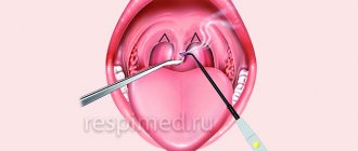
Root resection
Elimination of granuloma occurs in several stages:
- a passage to the top of the tooth opens by peeling off the gum shell;
- the root canals are cleaned and subsequently filled with medicinal substances;
- excision of the granuloma is carried out;
- the area is filled with synthetic fabrics;
- the tooth is filled.
On average, the operation takes about an hour.
Hemisection
If a tooth has many roots, then this procedure is prescribed (if there are complications that prevent the root from being saved).
The treatment procedure includes:
- removal of roots under the crown;
- filling the empty space between the roots with a special dental product;
- installation of a crown;
- monitoring the condition of the tooth using x-rays.
The method is simple, while the functionality of the tooth is preserved. If necessary, some time after the procedure, the patient can resort to prosthetics, provided that the root system remains healthy.
Cystotomy
The method is used when removal of large granulomas is required.
To implement it:
- a channel is created between the affected area and the oral cavity, through which pus absorbed by tampons is removed;
- after cleaning, the cavity is treated with antibacterial agents;
- sutures are placed;
- the cavity, free from suppuration, is filled with bone cells.
Measures to prevent granuloma after an extracted tooth
Preventive measures are aimed at preventing the development of the disease. They include:
- high-quality cleaning of teeth and gums on a daily basis;
- timely treatment of bleeding gums;
- regular visits to the dentist at least once a year (it is better to do this twice);
- replacing toothbrushes (old brushes accumulate bacteria that can provoke the development of the disease);
- contacting the dentist at the slightest pain associated with the gums or teeth;
- treatment of caries, periodontitis and pulpitis - quite often these diseases are the catalyst for the development of granuloma;
- the use of medicated toothpastes and herbal decoctions for rinsing;
- eating foods rich in calcium and other trace elements.
Complications after granuloma at the site of an extracted tooth
Lack of timely treatment and hope that the disease will go away on its own is fraught with serious consequences. Among the most common complications it is worth highlighting:
- development of periodontitis and further formation of a fistula;
- the occurrence of alveolitis is a consequence of the presence of an inflammatory process;
- formation of purulent flux;
- suppuration of the perimaxillary tissue;
- entry of pathogenic bacteria into the lymph nodes, from where they penetrate the cardiac system and internal organs (kidneys, liver and brain);
- development of facial asymmetry;
- the emergence of new foci of infection;
- infection of the body due to the penetration of pathogenic microorganisms into the blood vessels.
Timely removal of granuloma is a guarantee to avoid unpleasant health consequences.
Causes
There are several causes of the disease:
- Traumatic. The disease occurs due to a bruise, sharp biting on a hard object, or a blow to one tooth. Another possible cause is injury during root canal treatment with dental instruments.
- Infectious. It develops due to the penetration of microorganisms into the tissues surrounding the tooth root. Typically, periodontitis occurs due to Staphylococcus aureus, Streptococcus.
- Medication. Occurs due to the effect of potent drugs on the peri-apical tissues of the tooth. Signs of the disease appear within a few hours after exposure to the drug.
The inflammatory process can occur slowly or very quickly. This does not depend on the cause of its occurrence.