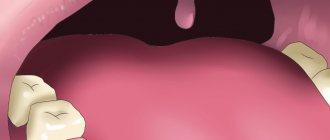
After tooth extraction, patients closely monitor the healing process of the hole. And usually everything goes well - the wound heals, new tissues form, redness in the area of the injured gum disappears. But sometimes frightening formations appear - lumps, hematomas, compactions. Of particular concern is the white growth after tooth extraction. In this case, consulting a doctor is advisable, since the growths can be both safe and requiring treatment.
Signs of alveolitis
The following symptoms indicate alveolitis of the socket:
- Socket pain that continues or renews on the 3-5th day after tooth extraction. In an uncomplicated postoperative period, it resolves in 1-2 days. At the beginning of the disease it is aching and becomes more intense after eating. At a later stage, the pain is constant and cannot be tolerated; painkillers help for a short time. It can radiate into the ear, into the temple on the side of the sore hole.
- The appearance of a bitter taste and putrid odor from the mouth.
- Enlargement and tenderness of the submandibular lymph nodes.
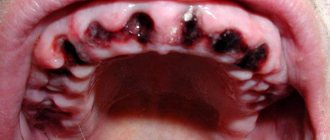
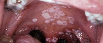
Upon examination, when alveolitis has just begun, the dentist discovers:
- redness of the gums,
- contents in the form of remains of a loose blood clot, food particles, saliva in the wound,
- that the hole is “dry”;
- in the midst of inflammation:
- swollen red mucous membrane along the edges of the hole,
- soreness when touched,
- there is a gray or yellowish coating on the walls.
What to do if bone loss occurs
If bone material comes out after a sinus lift, this should serve as an incentive for prompt action.
- The sooner the diagnosis is made and adequate treatment is started, the more favorable the further prognosis will be.
- If the start of treatment is delayed, pathological adhesions may develop with the implant being fixed in the wrong position.
In any case, a computed tomography scan is performed first . Allows you to determine the degree of “disaster” and not miss the onset of the inflammatory process:
- assess the condition of bone tissue;
- determine the quantity and placement of the remaining material;
- identify its presence or absence in the maxillary sinus.
If bone chips were released only from the oral cavity
Surgery is being performed. Sometimes it is possible to limit oneself only to plastic surgery of the bone material, filling the resulting gaps and re-applying sutures.
If osteomaterial gets into the sinus
Treatment can last for several months and will take place in several stages:
- Removal of a dislocated implant (if installed).
- Elimination of infectious complications - sinusitis, fistula (if present).
- After 1-2 months, in the absence of repeated complications and normal healing, a repeat sinus lift may be performed.
- Implantation is performed 3-6 months after the new bone is formed.
Our Center specializes in solving problems associated with complications in the maxillary sinuses
Patients often come to us after a failed sinus lift. The Center has a full-fledged ENT department to provide surgical care in complex clinical situations. A modern ENT unit includes all the necessary equipment for carrying out operations, including the removal of foreign bodies that provoked inflammation.
Levin Dmitry Valerievich
Founder and Chief Doctor of the Center
Causes of alveolitis
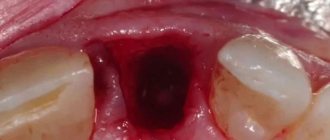
Normally, after a tooth is removed, a clot of blood cells and fibrin, the protein from which a blood clot is built, forms in the socket. It almost completely covers the bottom and walls of the alveoli. The clot serves as a mechanical barrier, biological protection against infection and additional injury to the wound surface. In this case, healing occurs by primary intention. The wound is slowly filled first with loose tissue, then with denser connective tissue, and later with young bone.
But there are situations when the healing process is disrupted.
Inflammation of the socket occurs if:
- The operation was highly traumatic, which reduced the protective capabilities of one’s own tissues;
- The blood clot, as the main defense, is broken or completely absent (“dry socket”):
- prolonged bleeding;
- early destruction of the clot;
- non-compliance with the rules of care (for example, increased rinsing, eating hard or too hot food soon after surgery);
- Tooth extraction was carried out for emergency reasons due to an acute infectious process: periodontitis or periodontitis with complications;
- There are common reasons:
- reduced immunity;
- blood clotting disorder;
- elderly age;
- lack of proper dental hygiene.
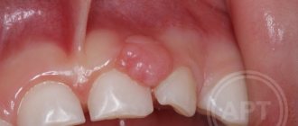
Reasons for the release of osteomaterial
The reasons for the appearance of osteomaterial differ depending on the location of its “loss”.
When appearing in the oral cavity
Bone chips enter the oral cavity as a result of suture divergence, disruption of the location of the bone material or the membrane holding it.
This complication appears in the first 2-7 days, when regeneration processes have not yet begun and the material has not been fixed by connective tissue (a single bone mass has not begun to form).
After at least 10-14 days from the moment of intervention, such a complication is rare - new bone tissue is formed, the granules bind and are not able to come out.
Dehiscence of sutures and displacement of the graft are often combined with the appearance of mobility of the dental implant (if installed) and bleeding.
Most often occurs due to errors by the patients themselves as a result of:
- playing sports (especially running, diving, cycling);
- head injuries;
- visiting the sauna;
- non-compliance with the rules for caring for seams.
Less commonly, the complication is associated with surgeon errors:
- use of low-quality suture material;
- errors in suturing;
- ignoring the use of membranes.
We recommend contacting trusted clinics that do not skimp on materials or doctors’ salaries.
When exiting through the nasal passages or nasopharynx
The appearance of bone material in the nasal passage on the operated side or exit through the nasopharynx is associated with its entry into the maxillary sinus, which communicates with the nasal cavity. This is possible if the sinus lining is damaged during surgery.
It is better to entrust such operations to an experienced doctor. Moreover, not just a dental surgeon, but an oral and maxillofacial surgeon with ENT training.
The situation can also occur with injuries leading to dislocation of a dental implant (in the case of simultaneous implantation and sinus lift). As a result of displacement of the implant, there is a possibility of compromising the integrity of the sinus mucosa.
As a result, bone material leaves the cavity through the nasal passage or nasopharynx. In the worst case, it remains in the sinus, which leads to the development of inflammation and unilateral sinusitis.
Any procedures carried out in the area of the maxillary sinuses must necessarily end with X-ray control. If foreign bodies are detected, removal must be immediate.
Diagnosis and treatment of alveolitis of the socket after tooth extraction
If you have had a tooth removed, but after 2 days the pain in the socket has not gone away, or additional signs of inflammation have appeared, you should not postpone your visit to the doctor. Self-medication can lead to complications such as periostitis, osteomyelitis, and damage to adjacent teeth. The sooner treatment begins, the faster and with less risk of unpleasant consequences the wound will heal.
Even if your tooth was removed in another clinic, our specialists will help you.
Typically, diagnosing alveolitis of the tooth socket is not difficult. A medical history and examination is sufficient. But if necessary, to clarify the depth of the lesion, you may be prescribed additional studies: radiography or radiovisiography.
SM-Dentistry uses only high-quality materials; all instruments are sterilized using powerful sterilizers that eliminate the possibility of infection.
Treatment measures for alveolitis at SM-Dentistry:
- Local anesthesia will relieve acute pain and help you endure treatment.
- Complete cleansing of the wound from parts of the destroyed blood clot, food debris, plaque - a clean wound surface should remain.
- Treatment with disinfectants, antimicrobial and anesthetic agents.
- Application of a special turunda (tampon with medications).
The doctor will give recommendations for further care:
- special baths with antiseptic;
- painkillers;
- gentle diet.
It is possible that treatment of socket alveolitis will need to be supplemented with physiotherapeutic methods.
Preventive measures in our Center
To avoid complications, our Center has developed a number of preventive measures - Uniform quality standards that all specialists follow.
- Ultrasonic formation of access to the maxillary sinus is carried out using a low-traumatic piezo installation NSK VarioSurg. The device does not injure the bone, gently softens it, and when it touches the sinus mucosa it turns off, which prevents its rupture.
- Lift-Control micro-instruments for lifting the sinus lining A coordinated set allows you to lift the mucous membrane as gently as possible, without damaging it.
- The use of barrier membranes is mandatory to prevent material from entering the sinus. First, the membrane is fixed, then bone material is introduced.
- Use of biocompatible bone materials We use bone materials based on inorganic tricalcium phosphate without the risk of allergies and rejection. We do not use donor material from other people, or synthetic substitutes that have not proven their effectiveness and reliability.
- The use of bone growth stimulants (BMP) Morphogenetic protein accelerates the maturation of new bone tissue due to the body's reserve forces. Prevents the risk of material shifting.
- Application of PRF membranes The top layer in the sinus lift sandwich technique is fixed with a PRF membrane made from blood plasma. Due to the high platelet content, it ensures the germination of all layers with blood vessels and the rapid maturation of new bone.
- Postoperative X-ray control All operations in our Center always end with a control CT scan on a high-precision 3D tomograph SIRONA GALILEOS with ENT mode settings. This eliminates the possibility of undetected perforation and penetration of bone material into the sinus.
Sinus lifting in our Center is performed only by maxillofacial surgeons
These are experienced doctors with ENT training and 7 years of work experience, excellent clinical thinking and well-developed manual skills.
Levin Dmitry Valerievich
Founder and Chief Doctor of the Center
Prevention of alveolitis at SM-Dentistry
If the operation is not an emergency, then dentists first treat caries and other inflammatory diseases in the oral cavity, which can become a source of infection.
After the operation, you will receive detailed recommendations for caring for the socket so that the blood clot forms correctly and does not break down prematurely. To do this, you must follow a diet (do not eat hard or hot foods), do not use intensive rinsing and cleaning in the area of the injured hole, and use antiseptic and painkillers.
Sign up for a consultation with a dental surgeon by calling 24/7 in Moscow +7 (495) 777-48-06.
Dentist consultation Treatment of caries
Dentists' recommendations
After surgery, it is important to monitor the condition of the wound and follow the doctor's instructions. The hole filled with blood gradually overgrows and is completely covered with granulation tissue 14-20 days after the operation. The first three days after tooth extraction you cannot:
- rinse the mouth,
- eat solid food
- drink through a straw
- smoking and drinking alcohol,
- use a brush with stiff bristles,
- take a hot bath and go to the sauna.
If possible, you should avoid physical activity and stop taking medications that prevent blood clotting. Compliance with the listed recommendations contributes to the speedy healing of the wound.
Prices
| Name of service | Price, rub.) * |
| Initial consultation with a dentist-therapist | 1,500 rub. |
| Repeated consultation with a dentist-therapist | 800 rub. |
| Treatment of caries (imaging, anesthesia, isolation of the oral cavity, installation of an insulating lining and filling made of composite light-curing imported material) | from 4,350 rub. |
| Treatment of alveolitis with socket revision | 3,000 rub. |
We accept VISA, MASTERCARD, MAESTRO bank cards for payment.
Would you like us to call you back?