A panoramic photograph of teeth or orthopantomogram (OPTG) is a radiographic examination technique that makes it possible to study in detail the anatomical features of both jaws, the condition of soft and bone tissue, dental roots, maxillary sinuses and joints. The study is carried out using equipment with reduced radiation exposure and does not pose any threat to the health of the body. Today we’ll talk about what this is, why a panoramic shot is needed and how often it can be taken.
What is OPTG and why is it performed?
Unlike a standard targeted photograph, an orthopantomogram allows you to obtain a fairly clear and detailed image of both jaws at once with the ability to study not only the condition of the teeth and their root system, but also the jaw bones, paranasal sinuses and other important anatomical structures. To understand what this diagnostic technique is, you first need to answer the questions: what does it do and why is it carried out. So, here's what the x-ray shows:
- deviations in the formation of occlusion - defects in bite, position, shape and size of teeth;
- early stages of caries and hidden foci of infection;
- periodontal pockets;
- neoplasms: cysts, granulomas, fibromas;
- hidden inflammatory processes and the extent of their damage;
- condition of the temporomandibular joint;
- impacted teeth that have not yet erupted - including wisdom teeth;
- condition of the root canals after treatment;
- diseases of the paranasal sinuses, for example, sinusitis.
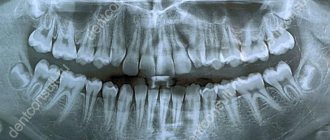
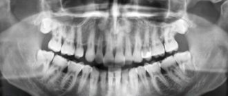
The procedure is mandatory before orthodontic treatment, prosthetics and, of course, implantation. In the latter case, the image gives the doctor the opportunity to carefully examine the condition of the jawbone, select suitable locations for implants, or prescribe a bone tissue augmentation procedure. To understand what the result of OPTG looks like, take a look at the example image below. Important! It should be noted that today, within the framework of one-stage implantation techniques, the preparatory stage involves undergoing a computed tomography scan. This is a more modern X-ray method that allows you to obtain a three-dimensional image of the patient’s jaws with the possibility of layer-by-layer study of tissues, without distorting the actual sizes and shapes of the teeth. OPTG is also performed before treatment or removal of wisdom teeth or “eights”. The image will allow you to identify the exact location and position of the elements, detect hidden carious lesions and make a decision on the need for removal. In general, an orthopantomogram helps to carefully examine the clinical picture, make an accurate diagnosis and prescribe adequate treatment.
Radiology
Radiography has two significant unavoidable disadvantages. Every doctor must understand the limits of radiography and, when taking and analyzing x-rays, take into account the limitations that this diagnostic method cannot overcome.
Firstly, an Rg image, like a regular photograph, is a two-dimensional projection of three-dimensional space. In the scientific literature, the term “3D object” is used to refer to the object being photographed, and “2D views” are used to refer to the resulting images. From a school drawing course or from a university course in engineering graphics, you may remember that in order to fully represent an object, it is necessary to depict three of its projections on paper. A tube of toothpaste will look like a circle in one projection, like a triangle in another, and like a rectangle in a third.
The anatomical shape of teeth and their internal structure are much more complex than a tube. Channels can meander in three-dimensional space, bifurcate and converge. It is impossible to reliably judge the structure of a tooth from one photograph. If there are two channels in the path of the beam, in the projection we will see them as one object. In order to see all the richness of the tooth structure, it is necessary to take at least two photographs from different angles.
The second disadvantage of radiography is the mixing in the resulting images of the projections of all objects located in the path of the X-rays. The image appears dirty. The doctor analyzing the X-ray image must have special knowledge and experience in order to highlight the objects he needs on it, without confusing them with others. The most revealing illustration of this deficiency is a teleradiographic photograph of the skull in profile. Teeth, branches of the lower jaw, nasal sinuses and other objects appear double in the image. It is impossible to reliably determine on which teeth the fillings are installed from this image; for this, other diagnostic measures must be carried out.
Sighted intraoral photographs of individual teeth appear clearer, but this appearance is deceptive. As we have already said, the tooth has a complex structure. Two channels located far from each other, when projected onto a plane, can coincide and look like one object with a fuzzy outline. In this case, it is not possible to determine whether both canals are sealed, and if not, which one.
Taking into account the shortcomings of the method, developers of X-ray technology and radiologists are developing special X-ray techniques. For example, the location of the sensor (sensor) directly in the oral cavity, filming one tooth from different angles, and others. In most cases, this is enough for successful dental treatment.
But there are difficult cases. Patients have problems that patients and doctors do not even always associate with dentistry. For example, a chronic runny nose or frequent sinusitis is a matter of concern for an otorhinolaryngologist (ENT doctor), but the cause of these diseases can be the tips of the roots of teeth or implants sticking out in the maxillary sinus. By the way, if you are really concerned about such problems and treatment by an otolaryngologist does not help, consult with him about sending you for an x-ray of the nasal sinuses.
The task of X-ray examination can be compared to determining the internal structure of an apple. Even in a smooth, beautiful fruit, the core may not be centered. The seeds can be either small or large, immature, eaten by worms. The entrance of a worm into an apple can be almost imperceptible, but the passages it digs are extensive and winding.
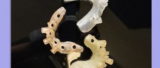
Get a complete picture of the structure of the bones of the facial skeleton, the location of the teeth, the condition of the temporomandibular joints, etc. allows computed tomography (CT) - a unique x-ray research method that involves obtaining a series of images on a special device - a tomograph - and further analysis of these images using a computer. This is a complex type of examination. The patient's head is fixed in the tomograph, after which the device takes a series of images parallel to each other, with an interval of 0.1 millimeters. It's like he's cutting an apple into thin slices. Each snapshot shows the status of the corresponding slice. For the study and diagnosis of the maxillofacial area, a special type of tomography has been developed - cone beam computed tomography (CBCT).
Subsequently, with the help of special computer programs, a complete picture can be assembled from the resulting images - a three-dimensional virtual model. It can be rotated in 3D space, displayed on the screen from different angles. You can examine not only the resulting sections; the assembled model can be cut in any other planes. Moreover, the doctor can draw a curve and obtain an image that is not available with any other examination methods. For example, by placing a curve along the arch of the teeth, you can get an ideal, interference-free panoramic image. By moving the curve from the vestibular (outer, cheek) surface of the teeth to the lingual (inner, palatal or tongue), the doctor can get a complete picture of the condition of the teeth and bones. Computed tomography provides the doctor with maximum information; all the anatomical features of the patient are available to him, defects and anomalies are visible, every “wormhole”.
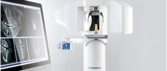
What equipment is used as part of the study?
This is not a new method - by now, OPTG has been actively used in dentistry for decades. And if earlier film cameras were used to obtain a panoramic image, today they have been replaced by more modern digital devices - orthopantomographs. The image is generated in a special computer program and, if necessary, can be printed on film. Images taken using digital equipment are more accurate and detailed, which virtually eliminates the risk of making an incorrect diagnosis. Answering the question about how long a panoramic photograph of teeth is valid, it must be said that the information received will remain relevant for several months - a maximum of six months. If more time has passed since the examination, the procedure must be repeated.
Radiation doses during computed tomography
Did you know that the X-ray machine does not have a built-in dosimeter to monitor the radiation dose the patient actually received during the examination?
How then can we find out the radiation dose received during the study?
To assess radiation risks from diagnostic x-ray examinations, there is a characteristic called Radiation AP (DAP). It is defined as the absorbed dose multiplied by the flow area and is expressed in [Gy cm2]. DAP reflects not only the dose in the radiation field, but also the area of irradiated tissue. Therefore, it may be a better indicator of the overall risk of malignancy than simply absorbed dose information. For example, an X-ray machine has several parameters that can be controlled. These are voltage (kV), current (mA), exposure time and irradiation field area. If the input dose is 1 mGy and the field area is 5x5 cm, then the DAP will be 25 mGy cm2. When the field increases to 10 cm x 10 cm with the same input dose, the DAP increases to 100 mGy cm2. This is 4 times higher than the previous value.
Today, medicine is guided by the ALARA principle (abbr. As Low As Reasonably Achievable) - one of the main criteria formulated in 1954 by the International Commission on Radiological Protection in order to minimize the harmful effects of ionizing radiation. This principle involves keeping doses as low as possible and achievably below the limits established by current regulations). Moreover, screening X-ray examinations are prohibited in dentistry. The doctor prescribes a test for the patient only if the risks of not receiving diagnostic information exceed the risks of radiation exposure. Based on the DAP value, the clinician can either reduce the current and voltage to reduce the flow rate, or select a smaller field of view (FOV) to reduce the irradiation area.
So what are the radiation doses received during the study, and what do they depend on?
Information about the DAP level is stored in DICOM files and can be read in the EzDent-i and Ez3D-i programs. More accurate information is provided by dosimetrists when licensing an x-ray room.
What is the radiation dose for OPTG and TRG images?
When using a Vatech PaX-i3D computed tomograph with a field of view of 12x9 cm, the upper limit of the effective dose will be 60 μSv. For a field of 5x5 cm 40 μSv. The upper limit of the panoramic image will be 10 μSv. Cephalometric image (TRG) – 5 µSv. Let's turn to SanPiN 2.6.1.1192-03 and SanPiN 2.6.1.2523-09 to find out the permissible radiation dose per year.
What is the permissible radiation dose per year?
For group A personnel (working directly with sources), the following dose limits are established: On average, 20 mSv per year for any consecutive 5 years. In this case, the annual radiation exposure should not exceed 50 mSv. For Group B personnel (do not directly work with sources, but are exposed to them): On average 5 mSv per year for any consecutive 5 years. In this case, the annual radiation exposure should not exceed 12.5 mSv.
What does the radiation dose depend on?
From the control parameters of the X-ray installation (current strength, voltage, exposure time) and from the irradiation area. And of course, it depends on what tissues and organs were exposed to this irradiation. Some human organs and tissues are more sensitive to the effects of the study than others: for example, at the same equivalent dose, cancer is more likely to occur in the lungs than in the thyroid gland, and irradiation of the gonads is especially dangerous due to the risk of genetic damage. *(1 mSv =1000 μSv)
Indications and restrictions
The procedure is completely painless, carried out as quickly as possible and without any discomfort for the patient. OPTG is prescribed in the following cases:
- as part of implantation – carried out to study bone tissue, select implant models and places for their implantation. Based on the data obtained, the doctor will be able to draw conclusions about the advisability of building up the jawbone;
- to assess the quality of dental canal treatment and the condition of the roots - before prosthetics;
- before starting orthodontic treatment - before installing braces and other corrective structures;
- before an upcoming operation, complex removal, including wisdom teeth;
- to examine the dental system in children, assess the correctness of its formation and identify possible deviations;
- in order to determine the extent of damage to periodontitis and periodontal disease;
- to identify neoplasms in hard and soft tissues.
This procedure may be required in various clinical cases. X-ray diagnostics is not advisable in the first and last trimester of pregnancy - it is carried out only in the presence of emergency indications and with the permission of a gynecologist.
Features of conducting OPTG for children
We figured out what is visible in panoramic photographs and why they are taken, and found out that this is a completely safe diagnostic method. Now it should be noted that an orthopantomogram for children is carried out taking into account some nuances. A radiation exposure of 55 μSv (1 shot) is completely safe for a child, so a similar procedure is allowed from 5-6 years of age. The examination assumes that the patient must stand still for some time, but for young children this task can be quite difficult . When it comes to small patients, the procedure is carried out in exposure mode - the area of exposure is reduced, and the sensor is adjusted to a trajectory of movement corresponding to the small size of the jaws.
“My son also recently had a panoramic photo taken. He's 10, everything went great. Now there are new technologies everywhere, everything is safe. The procedure took less than a minute, and the doctors assured me that there was nothing wrong with it. How else can you start correcting your bite? The orthodontist prescribed OPTG for us. So far I’ve made the plate, but in a couple of years we’ll get braces. There’s no place here without pictures!”
In pediatric dentistry, OPTG is used to detect the rudiments of permanent teeth, assess the correctness of their formation, to diagnose adentia and identify malocclusions. Most often, this procedure is prescribed by an orthodontist before starting to correct defects in bite and teeth position. The procedure is completely safe for the child
Harm and degree of radiation exposure
Let's start with the fact that radiation exposure is measured in micro- or millisieverts - μSv or mSv, respectively. According to the resolution of the Ministry of Health of the Russian Federation, the permissible radiation dose within the framework of OPTG for an adult patient should not exceed 55 μSv, and for children under 15 years of age - 24 μSv 1 . It should be noted right away that only digital X-ray equipment meets these requirements, while film devices provide a slightly higher load. To determine how many times you can take a panoramic photo, you need to take into account that the maximum permissible radiation dose for an adult patient per year corresponds to 1000 μSv. However, it is still not worthwhile to partake in such procedures, even despite their obvious safety and harmlessness . X-ray irradiation has a negative effect on a weakened body, especially with repeated examinations. The children's body is more susceptible to this kind of influence, therefore it is strictly forbidden to exceed the permissible dosages when examining small patients. When contacting the clinic for OPTG, be sure to clarify what type of equipment is used and what the degree of radiation exposure from one image will be. Some private dentistry purchases cheap, outdated equipment, which can produce 2-3 times the recommended dose. The best option where you can take a panoramic photograph of your teeth is a specialized diagnostic center equipped with modern high-precision equipment.
Features of irradiation during CT and not only
The operation of the tomograph is based on the effect of x-rays. Without knowing all the features of the procedure and equipment, some still believe that dental CT is harmful to health, since the body is exposed to radiation. At the same time, few people remember from a school physics course that a person is exposed to radiation everywhere, even outside the X-ray room, for example:
- Outdoors on clear days. The sun is the largest natural source of radiation.
- At home, being near switched on household appliances: TV;
- refrigerator;
- various kinds of gadgets, etc.
To understand what kind of radiation exposure a person receives during a CT scan of the jaw and whether it is harmful, it is worth learning a little more about the radiation itself. It is measured in sieverts (Sv), millisieverts (mSv) and microsieverts (µSv). According to SanPiN standards, the following are safe for a person undergoing preventive medical examinations:
- 1000 μSv per year for an adult;
- 300-400 µSv per year for children under 15 years of age.
Taking into account the fact that one x-ray examination is performed with a radiation dose from 2-3 μSv (sighting shot) to 16-18 μSv (panoramic shot of the jaw), it is permissible to take 300-500 targeted shots or 60-70 shots of the jaw per year. These figures reflect average data without reference to any specific type of equipment. But they also show that it is not harmful to the body to receive only a few microsieverts for a detailed diagnostic picture.
You might be interested in:
Dental diagnostics
Computed tomography of teeth and jaws
Orthopantomogram of teeth
Digital OPTG - description of the process
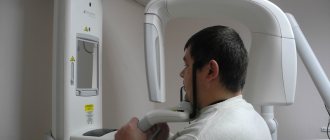
Now let's move on to consider the question of how a panoramic photograph of teeth is taken. To begin with, the patient is asked to remove all metal products from himself so that they do not affect the operation of the equipment and the final result. Next, the patient stands in the place indicated by the specialist, bites down on a special plate that ensures the immobility of the jaws, and after turning on the orthopantomograph, its moving part begins to rotate around the head. The procedure takes literally half a minute - the exact duration will depend on the type and newness of the equipment. Next, the resulting image is formed in digital form, that is, in a special computer program, or on film media (outdated version). A digital photograph can also be printed onto film if necessary.
Information content of the image and reading (deciphering) technique
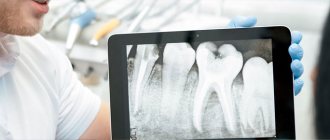
If you undergo an examination at a specialized diagnostic institution, the finished image is usually accompanied by an explanation from a specialist with a list of detected problems. Many people are interested in how to decipher an image on their own, how to read it correctly, and whether it can be understood at all. It should be noted here that in order to correctly explain the results of OPTG, you need to have a good understanding of the anatomy of the maxillofacial apparatus, and without the appropriate education, little can be understood. It is better to entrust this task to an experienced, qualified doctor.
Orthopantomogram during implantation
As part of implant treatment, a panoramic image is taken more than once. First, an X-ray examination is carried out at the preparatory stage - the doctor carefully examines the resulting image, assesses the condition of the bone tissue, selects places for implants, and makes a verdict on the need for bone grafting. If we are talking about one-stage immediate loading protocols, then patients are usually sent for a computed tomography scan of the jaw - a 3D image provides more detailed information about the clinical picture.
3D diagnostics makes it possible to study a more detailed three-dimensional image
You will have to undergo a repeat examination after implantation. In this case, OPTG will give the doctor the opportunity to assess the quality of the operation performed, monitor the process of engraftment of artificial roots, and recognize possible complications in the later stages of their development. Several photographs may be required during the osseointegration period of the implants.
Why do you need a panoramic shot of the jaw?
A panoramic photograph of the teeth gives the dentist the opportunity to assess the condition of all teeth, their relationship to each other, and determine changes in the roots of the teeth and in the bone tissue around the teeth. The visible part of the tooth makes up less than 50% of the entire tooth. Your dentist can only assess what is happening inside the gums and bones using x-rays.
Panoramic dental x-rays help dentists of all specialties make an accurate diagnosis, develop the correct treatment plan and monitor the outcome of treatment.
A panoramic photograph (orthopantomogram) is necessary in almost all areas of dentistry. Digital orthopantomography also provides the opportunity for a detailed examination of individual areas of the jaw for a more accurate diagnosis. Without X-rays, it is impossible to imagine diagnosing caries under crowns and fillings, as well as in hard-to-reach places, root canal treatment, dental prosthetics, dental implantation, periodontal disease treatment, bite correction, and wisdom teeth removal. The orthopantomogram is used in both adult and pediatric dentistry.
More information about the use of panoramic photographs of teeth for diagnostics in dentistry can be found here.
What are the advantages and disadvantages of OPTG
An orthopantogram can significantly improve the quality of diagnosis, and today dental treatment is practically not carried out without radiography, except for minor problems. Among the undeniable advantages of the technique, experts highlight the following points:
- speed of the procedure - literally in a couple of minutes the image is ready for study;
- modern equipment makes it possible to adjust the height and trajectory of the emitter, which made diagnostics possible for children and wheelchair users;
- minimal radiation exposure and absolute safety for the body;
- high quality and accuracy of the obtained images;
- the ability to zoom in on the area under study and study it in detail on a computer monitor.
With an orthopantomogram, minimal radiation exposure is used. If we talk about the disadvantages, the main disadvantage of OPTG is obtaining a flat two-dimensional image. As a result, the image may slightly distort the dimensions of teeth, root canals and bone tissue. Such deviations can vary from 15 to 30%. To obtain a more accurate image, it is better to resort to computed tomography.
What is the advantage of digital panoramic dental photography?
- Reduced exposure time, since the digital camera takes less time to create an image. This results in a reduction in radiation exposure to the patient by up to 90%.
- There is no need to develop films, which ensures fast image acquisition, eliminates the need to work with chemicals, and also eliminates the need for repeated x-rays in case of poor-quality development.
- It becomes possible to further examine the resulting images by enlarging individual areas using software.
- Possibility of convenient archiving and quick access to the database with images.
- Immediate transfer to the attending physician located outside the clinic.
- Convenient storage in the form of files.
Approximate prices
How much a panoramic dental photograph will cost directly depends on whether the study will be carried out using film or digital equipment - in the latter case, the cost will be an order of magnitude higher. Approximate prices are given below:
- a photograph on a film camera – about 700 rubles;
- digital photo - from 1000 rubles and 150-200 rubles for printing on film.
We have already mentioned above that today, for a more detailed layer-by-layer study of the tissues of the jaw apparatus, computed tomography (CT) is performed. Essentially, this is the same panoramic photo, but much more detailed and in 3D format. An important advantage of this technique is the accurate transmission of the sizes and shapes of all elements of the dental system.